4791
Determining the Relationship between DTI and MR Elastography Metrics in Highly Anisotropic White Matter Structures at 7T1Mechanical Engineering, University of Washington, Seattle, WA, United States, 2Biomedical Engineering and Imaging Institute, Icahn School of Medicine at Mount Sinai, New York City, NY, United States, 3Dartmouth College, Hanover, NH, United States, 4Icahn School of Medicine at Mount Sinai, New York City, NY, United States
Synopsis
Keywords: Elastography, Brain
Motivation: Changes in the relationship between MRE and DTI metrics in small white matter structures could indicate alterations in brain microstructure due to white matter damage.
Goal(s): This study aims to determine correlations between metrics measured by MRE and DTI at 7T in the healthy human brain.
Approach: MRE and DTI acquisitions were performed on 14 young, healthy volunteers at 7T, and Shear Stiffness, Damping Raio, FA and RD were calculated for each person.
Results: Significant correlations were found in small, highly anisotropic, brain regions between Shear Stiffness or Damping Raio and FA or RD.
Impact: The high resolutions achieved at 7T for both MRE and DTI allow us to investigate microstructural relationships in small, highly-anisotropic, brain regions. Changes in these metrics or relationships between these metrics could indicate alterations in microstructure integrity, suggesting potential damage.
Introduction
White matter microstructure has been known to change due to inflammation, demyelination, and neurological diseases and disorders. The most common imaging technique for investigating white matter microstructure integrity is diffusion tensor imaging (DTI), which measures restricted diffusion of water in tissue1. Leveraging ultra-high field 7 Tesla (7T) MRI, with increased signal-to-noise ratio and improved soft tissue contrast allows us to accurately map tissue microstructure. Previous studies at 3T have found correlations between MR Elastography (MRE) and DTI metrics in small white matter structures2,3, but low imaging resolution of the limited specificity small regions due to partial volume effects. In this study, we investigate the relationship between Shear Stiffness or Damping Raio as measured by MRE and Fractional Anisotropy (FA) or Radial Diffusivity (RD) as measured by DTI, both at high resolution at 7T in the human brain.Methods
Full brain coverage MRE (using a custom SE-2D-EPI-based sequence4) was performed on 14 healthy human subjects (Avg. age 27±3 years) at 1.1mm isotropic resolution and 50Hz vibration frequency5, using a 32-channel head coil (Nova Medical) on a 7T Siemens Magnetom MRI scanner (TR/slice=140ms, TE=65ms, GRAPPA=3, Partial Fourier 7/8). Raw data were collected for each of these scanning sessions, and images were reconstructed post-hoc using Gadgetron6 to reduce the occurrence of phase singularities often found in standard reconstructions of this type. Images were denoised using an MP-PCA algorithm7 and unwrapped using Segue 4D unwrapping8. The resulting unwrapped displacement data, were used to calculate Shear Stiffness and Damping Ratio using an iterative nonlinear viscoelastic inversion of the time-harmonic Navier’s equation9.We also performed a high-angular-resolved (HARDI) diffusion-weighted imaging sequence (dMRI) that allows us to acquire brain images with 1.05 mm isotropic resolution (b=1500 s/mm2, reversed-phase encoding in anteroposterior and posteroanterior directions for paired acquisition in 68 directions) with a total acquisition time of 20 minutes. The dMRI pre-processing was performed using the human connectome project (HCP) pipelines, adjusted to account for significant eddy currents. The pre-processing pipeline included skull-stripping, eddy current correction using FMRIB Software Library (FSL) (www.fmrib.ox.ac.uk/fsl), correction of gradient non-linearities using the HCP, B0-field inhomogeneity correction, and co-registration to the structural images from the Freesurfer pipeline10. This allowed estimation of whole brain maps of RD and FA.
Segmentation of individual structures in the white matter, specifically the body, genu, and splenium of the corpus callosum, the anterior, posterior, and superior corona radiata, and the superior longitudinal fasciculus, was performed using the ICBM-DTI-81 white matter atlas and parcellation map11. Registration of the ICBM-152 template to the DTI and MRE image spaces was performed using SPM1212 and the same transformation was applied to the masks generated by the white matter atlas. Region-wise correlations between Shear Stiffness or Damping Ratio to FA or RD were performed to determine the relationship between MRE metrics and DTI metrics of these highly anisotropic brain regions.
Results
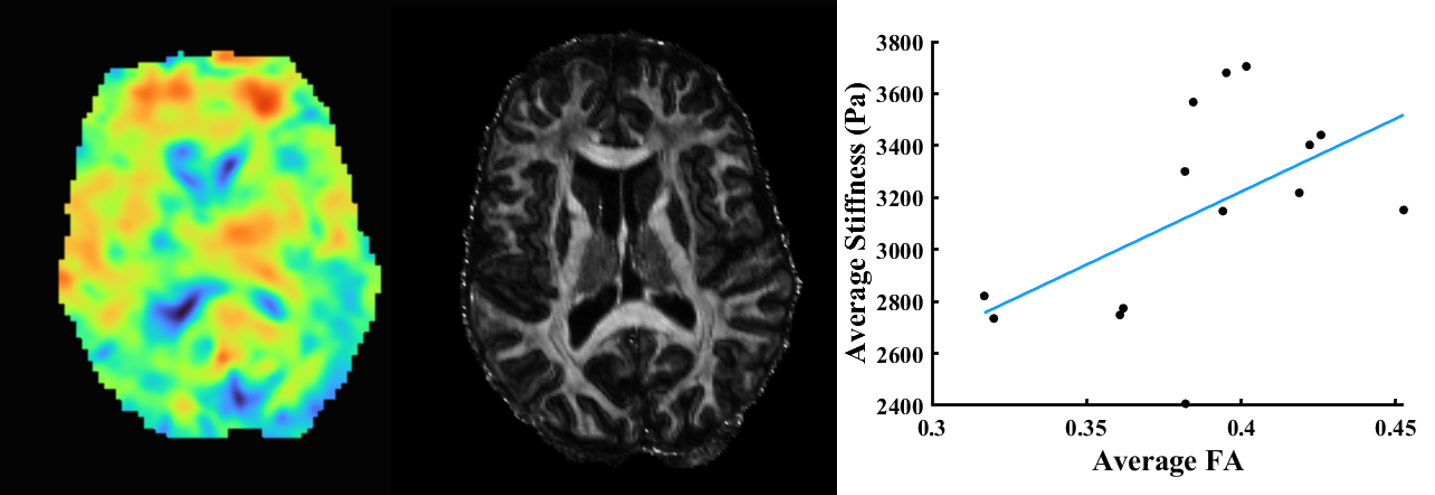
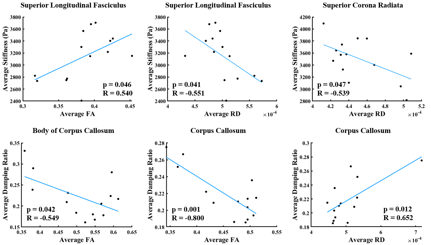
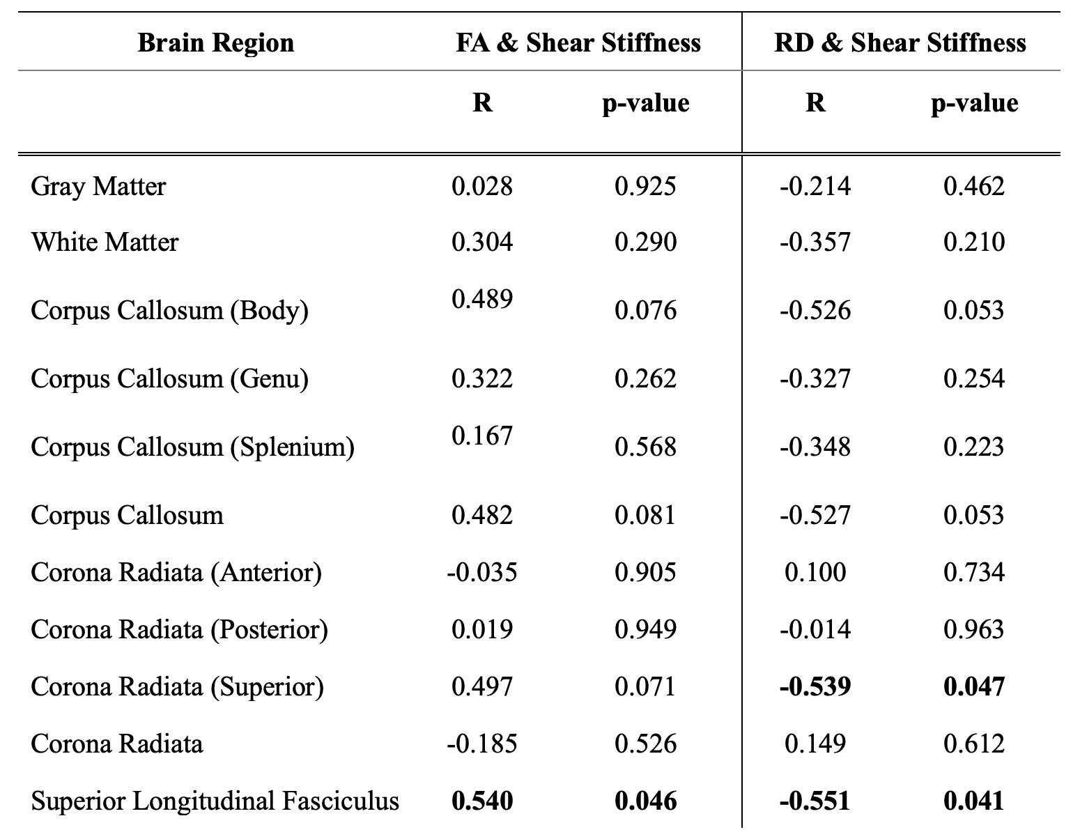
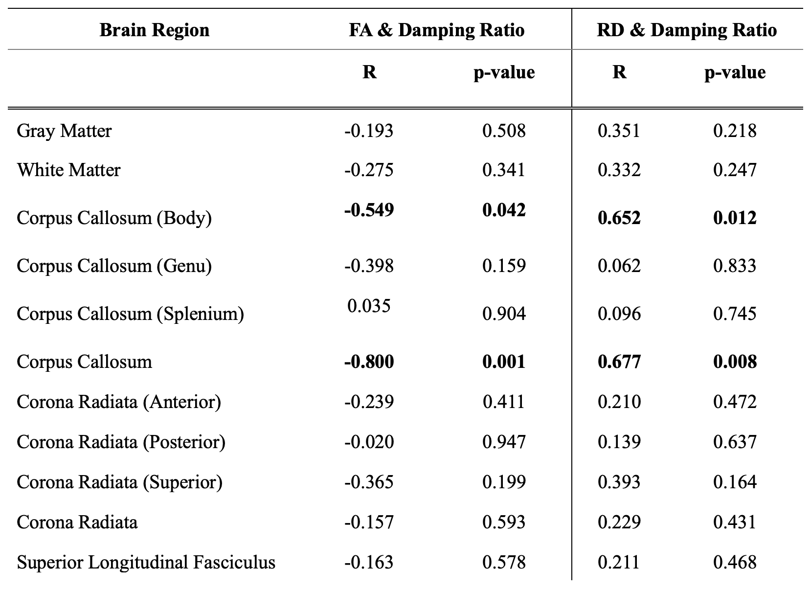
We found a significant positive correlation between FA and Shear Stiffness in the superior longitudinal fasciculus, with trending correlations in the body of the corpus callosum, the full corpus callosum, and the superior corona radiata. We also found significant negative correlations between FA and Damping ratio in the body of the corpus callosum and the full corpus callosum. We found significant negative correlations between RD and Shear Stiffness in the superior corona radiata and the superior longitudinal fasiculus, as well as significant positive correlations between RD and damping ratio in the body of the corpus callosum and the full corpus callosum.Discussion
Our correlation between FA and MRE metrics implies greater stiffness and lower energy dissipation across the volume in small regions where the uniform directionality of the white matter fibers is high. This indicates the need for anisotropic viscoelastic inversion specifically in these areas of the brain. Alterations in the correlation between metrics in these areas may be indicative of damage to the white matter microstructure. While it is possible that the correlation between RD and MRE metrics are related to axon density, this remains under investigation.Conclusion
While correlations between the DTI metrics of FA and RD, and MRE metrics of Shear Stiffness and Damping Ratio are not apparent in larger structures, the high resolution achieved at 7T for both MRE and DTI allows us to investigate these relationships in small, highly anisotropic, brain regions. Changes in these metrics or the relationships between these metrics could signal changes in microstructure indicative of damage to white matter microstructure.Acknowledgements
The authors would like to acknowledge Dr. Jelle Veraart for supplying the MP-PCA denoising algorithm. The authors would also like to acknowledge the support from NSF CMMI 1953323 and NIH funding R21AG071179.References
1. O’Donnell, L. J., & Westin, C.-F. (2011). An Introduction to Diffusion Tensor Image Analysis. Neurosurgery Clinics of North America, 22(2), 185-196. https://doi.org/10.1016/j.nec.2010.12.004
2. Curtis L. Johnson, et al., (2013). Local mechanical properties of white matter structures in the human brain, NeuroImage, 79, 145-152.
3. Aaron T. Anderson, et al., (2016). Observation of direction-dependent mechanical properties in the human brain with multi-excitation MR elastography, Journal of the Mechanical Behavior of Biomedical Materials, 59, 538-546.
4. Triolo, E. et al. Development and validation of an ultra-high field compatible MR elastography actuator. in Summer Biomechanics, Bioengineering and Biotransport Conference SB3C2021-325 (2021).
5. Triolo, E. R. et al. Design, Construction, and Implementation of a Magnetic Resonance Elastography Actuator for Research Purposes. Curr. Protoc. 2, 1–26 (2022).
6. Hansen, M. S. & Sørensen, T. S. (2013). Gadgetron: An open source framework for medical image reconstruction. Magn Reson Med 69, 1768–1776
7. Veraart, J. et al. (2016). Denoising of diffusion MRI using random matrix theory. Neuroimage 142, 394–406
8. Karsa, A. & Shmueli, K. (2019). SEGUE: A Speedy rEgion-Growing Algorithm for Unwrapping Estimated Phase. IEEE Trans. Med. Imaging 38, 1347–1357
9. McGarry MD, Van Houten EE, Johnson CL, Georgiadis JG, Sutton BP, Weaver JB, Paulsen KD. (2012). Multiresolution MR elastography using nonlinear inversion. Med Phys. 39(10):6388-96.
10. Fischl, B., Liu, A., & Dale, A. M. (2001). Automated manifold surgery: constructing geometrically accurate and topologically correct models of the human cerebral cortex. IEEE transactions on medical imaging, 20(1), 70-80.
11. Susumu Mori, et al., (2008). Stereotaxic white matter atlas based on diffusion tensor imaging in an ICBM template, NeuroImage, 40(2), 570-582.
Figures