4790
Regional Correlation of Stiffness and Perfusion in the Human Brain at 7T MRI through MR Elastography and Arterial Spin Labeling Techniques1Mechanical Engineering, University of Washington, Seattle, WA, United States
Synopsis
Keywords: Elastography, Elastography, Arterial Spin Labeling
Motivation: We are motivated to understand the impact of blood flow on the mechanical properties of brain tissue for applications in neurodegenerative pathophysiology.
Goal(s): Our goal was to establish a novel postprocessing framework for correlation of structural and functional properties characterized by MR elastography (MRE) and pulsed arterial spin labeling (PASL).
Approach: We obtained MRE and PASL in 8 healthy controls, segmented the brain, and conducted regional correlation analyses of elastograms and perfusion maps.
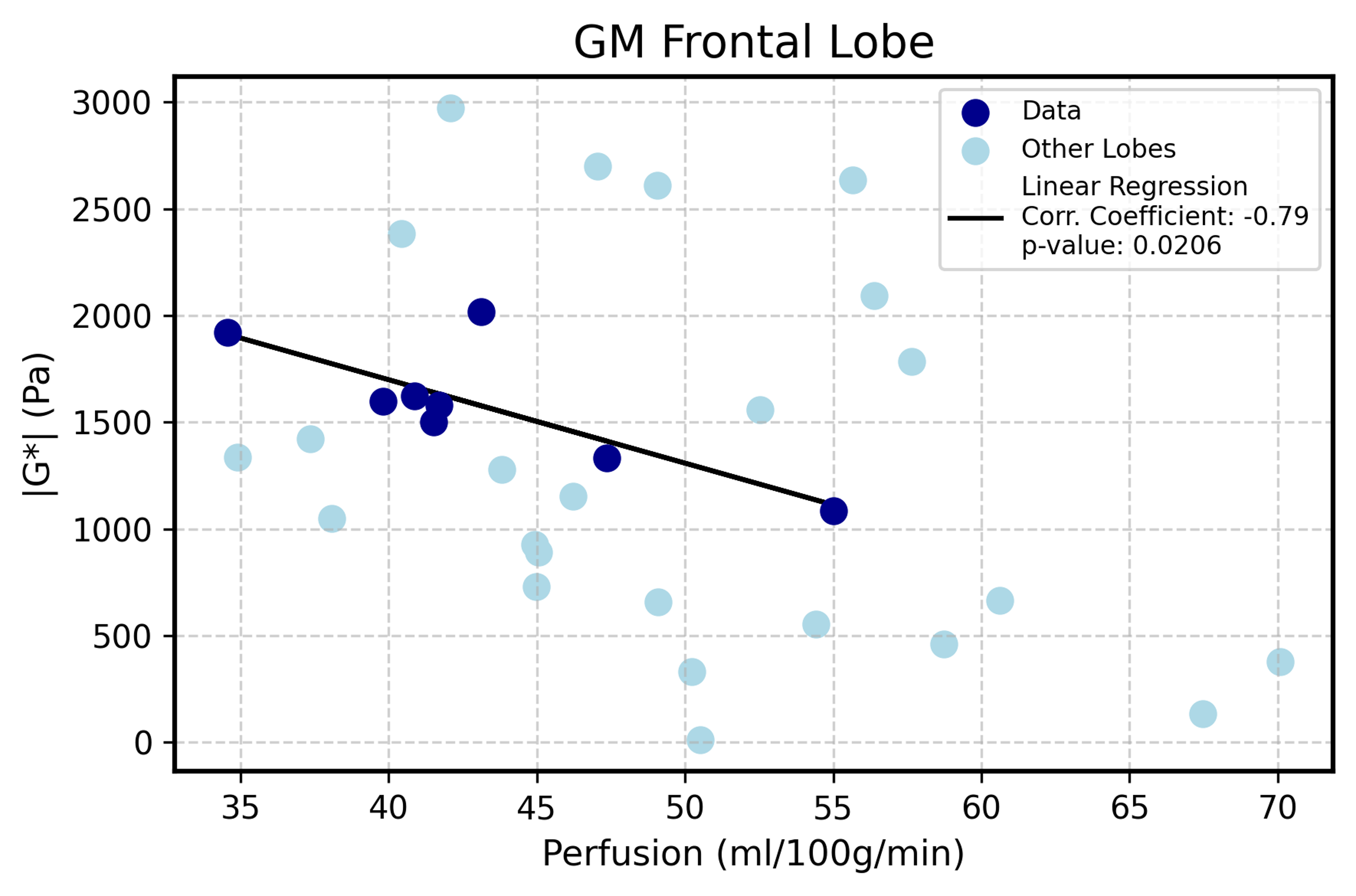
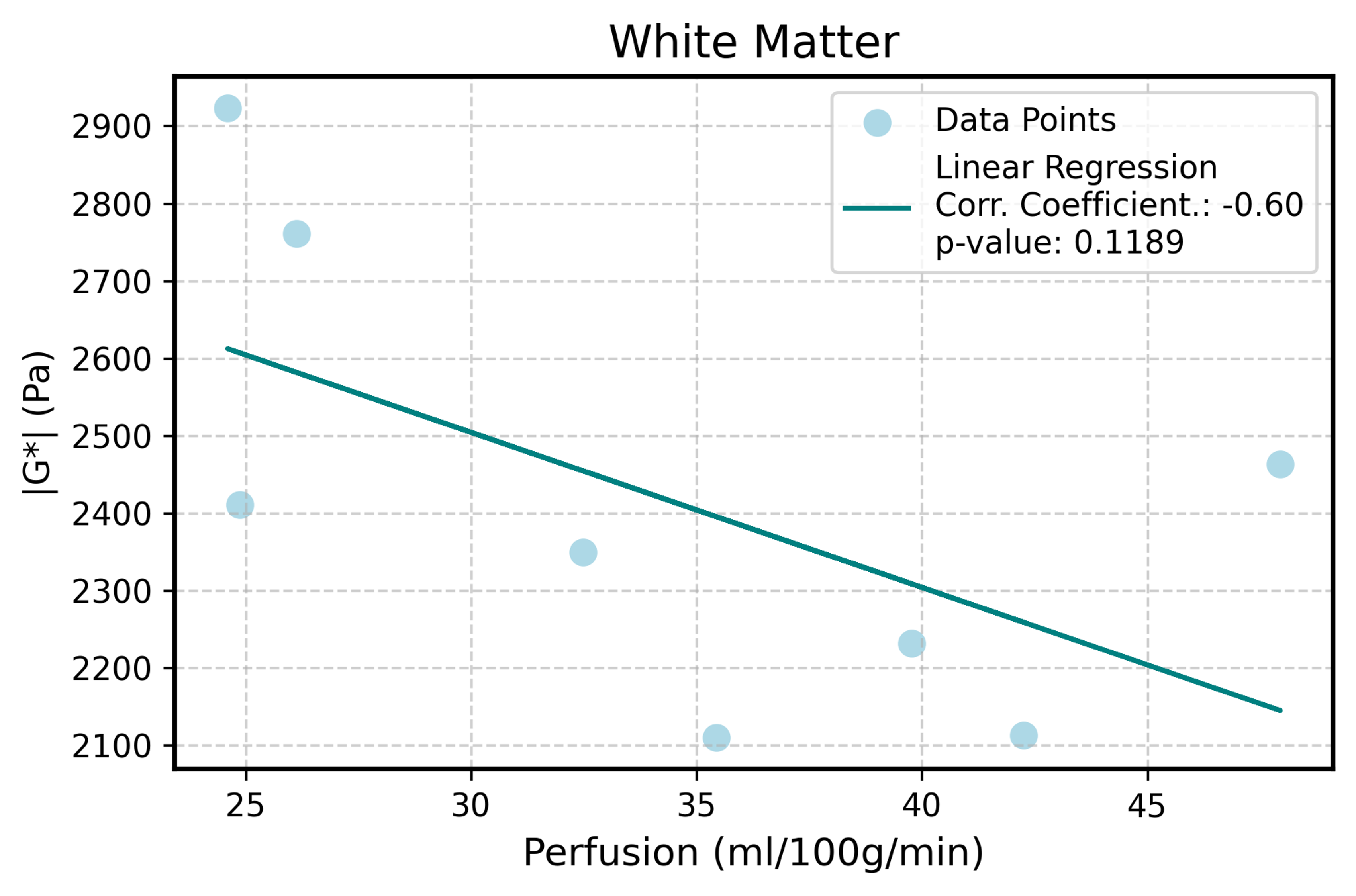
Results: After successful data processing and validation, we found significant inverse correlations in the cortical gray matter, some cortex regions, as well as a similar nonsignificant trend in other regions.
Impact: These study results, which show a perfusion–stiffness relationship in some brain regions, point to an underlying biological mechanism relating vasculature and viscoelastic properties; however, this research direction needs further investigation, more subjects, and improved ASL techniques to strengthen regional analysis.
Introduction
The mechanical properties of the brain give us insight into disease states and open avenues for new methods of medical diagnosis1. It is known that brain tissue gets softer as we age2, but it is unknown whether and how tissue property changes are related to the branched cerebral vascular system in health and disease. Research suggests that changes in perfusion may be correlated with cognitive deficits in mild cognitive impairment3, amyloid-ꞵ deposits in Alzheimer’s disease (AD)4, and even diseased liver tissue5. However, there is limited research that investigates the relationship between perfusion and tissue stiffness in the healthy brain. While It is unknown how the cascade of AD-associated events contributes to its pathogenesis, amyloid-deposits, tau tangles, tissue softening, hypoperfusion, and metabolism changes are disease correlates. It has been shown in one preliminary study that perfusion, stiffness, and flux rate are connected in the brain, due to higher intravascular pressure that is present in small vessels, coupled with the constriction of vessels in non-compliant tissue6. This suggests that perfusion, an indicator of cell metabolic activity and blood volume, will have an impact on the measurable mechanical properties of brain tissue and can be used as a biomarker of underlying pathology. The advanced neuroimaging tools of MR elastography (MRE) and arterial spin labeling (ASL) can be utilized to quantify brain stiffness and perfusion non-invasively by voxel. First, it is necessary to develop a modular processing pipeline for the correlation of stiffness and perfusion in a healthy cohort, quantified from MRE and ASL data, to establish a baseline understanding of these two metrics for future research in Alzheimer’s pathophysiology.Methods
In this study, we obtained pulsed ASL and MRE data from 8 healthy volunteers aged 20-35 on a Siemens Magnetom 7 Tesla scanner with a 32-channel head coil. The MRE sequence was an echo-planar spin-echo 2D pulse sequence with 3D motion-encoding gradients (TE=70ms, TR=5600ms, GRAPPA=3, 1.1mm isotropic resolution)7, and a custom pneumatic actuator applied vibrations at 50Hz8. The MRE phase magnitude images were masked using SPM129, denoised using a MP-PCA algorithm10 and unwrapped using Segue Phase Unwrapping11. The resulting unwrapped displacement data was used to calculate the magnitude of the complex shear modulus (|G*|) using an iterative nonlinear viscoelastic inversion of the time-harmonic Navier’s equation12. Also acquired at 7T, a PASL sequence was used with EPI readout (TE = 39ms, TR = 5000ms, 25 repeats, 3.5mm isotropic resolution). Arterial spins were labeled by a 10cm inversion slab proximal to the image slices, with the labeling method Q2TIPS13. Subtraction, Bayesian Inference, inversion of the kinetic model of label inflow, and equilibrium magnetization calculations from a proton-density (M0) image were used to acquire quantified perfusion in ml/100g tissue/min. FreeSurfer14 segmentation was used with a custom MATLAB script to calculate the correlation coefficient of stiffness and perfusion in gray and white matter regions. Only whole brain white matter was analyzed due to its low SNR resulting from high arterial transit time and relatively low perfusion15. During analysis, images were visually checked and regionally evaluated based on mean, standard deviation, and voxel number to determine inconsistencies. After this process, no subjects were removed as outliers.Results and Discussion
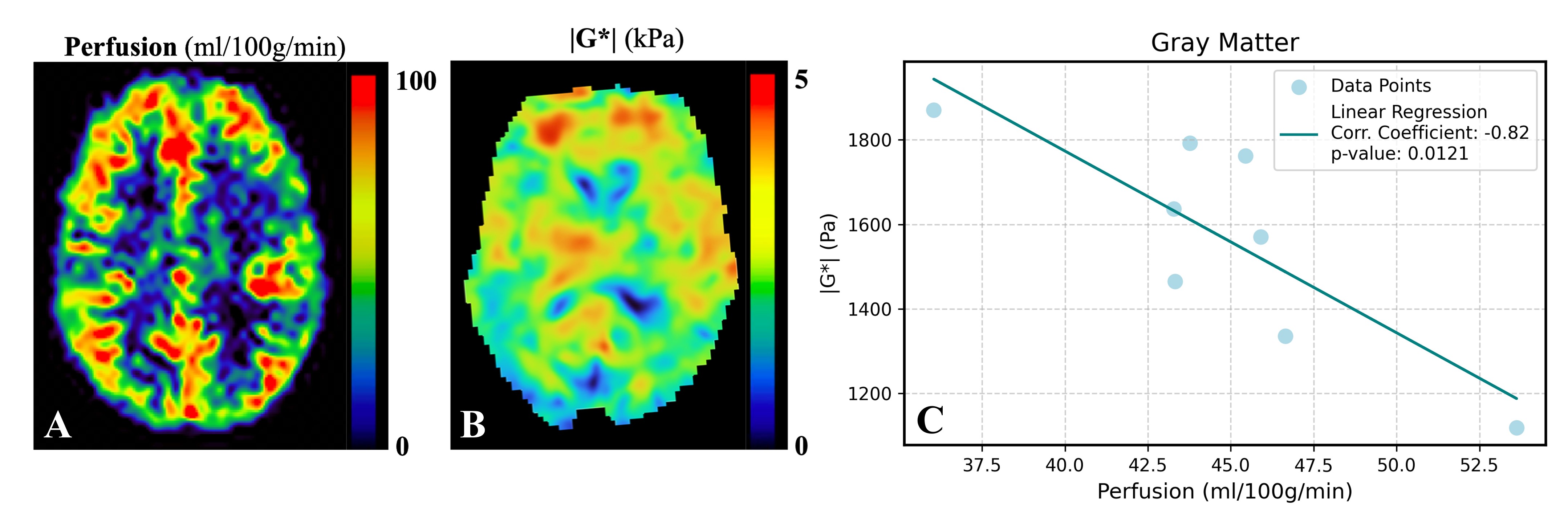
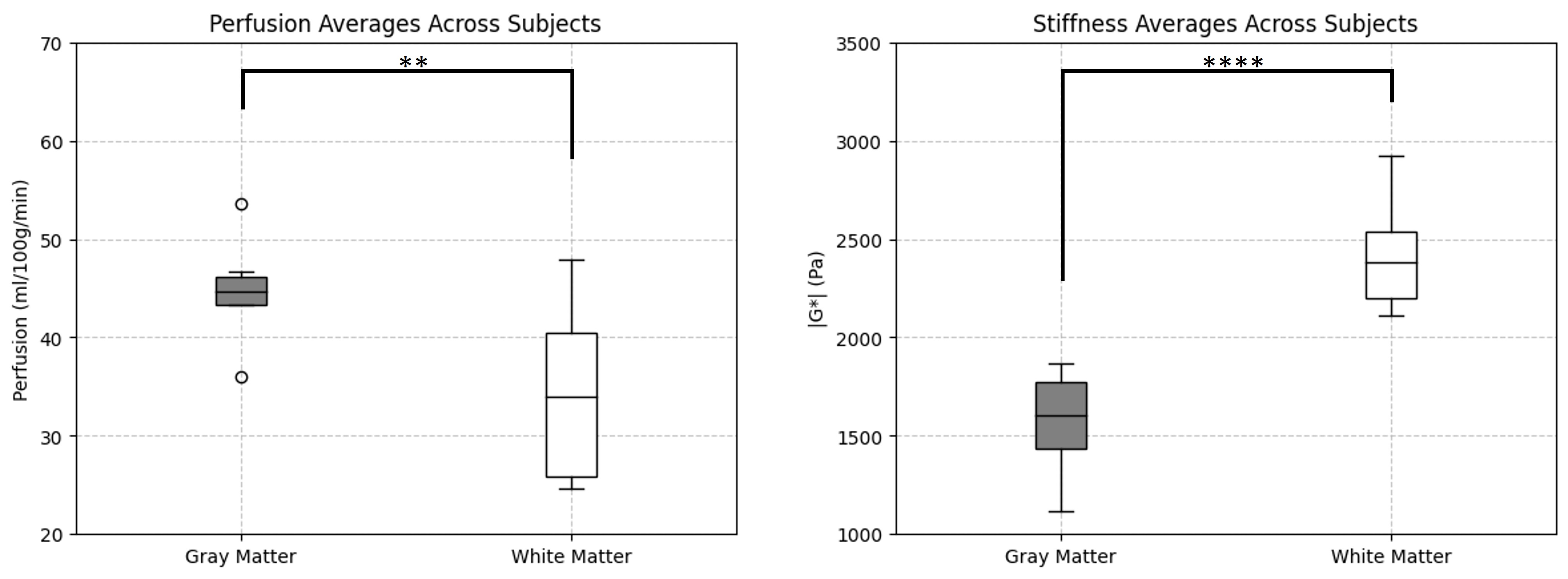
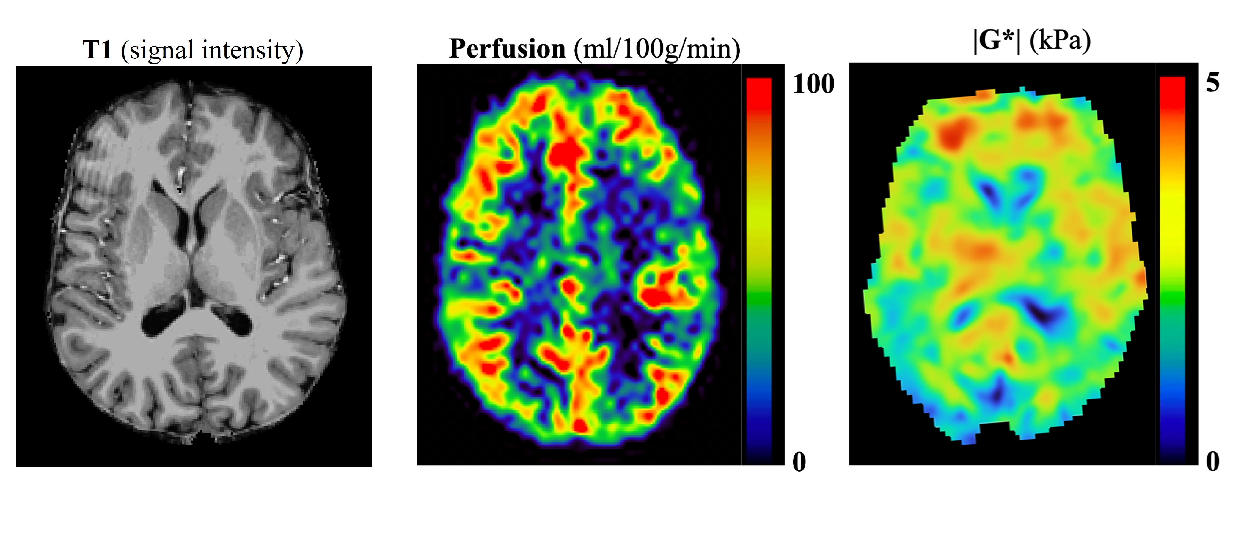
After both scans were coregistered to the matrix space of their respective T1 images using MRE and ASL magnitude volumes, we regionally correlated stiffness and perfusion across all 8 subjects. This analysis has shown varying strengths of inverse correlation between stiffness and perfusion in some gray matter regions of the brain. Within a cortical GM mask, stiffness and perfusion show a strong inverse correlation across subjects (p-value = 0.0121, r = -0.823). This result supports our hypothesis that increased blood flow is related to reduced stiffness due to an increase in relative size of vascular structures coupled with the constriction of vessels in non-compliant tissue6. This trend is also consistent with existing research showing reduced whole-brain stiffness following exercise16 (and therefore increased perfusion17).Conclusion
Our experimental results suggest that there is a measurable correlation between stiffness, a mechanical property of tissue, and perfusion, a measure of blood delivery within the tissue. Arterial spin labeling is unique in that by measuring delivery of blood to the brain tissue, it is a metric of brain health at the capillary bed level18. Unlike other vasculature scans, such as time of flight (TOF) angiography, ASL measures blood delivery rather than blood vessel characteristics. The establishment of correlations between stiffness and perfusion could enhance our understanding of disease pathology in Alzheimer’s Disease and other neurodegenerative diseases by highlighting the interplay between tissue mechanics and metabolic and neuroinflammatory changes.Acknowledgements
This work was supported by the National Science Foundation (NSF CMMI 1953323).References
1Bokkers et al. Whole-brain arterial spin labeling perfusion MRI in patients with acute stroke. Stroke, 2012.
2Arani et al. Measuring the effects of aging and sex on regional brain stiffness with MR elastography in healthy older adults. NeuroImage, 2015.
3Johnson et al. Pattern of Cerebral Hypoperfusion in Alzheimer Disease and Mild Cognitive Impairment Measured with Arterial Spin-labeling MR Imaging: Initial Experience. Radiology, 2005.
4Mattsson et al. Association of brain amyloid-ꞵ with cerebral perfusion and structure in Alziemer’s disease and mild cognitive impairment. Brain, 2014.
5Chouhan et al. Vascular assessment of liver disease–towards a new frontier in MRI. The British Institute of Radiology, 2016.
6Sack et. al., Journal of Cerebral Blood Flow and Metabolism, 2017.
7Johnson et al. 3D multislab, multishot acquisition for fast, whole-brain MR elastography with high signal-to-noise efficiency. Magnetic Resonance in Medicine, 2013.
8Triolo et al., Design, Construction and Implementation of a Magnetic Resonance Elastography Actuator for Research Purposes. Current Protocols, 2022.
9Penny et al. Statistical Parametric Mapping: The Analysis of Functional Brain Images. Psychology, 2011.
10Veraart et al. Denoising of diffusion MRI using random matrix theory. NeuroImage, 2016.
11Karsa, A., Shmueli, K. A Speedy rEgion-Growing Algorithm for Unwrapping Estimated Phase. IEEE Transactions on Medical Imaging, 2018.
12McGarry MD, Van Houten EE, Johnson CL, Georgiadis JG, Sutton BP, Weaver JB, Paulsen KD. (2012). Multiresolution MR elastography using nonlinear inversion. Med Phys. 39(10):6388-96.
13Luh et al. QUIPSS II with Thin-Slice TI1 Periodic Saturation. Magnetic Resonance in Medicine, 1999.
14Fischl B. (2012). FreeSurfer. NeuroImage, 62(2), 774–781.
15van Gelderen, P., de Zwart, J.A. and Duyn, J.H. (2008), Pittfalls of MRI measurement of white matter perfusion based on arterial spin labeling. Magn. Reson. Med., 59: 788-795.
16McIlvain, G. Acute Effects of High-Intensity Exercise on Brain Mechanical Properties and Cognitive Function. ISMRM, 2022.
17Ogoh, S., Ainslie, P., Cerebral blood flow during exercise: mechanisms of regulation. Journal of Applied Physiology, 2009.
18Jezzard et al. Arterial Spin Labeling for the Measurement of Cerebral Perfusion and Angiography. Journal of Cerebral Blood Flow & Metabolism, 2017.Figures