4785
Rapid 3D slab-selective MR elastography using interleaved motion encoding1School of Biomedical Engineering and Imaging Sciences, King's College London, London, United Kingdom, 2Siemens Healthineers AG, Erlangen, Germany, 3School of Engineering, Pontificia Universidad Católica de Chile, Santiago, Chile, 4Millennium Institute for Intelligent Healthcare Engineering, Santiago, Chile, 5Institute for Biological and Medical Engineering, Pontificia Universidad Católica de Chile, Santiago, Chile, 6INSERM U1148, LVTS, University Paris Diderot, Paris, France
Synopsis
Keywords: Elastography, Elastography
Motivation: Most current volumetric 3D MRE sequences are restricted to 2D slice acquisitions, but we propose a novel sequence for rapid 3D slab-selective MRE.
Goal(s): To demonstrate the feasilbility of a 3D slab-selective MRE sequence with an interleaved motion encoding scheme.
Approach: The proposed 3D MRE sequence allows the measurement of different wave offsets and motion encodings for a 3D slab in a single measurement with a constant repetition time, minimising total acquisition time.
Results: We show initial results of the proposed technique in a phantom and for a single breathhold 3D liver MRE in a healthy subject.
Impact: The proposed method can be applied for effiicient 3D high-resolution MRE. Applications include liver fibrosis and inflammation staging, but the sequence may also be used for brain MRE.
Introduction
Magnetic Resonance Elastography (MRE)1 enables non-invasive estimation of biomechanical tissue properties in-vivo, with applications including the assessment and staging of liver fibrosis2, and simultaneous assessment of liver fibrosis and inflammation from elasticity and viscosity has also previously been demonstrated with 3D MRE sequences 3,4. However, current volumetric 3D MRE sequences typically acquire in 2D slices, missing out on the higher SNR afforded by a 3D acquisition. We propose a 3D slab-selective rapid spoiled gradient-echo sequence with optimised timing, able to acquire a 3D MRE liver dataset in a single breathhold.Methods
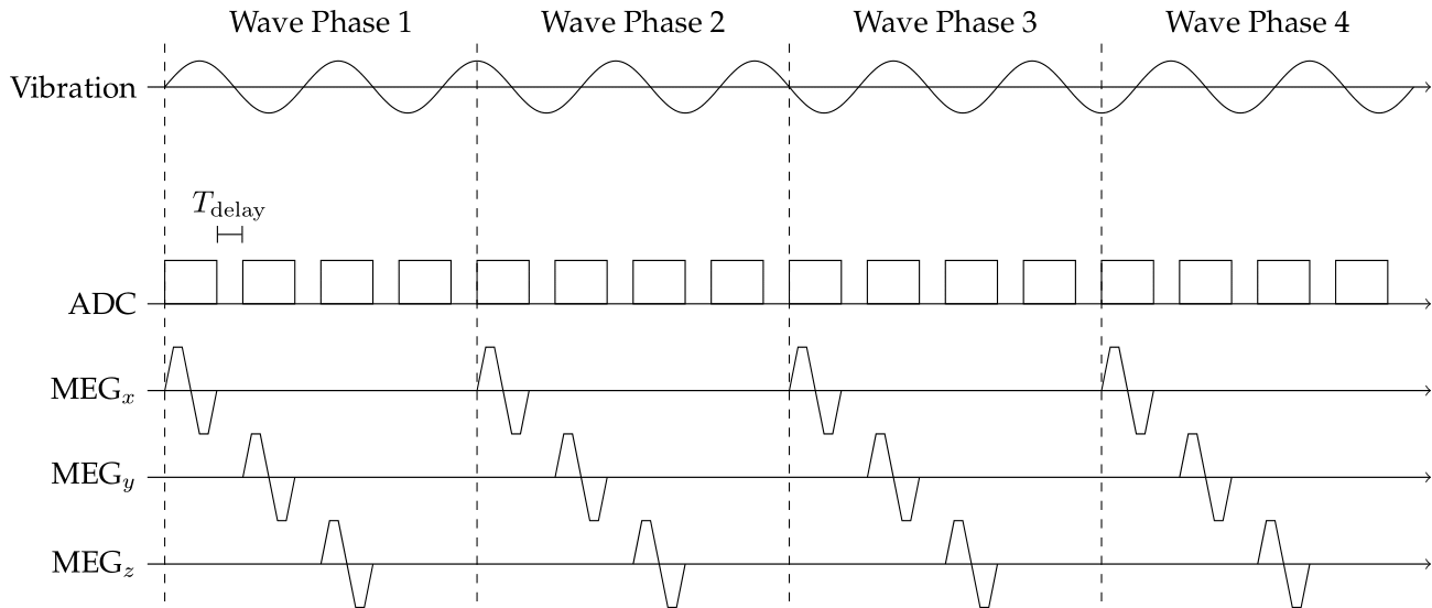
The design of the proposed 3D MRE sequence with 3D slab-selective phase-contrast spoiled gradient echo acquisition is depicted in Fig 1. The inner acquisition loop consists of the 3 motion encodings and the reference scan without motion encoding. A constant delay at the end of each imaging readout shifts the acquisition to the successive wave offset, similarly to the Ristretto MRE sequence5, minimising the total acquisition time. This is repeated till all wave offsets are acquired, then the next k-space point is acquired in a similar manner. Each k-space point acquisition is synchronised with the mechanical vibration. Importantly. for each motion encoding step, the design of the sequence allows to measure all k-space points in the 2D phase-encoding plane at the same wave motion state, thereby avoiding motion-related ghosting artifacts. The sequence can be combined with any two-dimensional phase encoding acceleration technique.The proposed sequence was implemented on a 3T system (MAGNETOM Vida, Siemens Healthcare, Erlangen, Germany) to realise a 3D slab-selective liver MRE measurement in a single breath-hold. Experiments were performed with 60Hz actuation frequency6 with four wave-phase offsets and a four-point unbalanced motion encoding scheme (gradient amplitude = 20 mT/m, total duration = 5.52 ms). Imaging parameters were 8 slices, 25% slice oversampling, 3mm isotropic resolution, FOV of 386 X 264 X 32 mm3, 2D CAIPIRINHA parallel imaging7 with a total acceleration factor of 4 (2x phase encoding and 2x partition encoding, partition shift = 1), elliptical shutter, TR= 9.4ms, TE=6.43 ms, flip angle = 10 deg, receiver bandwidth = 740 Hz/px, phase partial Fourier = 6/8. This resulted in a breath-hold duration of 22 seconds. This sequence was validated in an ultrasound gel phantom, and for a liver acquisition in one healthy subject.
The obtained phase images were unwrapped using a minimum cost flow technique8. The n-th motion encoding step (n = 1..3) in the proposed sequence is acquired at a different time with respect to the 0-th motion encoding step and therefore accumulates a phase $$$\phi_n=2\pi n \phi_\text{inc}$$$ where $$$\phi_\text{inc}=\frac{N_{T_\text{vib}}}{4 N_\text{WP}}$$$, where $$$N_{T_\text{vib}}$$$ is the number of vibration periods required for the acquisition of all wave offsets and motion encodings for one k-space point and $$$N_\text{WP}$$$ is the number of acquired wave offsets. Therefore, to obtain the 3D displacement field, a phase correction scheme is applied to each motion encoding after the temporal Fourier transform followed by the subtraction of the reference background phase. A 3D Gaussian filter of width σ = 2 pixels and a support of 3x3x3 pixels was applied to the displacement field. MRE reconstruction was performed using the curl operator to remove the compressional wave followed by a direct inversion of the complex wave equation9. The following parameters were retrieved: the magnitude of the complex-valued shear modulus (|G*| [kPa]), the shear wave speed (Cs [m/s]), and the loss modulus (G" [kPa]).
Results
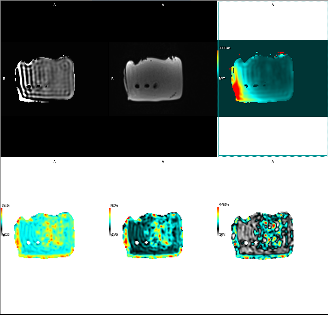
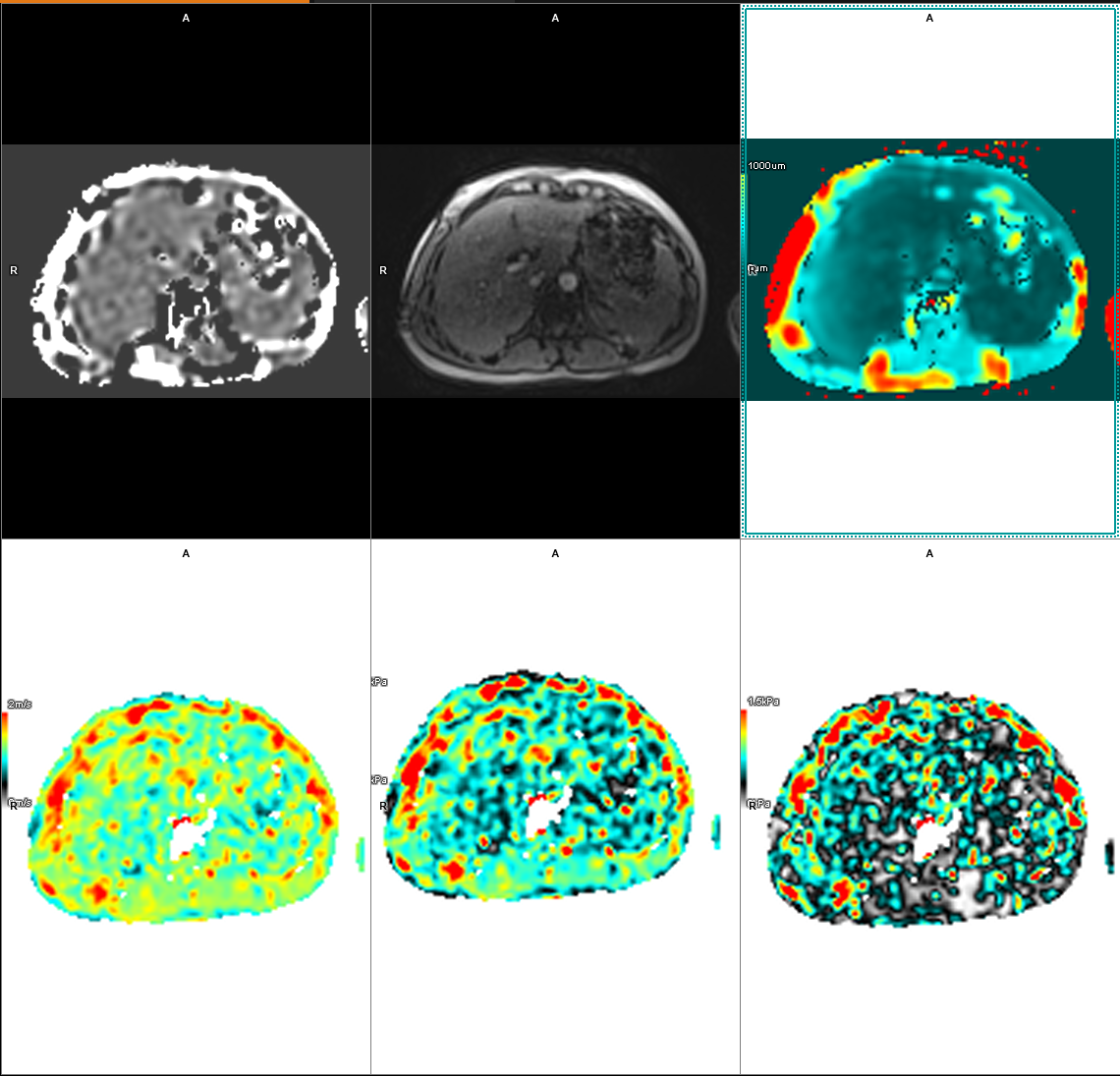
In the phantom (Fig. 2), the obtained values |G*| = 0.95 ± 0.08 kPa, Cs = 0.98 ± 0.05 m/s, and G’’ = 0.21 ± 0.08 kPa closely agree with the reference values for the phantom. The values obtained in the liver of the healthy subject (Fig. 3), |G*| = 1.61 ± 0.33 kPa, Cs = 1.3 ± 0.13 m/s, and G’’ = 0.64 ± 0.22 kPa, were also in agreement with literature values.Discussion
The proposed technique is more efficient than previously published 3D MRE techniques based on wave offset interleaving10, which requires a substantial increase in TR and an acquisition of 5 wave offsets instead of 4. The cartesian trajectory also reduces sensitivity to eddy currents and off-resonance effects compared to the spiral trajectory of other 3D fast MRE sequences11. Future research directions will include applications of this technique for high-resolution brain and cardiac MRE.Conclusion
We have introduced a novel sequence that enables rapid volumetric 3D slab-selective MRE, and showed an initial application for hepatic MRE in a single breathhold.Acknowledgements
The authors acknowledge financial support from: (1) BHF RG/20/1/34802 (2) EPSRC EP/V044087/1 (3) Wellcome EPSRC Centre for Medical Engineering (NS/A000049/1), (4) ANID Millennium Institute iHEALTH, ICN2021_004; Fondecyt 1210637 and 1210638; Basal Funding, IMPACT, FB210024 and (5) the Technical University of Munich – Institute for Advanced Study.References
1. Muthupillai R, Lomas DJ, Rossman PJ, Greenleaf JF, Manduca A, Ehman RL. Magnetic resonance elastography by direct visualization of propagating acoustic strain waves. Science. 1995 Sep 29;269(5232):1854-7.2. Venkatesh, S.K., Yin, M. and Ehman, R.L. (2013), Magnetic resonance elastography of liver: Technique, analysis, and clinical applications. J. Magn. Reson. Imaging, 37: 544-555.
3. , , , et al. Rheological determinants for simultaneous staging of hepatic fibrosis and inflammation in patients with chronic liver disease. NMR in Biomedicine. 2018; 31:e3956.
4. Darwish OI, Gharib AM, Jeljeli S, Metwalli NS, Feeley J, Rotman Y, Brown RJ, Ouwerkerk R, Kleiner DE, Stäb D, Speier P, Sinkus R, Neji R. Single Breath-Hold 3-Dimensional Magnetic Resonance Elastography Depicts Liver Fibrosis and Inflammation in Obese Patients. Invest Radiol. 2023 Jun 1;58(6):413-419.
5. Guenthner C, Sethi S, Troelstra M, Dokumaci AS, Sinkus R, Kozerke S. Ristretto MRE: A generalized multi-shot GRE-MRE sequence. NMR Biomed. 2019 May;32(5):e4049. doi: 10.1002/nbm.4049. Epub 2019 Jan 29. PMID: 30697827; PMCID: PMC6590281.
6. Runge JH, Hoelzl SH, Sudakova J, Dokumaci AS, Nelissen JL, Guenthner C, Lee J, Troelstra M, Fovargue D, Stoker J, Nederveen AJ, Nordsletten D, Sinkus R. A novel magnetic resonance elastography transducer concept based on a rotational eccentric mass: preliminary experiences with the gravitational transducer. Phys Med Biol. 2019 Feb 6;64(4):045007. doi: 10.1088/1361-6560/aaf9f8. PMID: 30566925.
7. Breuer FA, Blaimer M, Mueller MF, Seiberlich N, Heidemann RM, Griswold MA, Jakob PM. Controlled aliasing in volumetric parallel imaging (2D CAIPIRINHA). Magn Reson Med. 2006 Mar;55(3):549-56. doi: 10.1002/mrm.20787. PMID: 16408271.
8. Costantini M. A novel phase unwrapping method based on network programming. IEEE Transactions on Geoscience and Remote Sensing. 1998;36(3):813-821. doi:10.1109/36.673674
9. Sinkus, R., Tanter, M., Catheline, S., Lorenzen, J., Kuhl, C., Sondermann, E. and Fink, M. (2005), Imaging anisotropic and viscous properties of breast tissue by magnetic resonance‐elastography. Magn. Reson. Med., 53: 372-387.
10. van Schelt AS, Gottwald LM, Wassenaar NPM, Runge JH, Sinkus R, Stoker J, Nederveen AJ, Schrauben EM. Single Breath-Hold MR Elastography for Fast Biomechanical Probing of Pancreatic Stiffness. J Magn Reson Imaging. 2023 May 17. doi: 10.1002/jmri.28773. Epub ahead of print. PMID: 37194646.
11. Peng X, Sui Y, Trzasko JD, Glaser KJ, Huston J 3rd, Ehman RL, Pipe JG. Fast 3D MR elastography of the whole brain using spiral staircase: Data acquisition, image reconstruction, and joint deblurring. Magn Reson Med. 2021 Oct;86(4):2011-2024. doi: 10.1002/mrm.28855. Epub 2021 Jun 7. PMID: 34096097; PMCID: PMC8498799.
Figures