4777
Wall Shear Stress Measured with 4D Flow MRI Correlates with Biomarkers of Inflammation and Collagen Synthesis in Aortic Dilation1Department of Health, Medicine and Caring Sciences (HMV), Linköping University, Linköping, Sweden, 2Center for Medical Image Science and Visualization (CMIV), Linköping University, Linköping, Sweden, 3Department of Medicine, Karolinska Intitute, Solna, Sweden, 4Department of Clinical Physiology, and Department of Health, Medicine and Caring Sciences (HMV), Linköping University, Linköping, Sweden, 5Östersund hospital, Östersund, Sweden, 6Department of Cardiology in Linköping, and Department of Health, Medicine and Caring Sciences, Linköping University, Linköping, Sweden
Synopsis
Keywords: Flow, Velocity & Flow, Aortic disease
Motivation: Hemodynamics and circulating biomarkers could offer urgently needed improved risk-stratification in individuals with aortic dilation.
Goal(s): To explore correlations between hemodynamics markers measured with 4D Flow MRI and circulating biomarkers in aortic dilation individuals with tricuspid aortic valves.
Approach: Wall shear stress (WSS) parameters were assessed from 4D flow MRI in cases with dilation and controls. Blood plasma samples were used to quantify the inflammation biomarker IL-6 and several extracellular matrix biomarkers including collagen type I α1 chain (COL1α1).
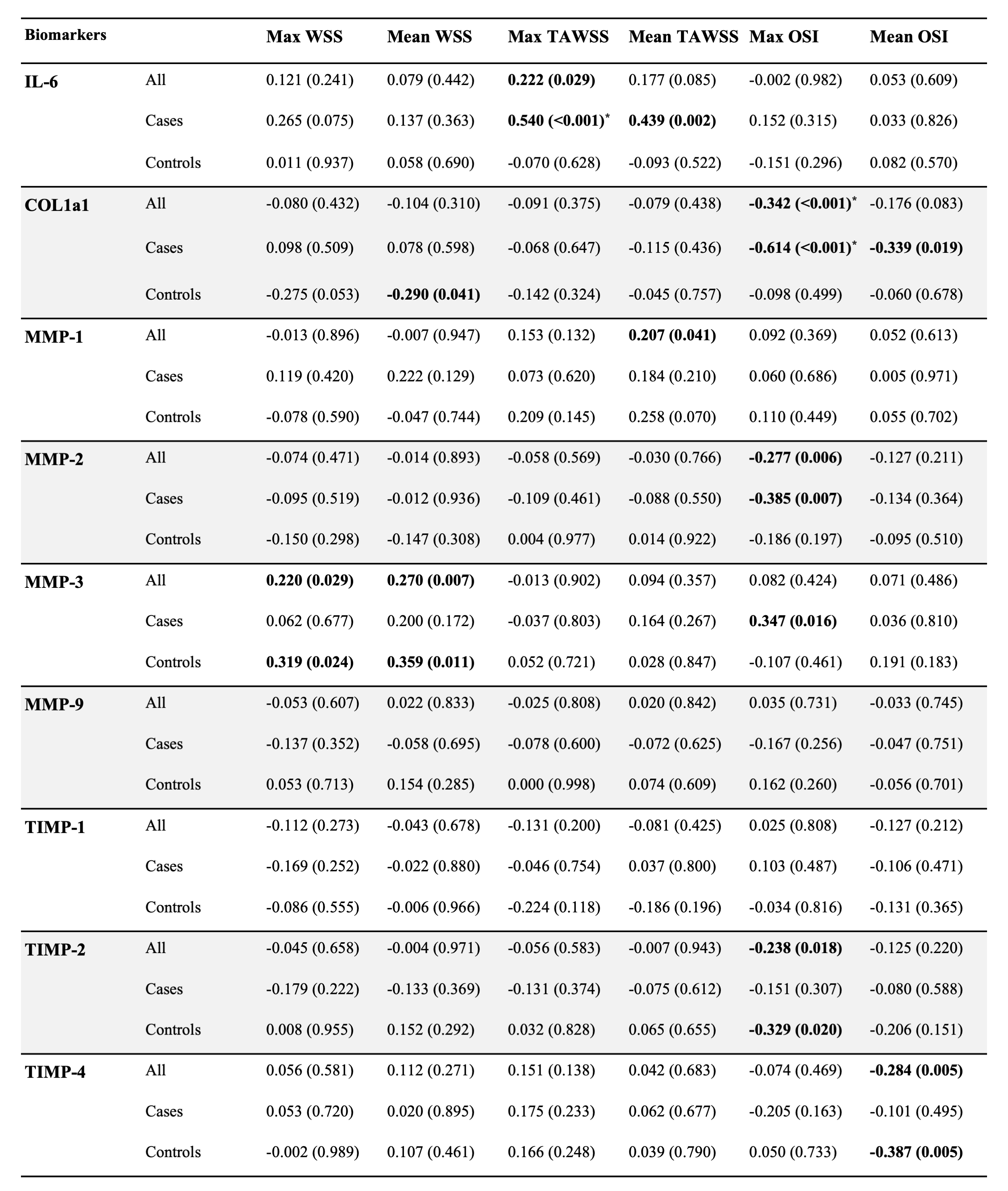
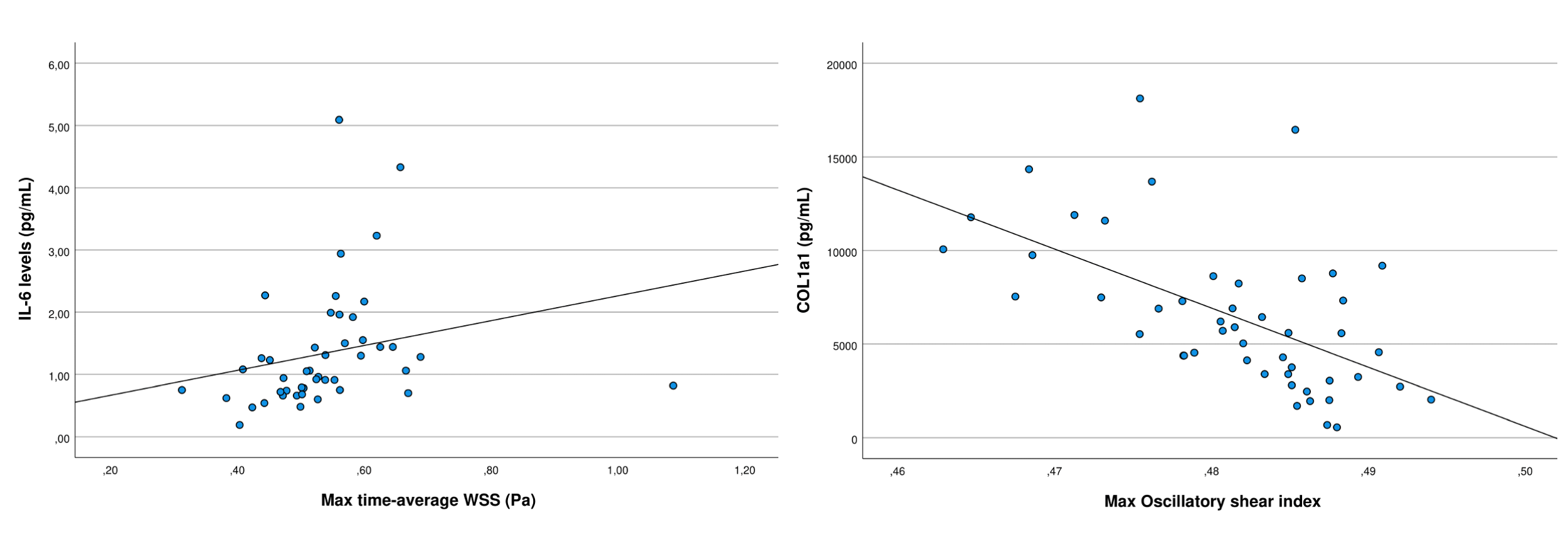
Results: IL-6 correlated to time-averaged WSS (r=0.54, p<0.001), whereas COL1α1 correlated to the oscillatory shear index (r=0.61, p<0.001).
Impact: The observed correlations between 4D Flow MRI-derived wall shear stress and circulating biomarkers of inflammation and collagen synthesis raises the intriguing possibility that altered aortic hemodynamics causes biomarker leakage from the aortic tissue into the circulation.
Introduction
Improved risk-stratification is needed for individuals with ascending aortic dilation. Current guidelines for asymptomatic patients suggest annual surveillance imaging until the ascending aortic diameter reaches a threshold for elective prophylactic surgery1. However, most aortas grow slowly, and a minority of patients suffer complications2. Moreover, most dissections that involve the ascending aorta occur when the diameter is below the threshold for surgery3. New clinical tools could permit reduction of unnecessary follow-ups in low-risk individuals, and intensified follow-ups and potentially earlier surgery in high-risk individuals.Mounting evidence supports the notion that altered hemodynamics measured with three-dimensional cine phase-contrast magnetic resonance imaging (4D Flow MRI) could improve risk stratification of aortic dilation4,5. Circulating biomarkers in venous blood samples represent another way to potentially improve risk stratification of aortic dilation. Investigating associations between hemodynamics and circulating biomarkers could improve our understanding of aortic dilation mechanisms and aid in the identification of biomarkers linked to adverse hemodynamics.
The aim of this study was to explore associations between established hemodynamic markers of ascending aortic dilation measured with 4D Flow MRI and circulating biomarkers in plasma in aortic dilation patients with tricuspid aortic valves (TAV).
Methods
Screening of aortic diameter in 5000 individuals in the Swedish CArdioPulmonary bioImage Study (SCAPIS) resulted in the identification of 74 individuals with mild-to-moderate aortic dilation ≥40 mm at computed tomography. Venous blood sampling and MRI were completed in 48 individuals with TAV and 50 TAV controls following written informed consent. Subject characteristics were available from other SCAPIS examinations6 (Table 1). 4D Flow MRI data were acquired at 1.5T (Philips Ingenia, Philips Healthcare, Best, the Netherlands). Plasma samples were used to quantify the inflammation biomarker IL-6 and several extracellular matrix biomarkers including collagen type I α1 chain (COL1α1), matrix metalloproteinases (MMP) and tissue inhibitors of MMPs (TIMP).Time-resolved segmentations of the aorta were generated with a semi-automated atlas-based method7. Hemodynamic markers derived from 4D Flow included peak WSS, time-averaged WSS (TAWSS) and the oscillatory shear index (OSI) reported as average and as maximum (99th percentile) in the ascending aorta8. Peak WSS was defined as maximum WSS in time, and TAWSS as the average WSS over the cardiac cycle. The OSI quantifies the degree to which WSS changes direction during the cardiac cycle and is used as a measure of disturbed WSS.
Results
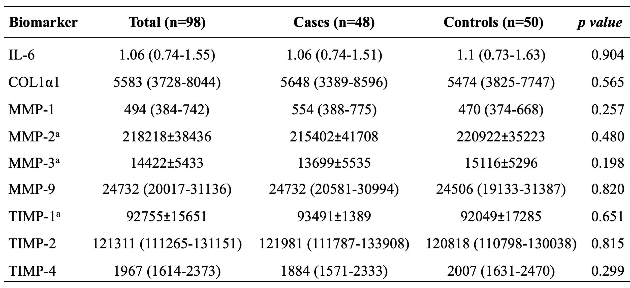
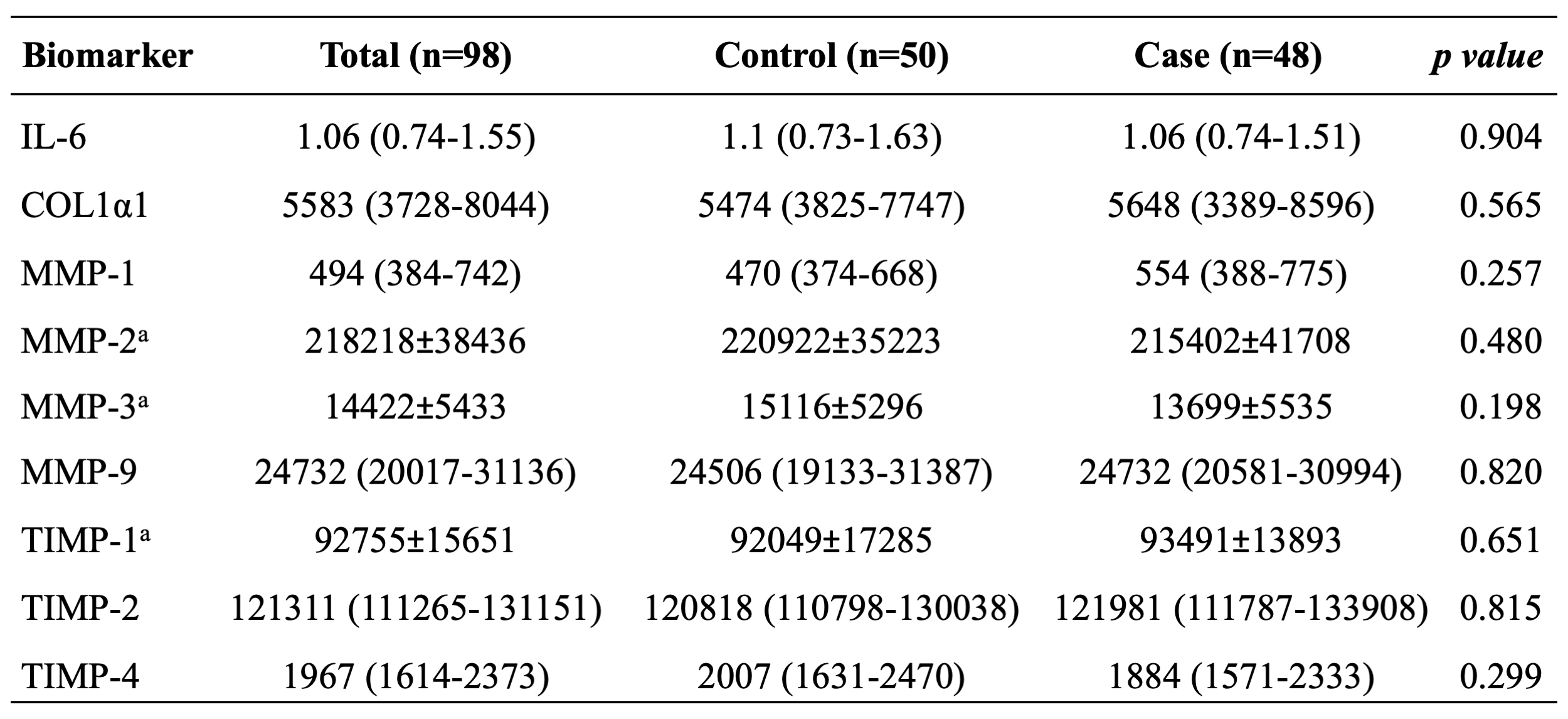
Hemodynamics differed significantly between cases with aortic dilation and controls, but biomarker levels did not (Table 2 and 3). Several hemodynamics markers correlated significantly with circulating biomarkers (Table 4). Correlations between COL1α1 vs. maximum OSI and IL-6 vs. maximum TAWSS remained after Benjamini-Hochberg correction for false discovery rate, and were higher in the cases vs. the whole population (Table 4, Figure1).Discussion
In exploring associations between hemodynamics markers and biomarkers, we have identified promising biomarkers of aortic dilation in TAV that correlate with adverse hemodynamics. Further, our results suggest that the combination of the hemodynamics markers and biomarkers can help in improving risk stratification of patients with aortic dilation.Our main findings were that the multifunctional cytokine IL-6 correlated positively with TAWSS. Several biomarkers related to the ECM correlated significantly with different aspects of WSS. Most notably, the type I collagen biomarker COL1α1, for which decreased tissue levels have been associated with increased risk of thoracoabdominal aortic aneurysms in mice9, correlated inversely with maximum OSI.
Whole-cardiac cycle WSS parameters, especially OSI, correlated with biomarkers to a larger extent than systolic WSS. These findings challenge the current paradigm in which systolic WSS is the most important hemodynamic marker of aortic dilation. Extending the analysis of 4D Flow to the whole cardiac cycle may open up for stronger and novel findings of the relationship between adverse hemodynamics and aortic dilation.
Conclusion
Several novel associations were discovered between 4D flow MRI derived hemodynamic markers of aortic dilation and circulating biomarkers in plasma. Patients with aortic dilation were hemodynamically different when compared to controls, but circulating biomarker levels were not different between groups. These results may facilitate the development of new tools for risk stratification of patients with aortic dilation. Further studies should validate these findings in similar as well as more diverse cohorts, and examine them in a longitudinal fashion.Acknowledgements
We acknowledge support received from Philips Clinical Science, Sweden.References
1. Aalbregt, E. et al. Quantitative Magnetic Resonance Imaging to Assess Progression and Rupture Risk of Aortic Aneurysms: A Scoping Review. J. Endovasc. Ther. Off. J. Int. Soc. Endovasc. Spec. (2023). doi:10.1177/15266028231204830
2. Guo, M. H. et al. Association of Mortality and Acute Aortic Events With Ascending Aortic Aneurysm: A Systematic Review and Meta-analysis. JAMA Netw. Open 1, e181281 (2018).
3. Pape, L. A. et al. Aortic Diameter ≥5.5 cm Is Not a Good Predictor of Type A Aortic Dissection: Observations From the International Registry of Acute Aortic Dissection (IRAD). Circulation 116, 1120–1127 (2007).
4. Guala, A. et al. Wall Shear Stress Predicts Aortic Dilation in Patients With Bicuspid Aortic Valve. JACC Cardiovasc. Imaging 15, 46–56 (2022).
5. Korpela T. et al. Flow displacement and decreased wall shear stress might be associated with the growth rate of an ascending aortic dilatation. Eur J Cardiothorac Surg 61,395-402 (2022)
6. Swahn, E., Lekedal, H., Engvall, J., Nyström, F. H. & Jonasson, L. Prevalence and determinants of dilated ascending aorta in a Swedish population: a case–control study. Eur. Heart J. Open 3, oead085 (2023).
7. Bustamante, M. et al. Automated multi-atlas segmentation of cardiac 4D flow MRI. Med. Image Anal. 49, 128–140 (2018).
8. Trenti, C. et al. Wall shear stress and relative residence time as potential risk factors for abdominal aortic aneurysms in males: a 4D flow cardiovascular magnetic resonance case–control study. J. Cardiovasc. Magn. Reson. 24, 18 (2022).
9. Rahkonen, O. et al. Mice with a deletion in the first intron of the Col1a1 gene develop age-dependent aortic dissection and rupture. Circ. Res. 94, 83–90 (2004).
Figures
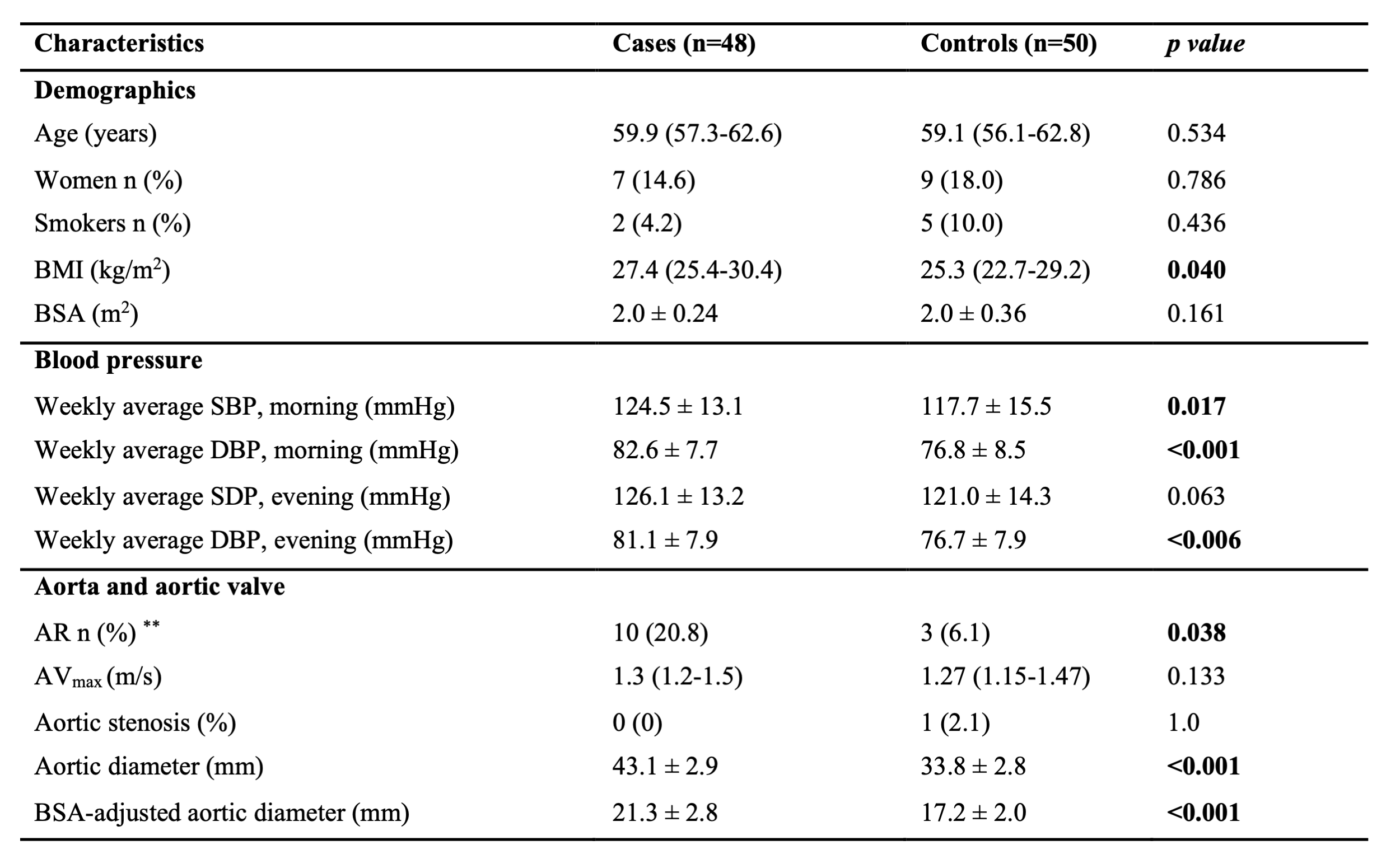
Table 1. Baseline Characteristics in the Study Population (n=98). Values presented as mean ± SD a or median (IQR). Valve characteristics examined by echocardiography. Aortic diameter measured by computed tomography. Bold indicates a p < 0.05 from Student T-test, Mann-Whitney or Fischer exact test. **All aortic regurgitations considered mild except one case who had moderate regurgitation. BMI = body mass index; BSA = body surface area; SBP = systolic blood pressure; DBP = diastolic blood pressure; AR = aortic regurgitation; AVmax = aortic valve maximum velocity.