4773
Accurate Peak and Mean Velocity with Deep Learning-Reconstructed highly Undersampled 4D Flow MRI using FlowVN1Department of health, medicine and caring sciences, Linköping University Sweden, Linköping, Sweden, 2Center for medical image science and visualization (CMIV), Linköping University Sweden, Linköping, Sweden, 3Department of thoracic and vascular surgery, and Department of health, medicine and caring sciences, Linköping University Sweden, Linköping, Sweden
Synopsis
Keywords: Flow, Cardiovascular, Image Reconstruction
Motivation: 4D Flow MRI is limited by long scan times. Accelerated imaging by compressed sensing leads to long reconstruction times.
Goal(s): The goal of this work was to evaluate a deep learning-based method (FlowVN) for reconstruction of pseudo-randomly heavily undersampled Cartesian 4D Flow.
Approach: In this study, we explored FlowVN for the reconstruction of different acceleration factors and did velocity analysis of 4D Flow MRI.
Results: The results show that FlowVN rapidly reconstructs undersampled 4D Flow images with good accuracy for average and peak velocity in the ascending aorta even at high acceleration factors.
Impact: High-quality rapid reconstruction of highly undersampled 4D Flow MRI with deep learning has the potential to substantially facilitate the use of 4D Flow MRI in the clinical routine.
Introduction
4D Flow MRI offers comprehensive assessment of cardiovascular hemodynamics but is limited by long scan times. Different techniques have been used to accelerate the acquisition, including SENSitivity Encoding (SENSE)1 and compressed sensing2. While these efforts into accelerated image acquisition have led to acceptable 4D Flow MRI scan times, a drawback of especially iterative reconstruction methods such as compresses sensing is that the reconstruction of the undersampled data becomes excessively complex and computationally expensive.Deep learning has the potential to offer very fast reconstruction times and overcome limitations of conventional reconstruction methods such as SENSE and compressed sensing3. While several networks have demonstrated encouraging results for phase-contrast MRI, an implementation of a deep variational network termed FlowVN has shown highly promising initial results for 4D Flow MRI with network training based only on 11 datasets and rapid reconstructions on the order of a few seconds3.
The aim of this study was to deploy FlowVN at a new site and evaluate its performance in terms of image quality and quantification of blood flow in the ascending aorta.
Methods
The original implementation of FlowVN was modified for TensorFlow 2.0. To train the network, 4D Flow MRI data were acquired in 10 patients status post mitral valve repair at a 1.5T Philips Ingenia scanner (Philips Healthcare, Best, the Netherlands). The 4D Flow MRI data were acquired with a free-breathing, respiratory navigator gated sequence. SENSE acceleration a total undersampling factor of 4 was used. Scan time was about 10-15 minutes including navigator gating. Reconstruction was performed using ReconFrame (Gyrotools LLC, Zürich, Switzerland) in MATLAB (Mathworks, Natick, MA, United States). The SENSE reconstructed, fully sampled data was used for FlowVN training. We used the Walsh method4 to generate the sensitivity maps needed by FlowVN. Network training on a computer equipped with an NVIDIA Quadro P6000 24GB GPU took approximately three days.To test the trained network, fully sampled 4D flow data was acquired using the same 4D Flow CMR sequence as for the acquisition of training data, but without undersampling in 2 healthy volunteers. Additionally, respiratory navigator gating was omitted. The scan time was 23 minutes. The data were retrospectively undersampled at acceleration factors ranging from 6 to 22 using cartesian golden angle under sampling5. The acquisition and processing of training and testing data is summarized in Figure 1.
For evaluation, the aorta was segmented using an automated atlas-based method6. The quality of the reconstructed velocity images was assessed quantitatively based on the relative error of velocity magnitudes in the aorta, the angular error (angular dissimilarity between velocity vectors), and the normalized root mean square error (nRMSE) of the magnitude images. Additionally, the average and maximum flow velocity in aorta were computed.
Results
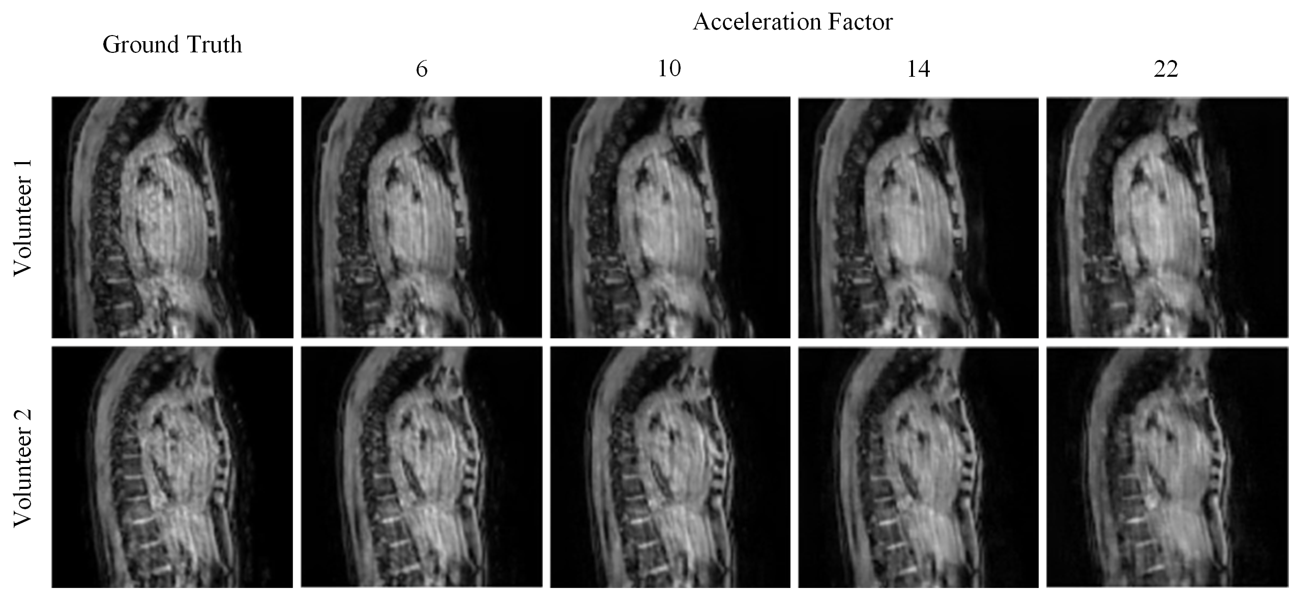
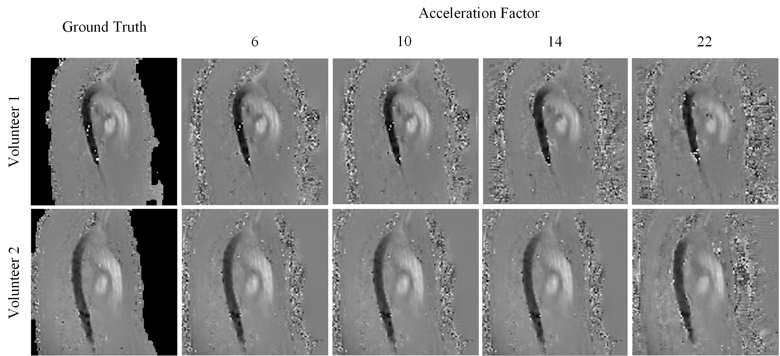
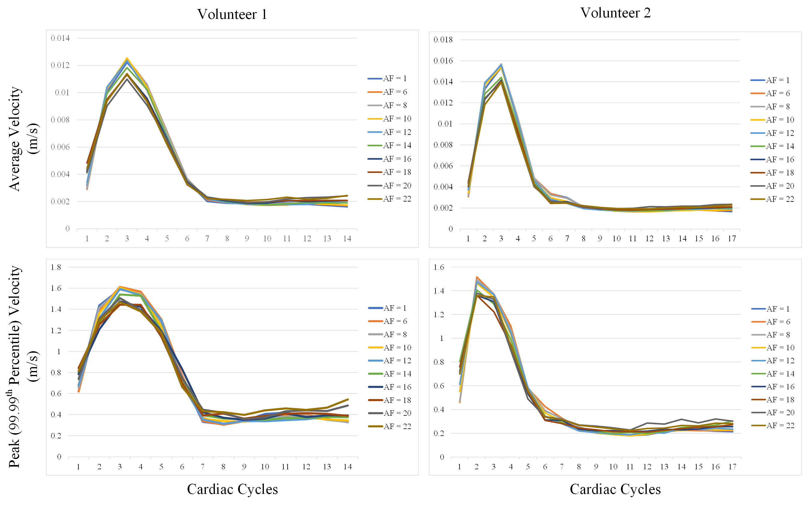
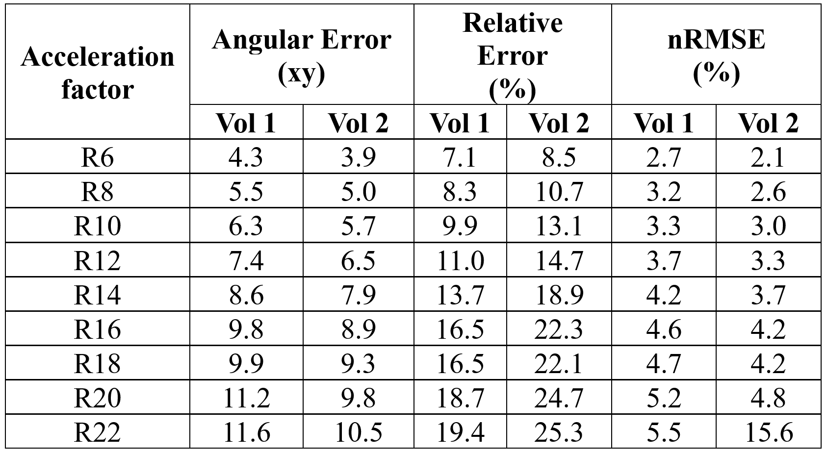
FlowVN reconstructions took 2 minutes and 25 seconds for each acceleration factor. Figure 2 shows ground truth and FlowVN-reconstructed magnitude images and Figure 3 shows ground truth and FlowVN-reconstructed phase images (in FH direction) at different acceleration factors. Overall, for FlowVN-reconstruction results there is good visual agreement between accelerated and ground truth images even at high acceleration factors (see Figure 2 and 3). The quantitative image quality evaluation showed that the errors for both the velocity and magnitude images increase with increasing acceleration factors (see Table 1). Plots of average and peak (99.99th Percentile) velocity over the cardiac cycle are shown in Figure 4. Peak (99.99th Percentile) velocity appears accurately resolved even for acceleration factors of 12.Discussion
In this study, we explored FlowVN for the reconstruction and analysis of 4D Flow MRI. The FlowVN network appears to be robust and is fast in terms of reconstruction time. The reconstructed images show some artifacts which we believe are largely explained by (1) the training data was acquired with 4x SENSE undersampling in patients post mitral valve repair, and 2) the data used for evaluation was acquired without navigator gating. However, in spite of these limitations in the training and test data, the network was capable of reconstructing highly undersampled 4D Flow MR images with remarkably good image quality and maintained accuracy in terms of mean and peak (99.99th Percentile) velocity in the ascending aorta.Conclusion
Reconstruction of highly undersampled 4D Flow MRI using FlowVN is fast and accurate. FlowVN shows good results for the measurement of blood flow in ascending aorta. Further studies will be carried out to improve the magnitude images and assess the performance of FlowVN for turbulence mapping.Acknowledgements
We acknowledge support received from Philips Clinical Science, Sweden.References
[1] Pruessmann K.P., Weiger M., Scheidegger, M.B, et al. SENSE: sensitivity encoding for fast MRI. Magnetic Resonance in Medicine. 1999; 42(5): 952-962.[2] Lustig M., Donoho D. and Pauly J.M. Sparse MRI: The application of compressed sensing for rapid MR imaging. Magnetic Resonance in Medicine. 2007; 58(6): 1182-1195.
[3] Vishnevskiy V., Walheim J. and Kozerke S. Deep variational network for rapid 4D flow MRI reconstruction. Nature Machine Intelligence. 2020; 2(4); 228-235.
[4] Walsh D.O., Gmitro A.F. and Marcellin M.W. Adaptive reconstruction of phased array MR imagery. Magnetic Resonance in Medicine. 2000; 43(5); 682-690.
[5] Winkelmann S., Schaeffter T., Koehler T. et al. An optimal radial profile order based on the Golden Ratio for time-resolved MRI. IEEE transactions on medical imaging. 2006; 26(1); 68-76.
[6] Bustamante M., Gupta V., Forsberg D., et al. Automated multi-atlas segmentation of cardiac 4D flow MRI. Medical image analysis. 2018; 49; 128-140.
Figures
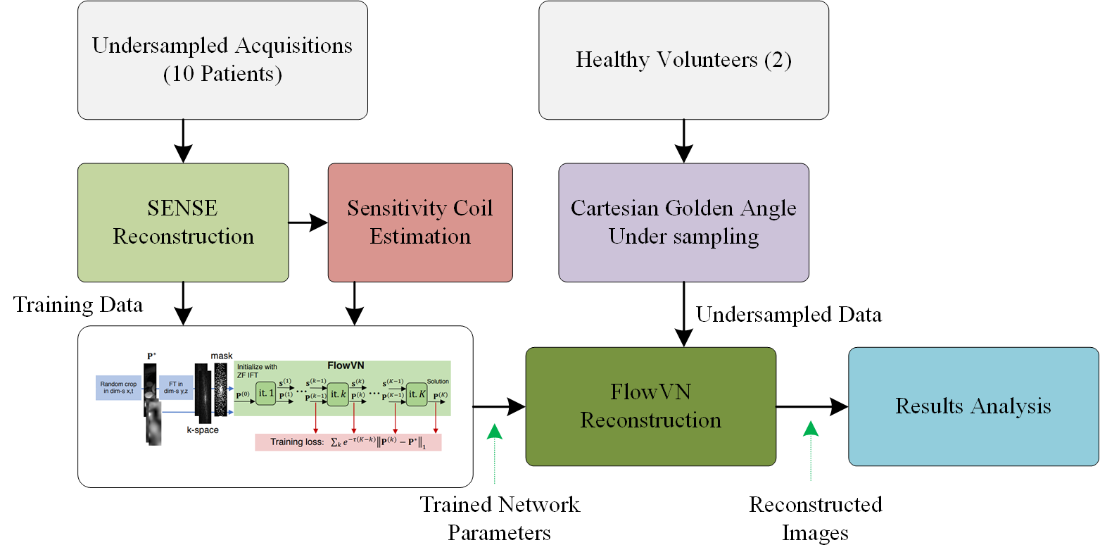
Figure 1: Flow diagram of the method used to assess the performance of the FlowVN3