4771
A Multi-Directional 4D PCMR Flow Phantom: Design and Experiments1CMRR, University of Illinois Chicago, Chicago, IL, United States, 2VasSol Inc., River Forest, IL, United States, 3Economics, Claremont McKenna College, Chicago, IL, United States, 4Center of Magnetic Resonance Research, University of Illinois Chicago, Chicago, IL, United States, 5Biomedical Engineering, University of Illinois Chicago, Chicago, IL, United States, 6Department of Neurosurgery, University of Illinois Chicago, Chicago, IL, United States, 7Radiology, University of Illinois Chicago, Chicago, IL, United States
Synopsis
Keywords: Flow, Validation, 4D PCMR, Flow, Phantom
Motivation: Accurate blood flow measurement is vital for the diagnosis of cardiovascular disorders. Existing phantoms focus primarily on validating flow using phase-contrast MR (PCMR) techniques along the three orthogonal directions. No phantom has been reported for the validation of flow in non-orthogonal orientations.
Goal(s): To validate flow quantification accuracy on a phantom in non-orthogonal directions using 4D flow MRI.
Approach: A multi-directional plexiglass phantom was designed and fabricated to evaluate accuracy of 4D flow PCMR using blood-mimicking flows within tubing controlled by a programmable pump.
Results: The phantom was successfully used to validate flow in orthogonal and non-orthogonal directions simultaneously with 4D flow MRI.
Impact: The flow phantom can be used to validate flow in 5 directions simultaneously with 4D flow MRI. The phantom has the potential to be used to optimize and standardize the 4D flow MR protocol parameters for clinical applications.
Introduction
4D flow MRI is increasingly used in the studies of cardiovascular diseases, cerebrovascular diseases, etc.1-3 Despite its remarkable promise, the validation of 4D flow MRI measurements in living subjects remains a formidable challenge. Current validation phantoms are predominantly designed for flow measurement in orthogonal directions4-7. Nevertheless, vessels in the body can be in any orientation. A phantom for validating flow in non-orthogonal direction is desirable.Phantom Design
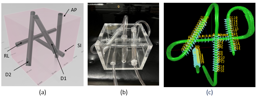
A 5-directional flow phantom was designed and optimized using 3D Builder and MATLAB. The phantom included flexible PVC tubing enclosed in a plexiglass cube. The tubing ran along five directions: three orthogonal directions(RL, AP and SI), one oblique direction parallel to a face diagonal(D1), and another oblique direction corresponding to the body diagonal(D2), shown in Figure 1(a). The phantom was optimized to meet the following criteria: 1) fitting within a 32-channel head coil, 2) maintaining maximum distances between adjacent tubings to prevent signal interference, 3) facilitating easy tubing connection locations on each side of the cube, 4) maximizing non-orthogonal tubing lengths, and 5) rounding length and location to 1/16” for easy fabrication. A cost function was defined and iteratively minimized until a plateau was reached. The cube phantom was constructed using plexiglass panels (½” thickness) and plexiglass tubes (ID ⅝”) shown in Figure 1(b). The cube’s edge length was 5½ inches. The minimum plexiglass tube length was 4½ inches.4D Flow Experiment Studies
During experiments, a plastic tubing (ID=¼") was threaded through the phantom channels sequentially, shown in Figure 1(b), and connected to a computer-controlled flow pump-CompuFlow1000MR8, located outside the scanner room. The pump was preloaded with a blood-mimicking fluid (40:60 glycerol: distilled water). Phantom flow experiments were conducted on an MR7503T MRI scanner involving both constant and pulsatile flow profiles, with flow rates from 500 ml/min to 1000 ml/min. The data were acquired using the GE HyperKat 4D flow protocol with parameters: Resolution (0.7x0.7x0.8mm3 to 1.4x1.4 x1.4mm3), TR/TE =(5.2/2.8ms, 6.0/3.2ms), Flip Angle =8o, NEX =4, phases =2 0, VENC =60-100cm/s, Scan Durations 15-24 minutes.Data Analysis
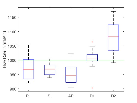
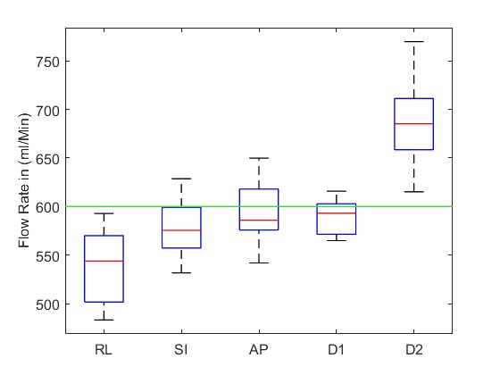
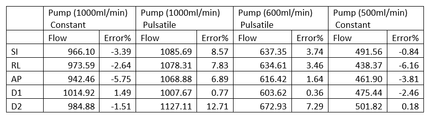
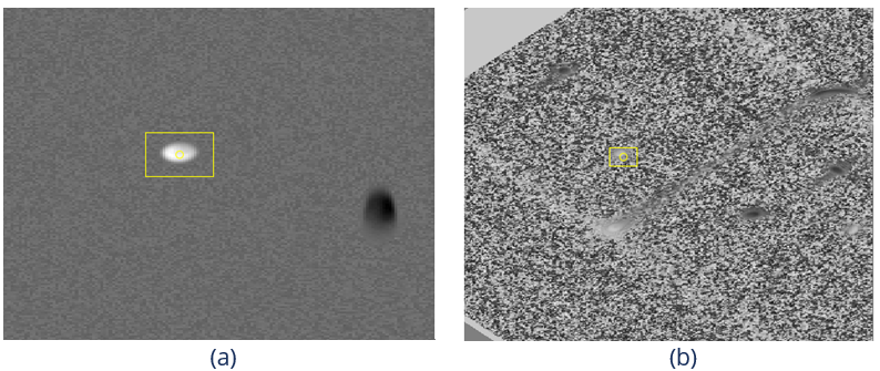
4D data was processed with an in-house software, and the measured flow rates with 4D data were compared to the pump settings. First, pseudo 2D PCMR series perpendicular to each tube centerline at the selected locations were created from 4D data. Each pseudo 2D PCMR series contains 20 magnitude and phase images. A pixel value of a pseudo 2D phase image was calculated by projecting the velocity of the pixel in the space to the normal direction of the 2D slice at a time point using equation: $$$ v=Cos(θ)√(v_x^2+v_y^2+v_z^2 )$$$, where θ is the angle between the pixel velocity and the slice normal, $$$v_x^2,v_y^2, v_z^2$$$ are the square of the velocities calculated from the 4D phase images. The magnitude pixel values were calculated by trilinear interpolating 4D magnitude images in the space at the same time point. Then the flow region of interest (ROI) was manually drawn on one magnitude image and then copied to all images. The flow rate of each phase was calculated by adding up the flow volume of each pixel inside the ROIs. The flow rate of the channel is the summation of flow rate of all time points. To minimize the measurement error, multiple measurement points evenly distributed along the centerline of each tube were selected as shown in Figure 1(c). Table 1 shows the average flow rate measured in each tubing direction of the phantom for 2 constant and 2 pulsatile flow experiments. The data shows that the maximum error for the constant flow experiment was less than 8%, while the maximum error of the pulsatile flow experiment was 12.71% in the D2 direction. The boxplots of two representative cases are shown in Figure 2 and Figure 3. Figure 4 shows a conventional 2D phase image and a calculated 2D phase image from 4D. 4D phase images exhibit notably higher noise levels when compared to their 2D counterparts. The signal-to-noise ratio (SNR) for a typical 2D PCMR image was around 300, and reduced to 54.42 or less in the 4D phase images. Furthermore, the flow measurement errors were generally noticeably pronounced in the non-orthogonal directions than in the orthogonal ones.Conclusions
The multi-directional flow phantom successfully validated 4D flow MRI measurements, providing insights into the potential for optimizing and standardizing 4D flow MRI protocol parameters for clinical applications. Despite its inferior performance in SNR and quantification errors when compared to conventional 2D PCMR, the time efficiency and extensive information content may make 4D flow an attractive alternative for quantitative flow studies that are relevant to cerebrovascular or cardiovascular diseases.Acknowledgements
This work is partially supported by NIH-1S10RR028898. Kezhou Wang is an employee of VasSol Inc.References
1. Markl M, Frydrychowicz A, Kozerke S, Hope M, Wieben O. 4D flow MRI. J Magn Reson Imaging. 2012 Nov;36(5):1015-36. doi: 10.1002/jmri.23632. PMID: 23090914.
2. Soulat G, McCarthy P, Markl M. 4D Flow with MRI. Annu Rev Biomed Eng. 2020 Jun 4;22:103-126. doi: 10.1146/annurev-bioeng-100219-110055. Epub 2020 Mar 10. PMID: 32155346.
3. Arshid A, MScPhilippe G, MDAudrey S, MDNadia C, MDGiorgios A, MDStephane S, MDDaniel S, MDVéronique M, MDMarc Z. Four-dimensional Flow MRI: Principles and Cardiovascular Applications. RadioGraphics, Volume 39, Number 3, 2019
4. Keenan KE, Ainslie M, Barker AJ, Boss MA, Cecil KM, Charles C, Chenevert TL, Clarke L, Evelhoch JL, Finn P, Gembris D, Gunter JL, Hill DLG, Jack CR Jr, Jackson EF, Liu G, Russek SE, Sharma SD, Steckner M, Stupic KF, Trzasko JD, Yuan C, Zheng J. Quantitative magnetic resonance imaging phantoms: A review and the need for a system phantom. Magn Reson Med. 2018 Jan;79(1):48-61. doi: 10.1002/mrm.26982. Epub 2017 Oct 30. PMID: 29083101.
5. Gadda G, Cocozza S, Gambaccini M, Taibi A, Tedeschi E, Zamboni P, Palma G. NO-HYPE: a novel hydrodynamic phantom for the evaluation of MRI flow measurements. Med Biol Eng Comput. 2021 Sep;59(9):1889-1899. doi: 10.1007/s11517-021-02390-2. Epub 2021 Aug 8. PMID: 34365590; PMCID: PMC8382656.
6. Aristova M, Vali A, Ansari SA, Shaibani A, Alden TD, Hurley MC, Jahromi BS, Potts MB, Markl M, Schnell S. Standardized Evaluation of Cerebral Arteriovenous Malformations Using Flow Distribution Network Graphs and Dual-venc 4D Flow MRI. J Magn Reson Imaging. 2019 Dec;50(6):1718-1730. doi: 10.1002/jmri.26784. Epub 2019 May 9. Erratum in: J Magn Reson Imaging. 2020 Nov;52(5):1571-1574. PMID: 31070849; PMCID: PMC6842032.
7. Radiological Society of North America. Quantitative imaging biomarkers alliance. Available from: https://www.rsna.org/en/research/ quantitative- imaging-biomarkers-alliance.
8. Shelley Medical Imaging Technologies. Compuflow 1000 MR, pro- grammable physiological flow pump & accessories. Available from: https://www.simutec.com/Products/ppfp.html
Figures