4760
Temporal profile block-matching denoising enables free-breathing dynamic 19F lung MRI with sub 0.5 second acquisition time1Biomedical Engineering Department, UNC Chapel Hill, Chapel Hill, NC, United States, 2Department of Radiology and BRIC, UNC Chapel Hill, Chapel Hill, NC, United States
Synopsis
Keywords: Lung, Lung, Free-breathing, 19F, denoising,cystic fibrosis
Motivation: 19F pulmonary MRI is often used to characterize abnormalities in case of cystic fibrosis but its long scan time requires patients to hold their breath, which is uncomfortable and complicates acquisition.
Goal(s): We introduce a denoising method to be used with fast 19F imaging that will enable free-breathing dynamic imaging.
Approach: 12 subjects underwent both conventional breath-hold and fast free-breathing imaging. Temporal block-matching denoising was done for free-breathing images.
Results: Our denoising method enables free-breathing dynamic 19F lung imaging. Images are recovered from the noise floor and show similar characteristics to conventional breath-hold imaging.
Impact: Our denoising method enables free-breathing 19F dynamic lung imaging with sub 0.5 second acquisition time. Free-breathing imaging will increase patient comfort and potentially extend ventilation MRI use to more patients, including those with severe lung disease.
Introduction
Multi-breath dynamic 19F MRI acquired with volumetric interpolated breath-hold examination (VIBE) can detect abnormal ventilation in cystic fibrosis patients with normal FEV1. However, this approach requires patients to perform ten to twelve 18-second breath holds upon instructions, making studies of young children and patients with advanced CF difficult. We can eliminate the discomfort of breath-hold by using a 0.45-second spiral MRI, essentially enabling free-breathing acquisition. The catch is: as we gain in acquisition speed, we lose in SNR. The noise level in the spiral MR image is 6 times that of VIBE. In this abstract, we proposed a denoising technique, leveraging redundancy in dMR images to recover the signal from under the noise floor. Our method groups non-local blocks with similar temporal profiles together, utilizing the highly correlated information from them for effective noise removal.Methods
Data8 participants with cystic fibrosis and 4 healthy participants underwent conventional breath hold (VIBE) and fast spiral 19F imaging. 1H 3D-UTE scans (TE/TR 0.05/2.42 ms, FA 5o, resolution 2.14X2.14x2.5 mm, acquisition time 17 seconds) and 19F VIBE scans (TE/TR 1.61/13ms, FA 74o, resolution 6.25X6.25x15mm, acquisition time 18 seconds) were acquired on breath holds. The 19F VIBE was immediately followed by a 4-arm spiral sequence (TE/TR 0.48/11 ms, FA 74o, resolution 6.25X6.25x15 mm, acquisition time 0.45 seconds). Wash-out portion was done when patient inhales room air and was run until no 19F signal was visible.
Denoising
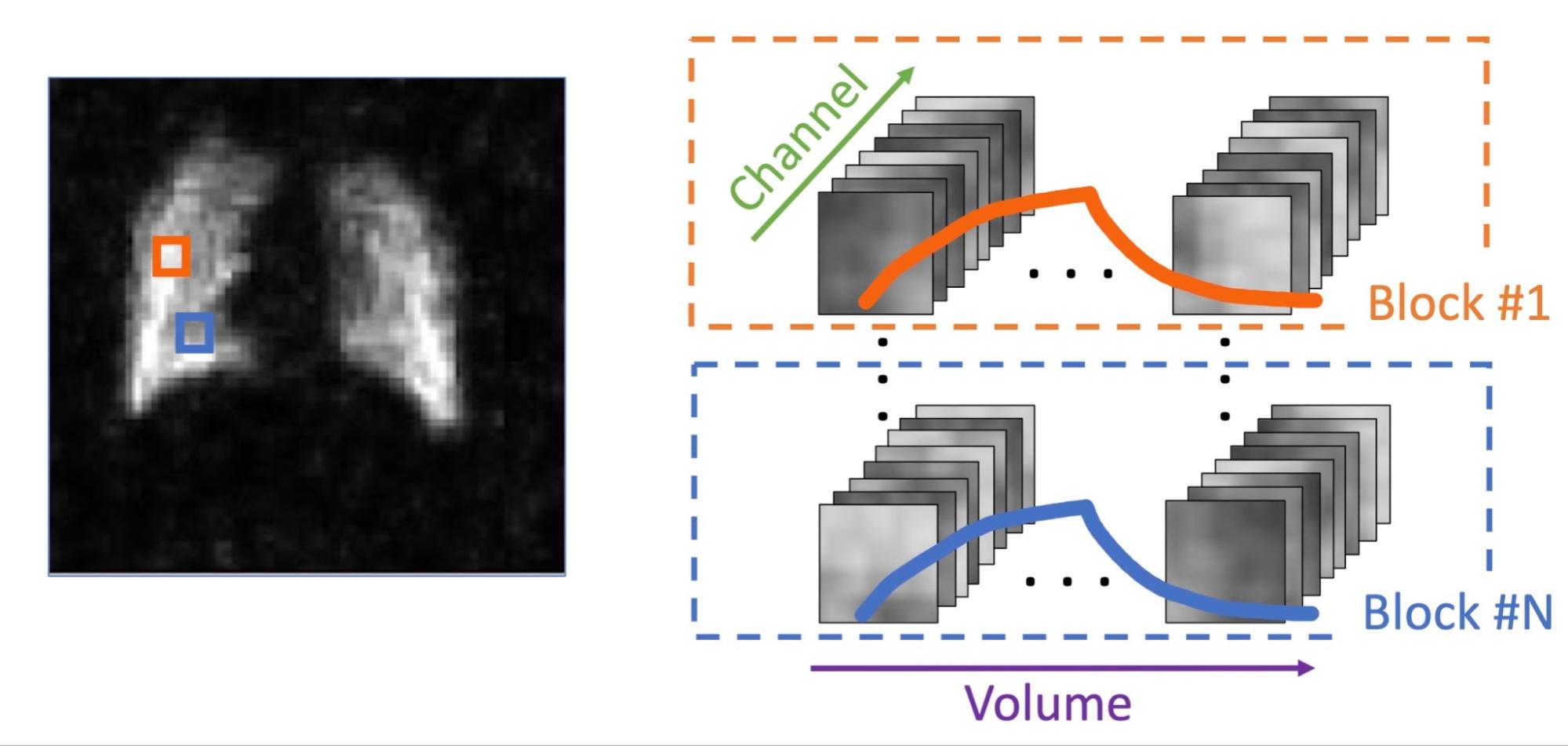
Due to short acquisition time (0.45s vs 18s VIBE, 40x faster), spiral images have significantly lower SNR. We employ temporal profile block-matching followed by optimal shrinkage of singular value to remove noise from spiral data.Information from MR images is redundant in a sense that the voxel signal across multiple receiving coil channels is highly correlated. This redundancy can be leveraged for denoising via random matrix theory (RMT) with optimal shrinkage of singular values [2]. In case of dynamic lung imaging, multiple spatial locations might share similar temporal signal curves depending on their biological characteristics. To increase the redundancy for better denoising, we employ a 4-D block-matching step to select groups of 3x3x3 blocks with similar signals. Information from these blocks across multiple channels are then concatenated to form a 2-D Casorati matrix. Signal from this matrix is highly correlated in three ways:
- Signals within a 3x3x3 block are similar and structural details are smooth;
- Signals at the same location across receiving coil channels are similar as we are imaging the same object; and
- Signals across matching blocks and timepoints are similar because we group blocks with similar temporal profiles.
Results
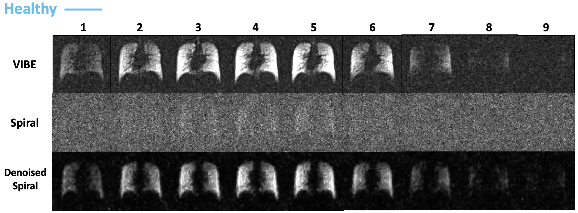
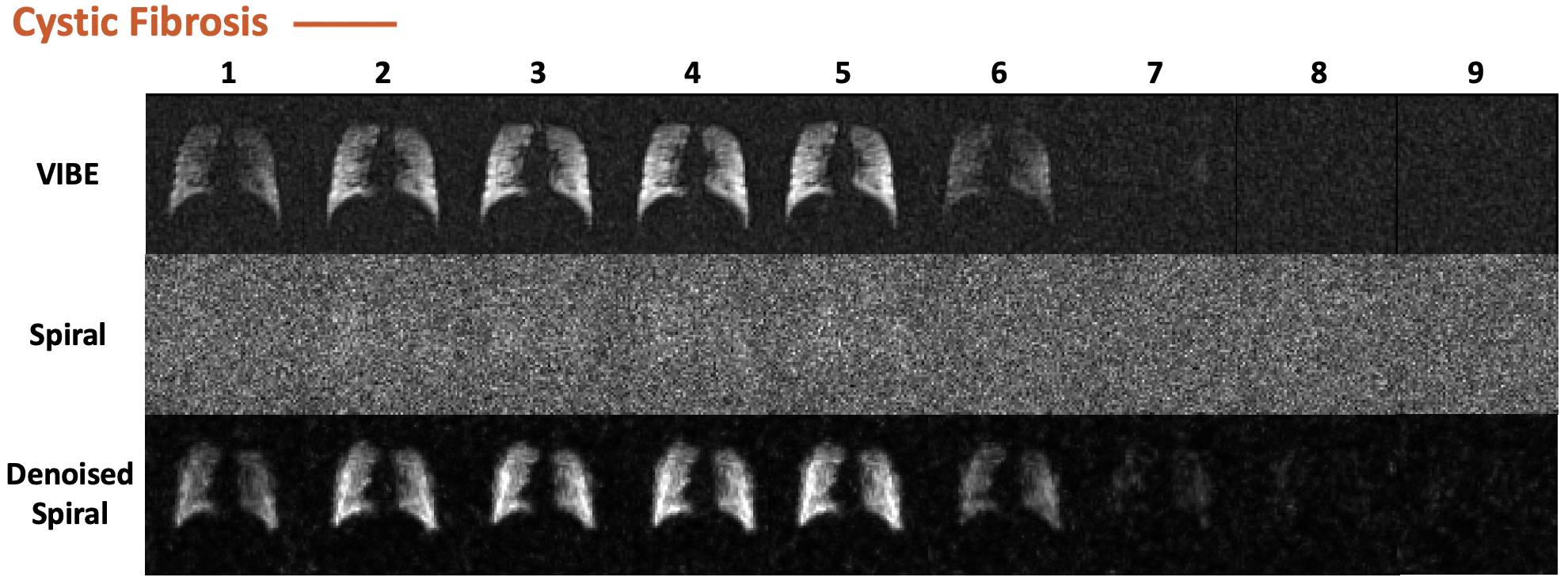
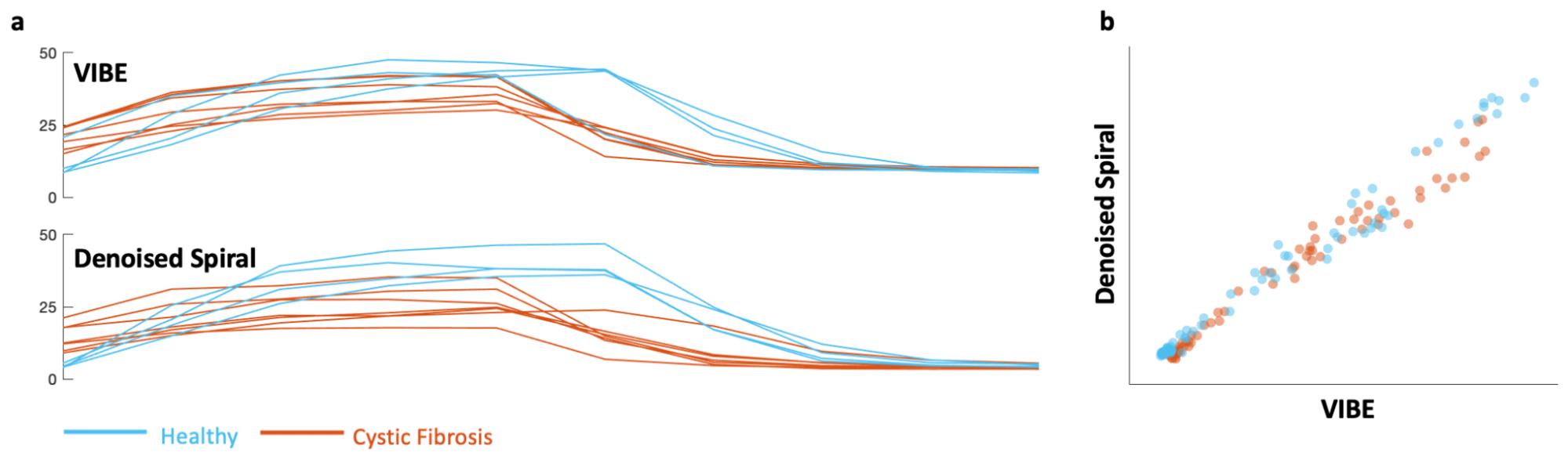
Without denoising, fast spiral data are unusable with structural details buried under the noise floor (average SNR of 2.5, Figs. 2 & 3). Our denoising method recovered the signal in both healthy and CF subjects (average SNR of 31.6). Images show clear structural details similar to VIBE results.We test the similarity between denoised spiral and VIBE signal by plotting the wash-in wash-out curves (Fig. 4a). Overall, denoised spiral results show wash-in wash-out curves similar to VIBE results in both healthy and CF subjects. The differences between healthy and CF curves are also retained after denoising. Fig. 4b shows the linear relationship (R2=0.99) between VIBE and the denoised spiral, confirming the effectiveness of our denoising method with minimal structural differences.
Conclusion
We leveraged the temporal profile of the free-breathing spiral 19F MRI data to recover the signal from under the noise floor with minimal bias to breath-hold VIBE. Our denoising method delineated the dynamics of free-breathing signal, showing high agreement between spiral sequence and VIBE. These dynamics results suggest that free-breathing 19F MR together with the time profile-based denoising approach can quantify lung ventilation.Acknowledgements
No acknowledgement found.References
1. Goralski JL, Chung SH, Glass TM, Ceppe AS, Akinnagbe-Zusterzeel EO, Trimble AT, et al. Dynamic perfluorinated gas MRI reveals abnormal ventilation despite normal FEV1 in cystic fibrosis. JCI Insight 2020;5(2):e133400.
2. Huynh KM, Chang WT, Chung SH, Chen Y, Lee Y, Yap PT. Noise mapping and removal in complex-valued multi-channel MRI via optimal shrinkage of singular values. International Conference on Medical Image Computing and Computer-Assisted Intervention. September 27, 2021; pp. 191-200. Springer, Cham.
Figures