4755
Comparison of dissolved xenon imaging methods: 2D CSI vs 4-echo flyback radial spectroscopic imaging.1POLARIS, Division of Clinical Medicine, University of Sheffield, Sheffield, United Kingdom
Synopsis
Keywords: Lung, Hyperpolarized MR (Gas)
Motivation: Several dissolved xenon imaging methods have been proposed to detect impaired gas transfer in the lung. However, none have been validated against a gold standard CSI sequence.
Goal(s): To validate quantitative xenon gas exchange metrics from a 4-echo radial EPSI implementation against those from CSI.
Approach: 4-echo radial EPSI results were compared to CSI in 2 healthy volunteers.
Results: We found good correlations between xenon gas exchange metrics from CSI and radial EPSI and minimal biases (0.4 to 5.8%) between the two methods for the main outcome parameter RBC:M, the ratio of dissolved xenon signals from the blood and in the membrane.
Impact: Our implementation of dissolved xenon imaging using a 4-echo radial EPSI is validated against the reference standard CSI sequence, providing confidence in the quantitative nature of the xenon gas exchange metrics from this method to assess lung diseases.
Introduction
Due to the solubility of 129Xe gas in the lung parenchyma, its diffusivity into the blood capillaries and the large chemical shift between 129Xe gas, 129Xe dissolved in the membrane and bound to the red blood cells (RBC), it is possible to generate regional maps of surrogate gas exchange metrics (ratio of 129Xe in the different compartments RBC:M, RBC:Gas and M:Gas). Several imaging methods have been developed to produce 3D images within a single ~15s breath-hold, ranging from adaptation of the standard CSI sequence (1), 1-pt Dixon (2) and multi-point imaging techniques (3-5). Results have mainly been validated empirically by showing sensitivity to regional gas transfer limitation and disease progression in interstitial and obstructive lung diseases (6-8), but the imaging ratios have to date not been quantitatively validated against the gold standard CSI method. The aim of this work was to compare our previously developed 4-pt radial echo planar spectroscopic imaging (EPSI) technique (9) against a reference CSI sequence and compare the imaging metrics regionally in healthy volunteers.Methods
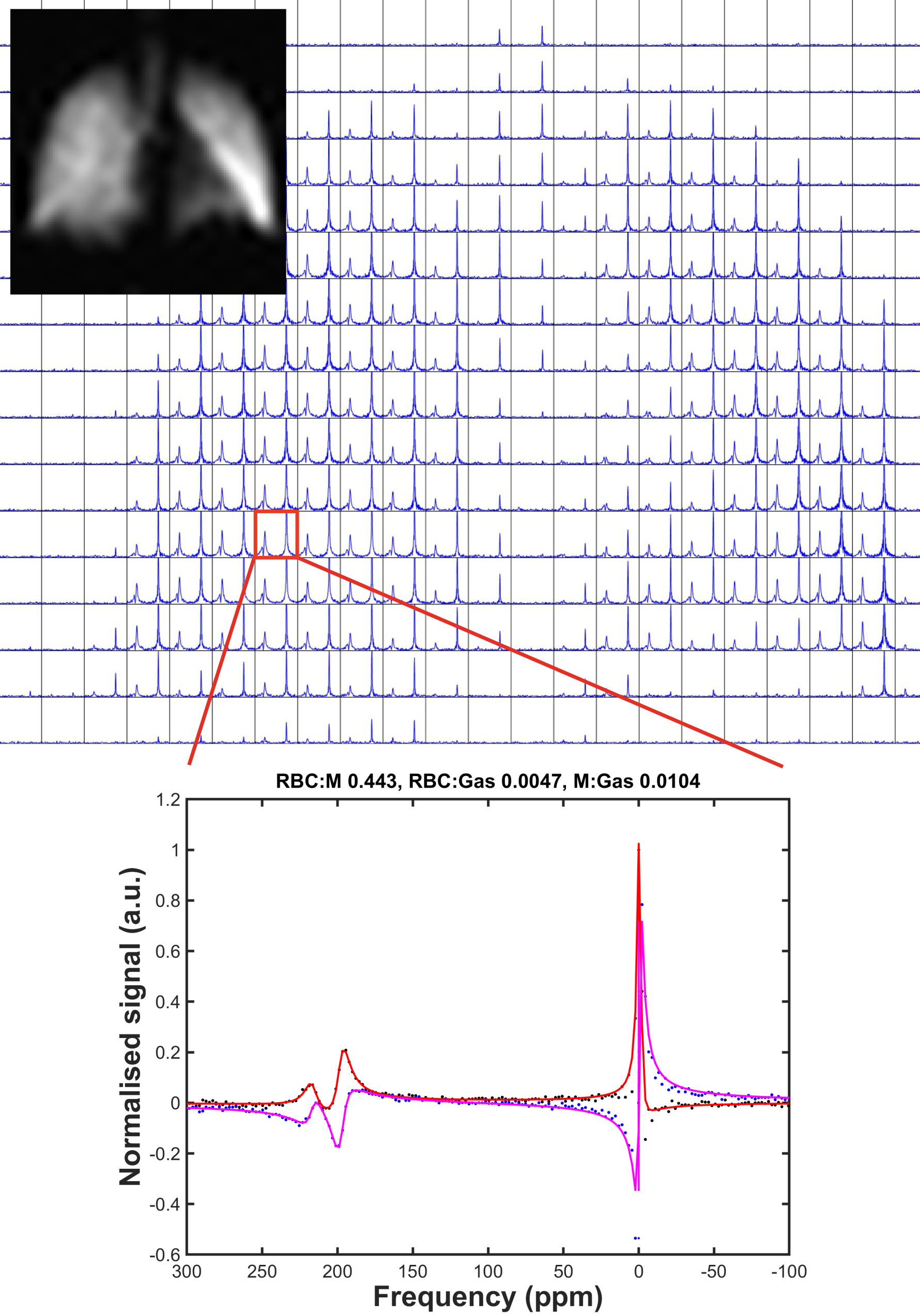
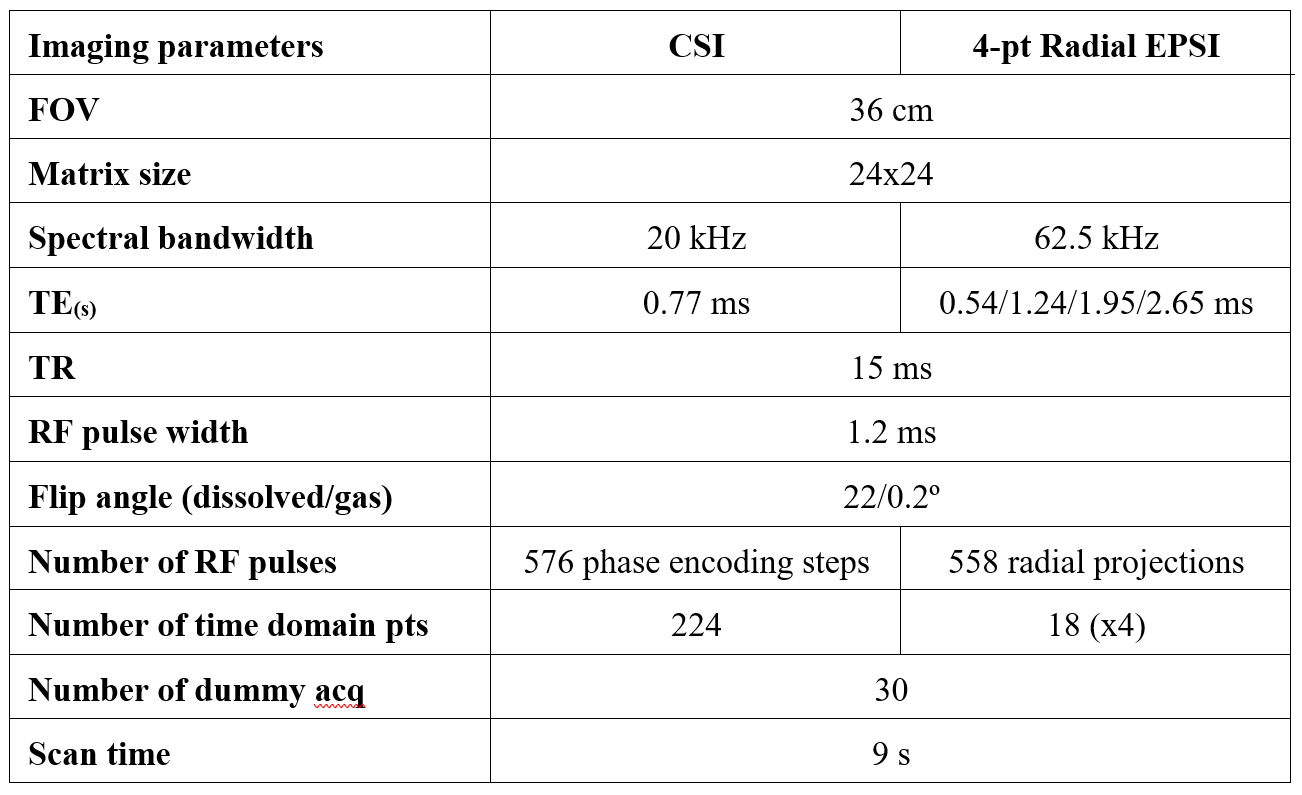
Two healthy volunteers with no known respiratory conditions were recruited. Imaging was performed on a 1.5T GE Artist scanner with 129Xe polarized to ~30% with a spin-exchange optical pumping polariser (POLARIS, Sheffield, UK) (10). Images were acquired with flexible quadrature transmit/receive vest coil during breath-hold after the inhalation of 1L of HP 129Xe from functional residual capacity. To compare both sequences in a reasonable breath-hold time (<10s) and with an appropriate spatial and spectral resolution, imaging was performed in 2D due to the relatively long CSI acquisition time. The 4-pt 3D radial EPSI sequence from (9) was adapted for 2D. Noticeable changes were an increased acquisition bandwidth and a high oversampling factor (~10) in the radial dimension, to obtain a similar acquisition time. Special care was taken to match imaging parameters (see Table 1) between the two sequences, using the same non-spatially selective RF pulse and 129Xe signal dynamics regime (FA/TR, see (11)), following the consensus recommended imaging parameters (12). 4-pt EPSI data were analysed as previously described (4). CSI data were reconstructed to a 24x24 matrix of 512-point frequency spectra. A triple Lorentzian fit of individual spectra (see (13)) was performed to derive peak amplitudes (see Figure 1) and produce ratio maps of RBC:M, RBC:Gas, M:GAS. Linear regression, Spearman correlation and Bland-Altman analysis were performed between the maps resulting from both imaging methods in Graphpad.Results
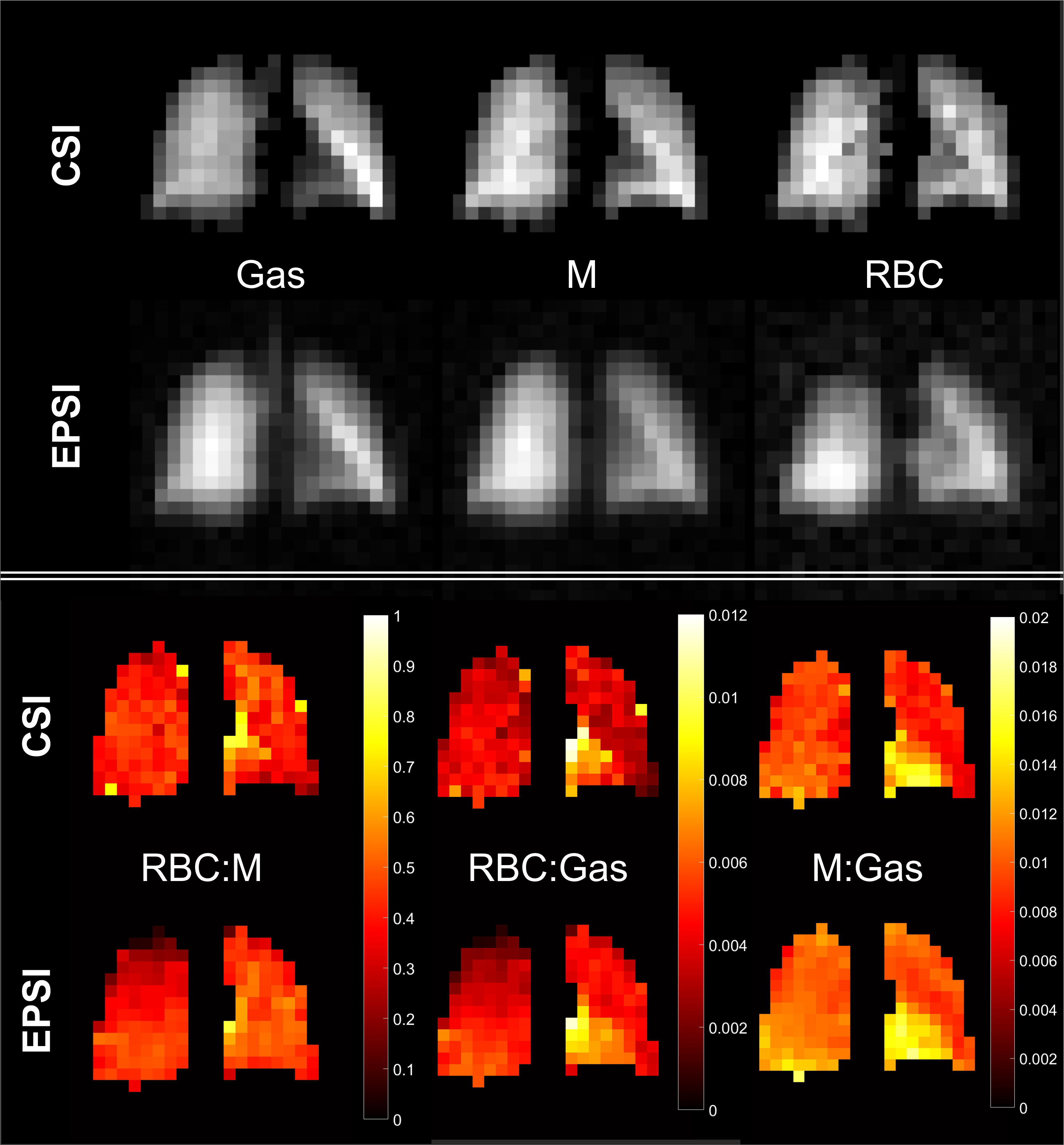
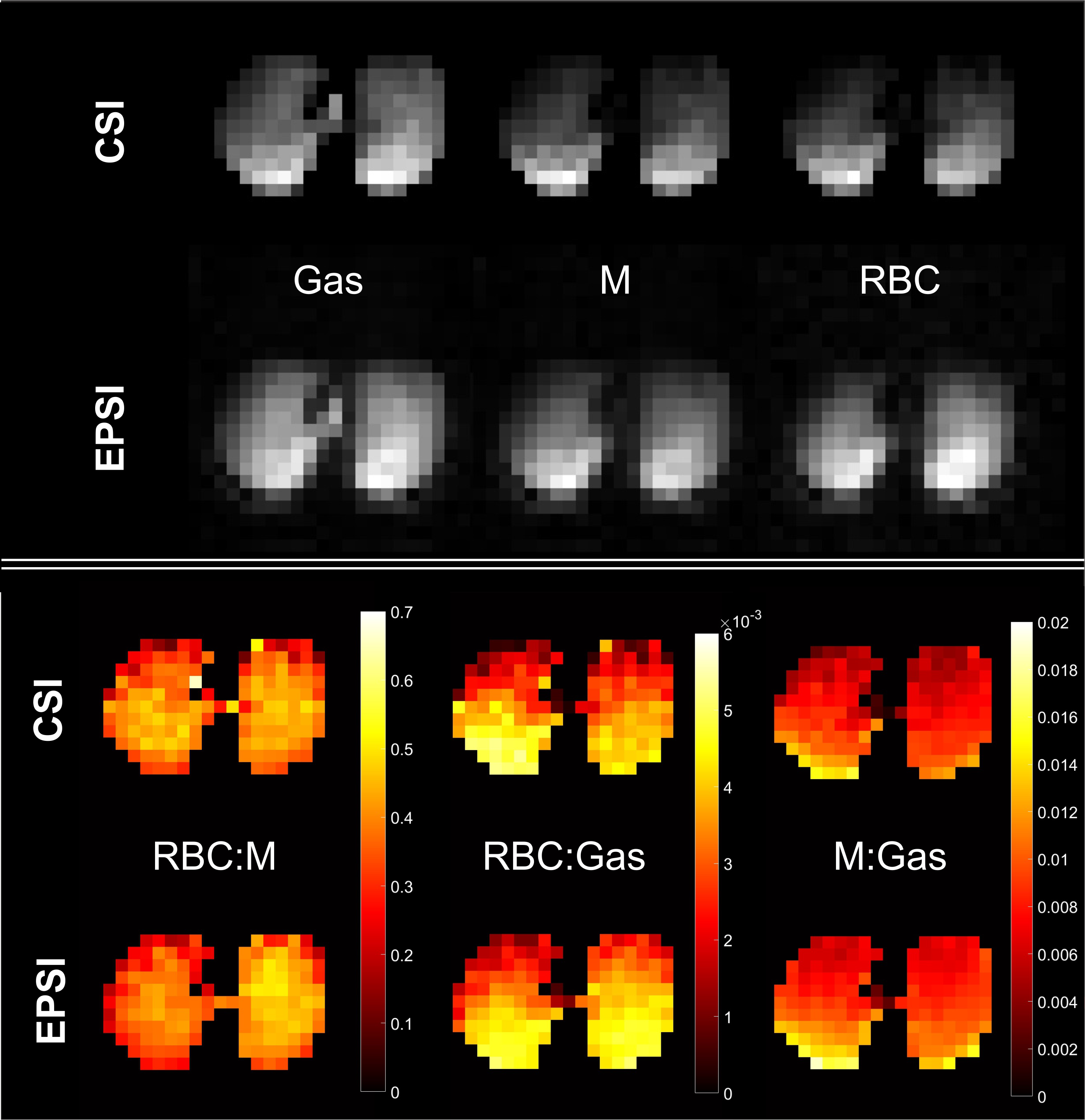
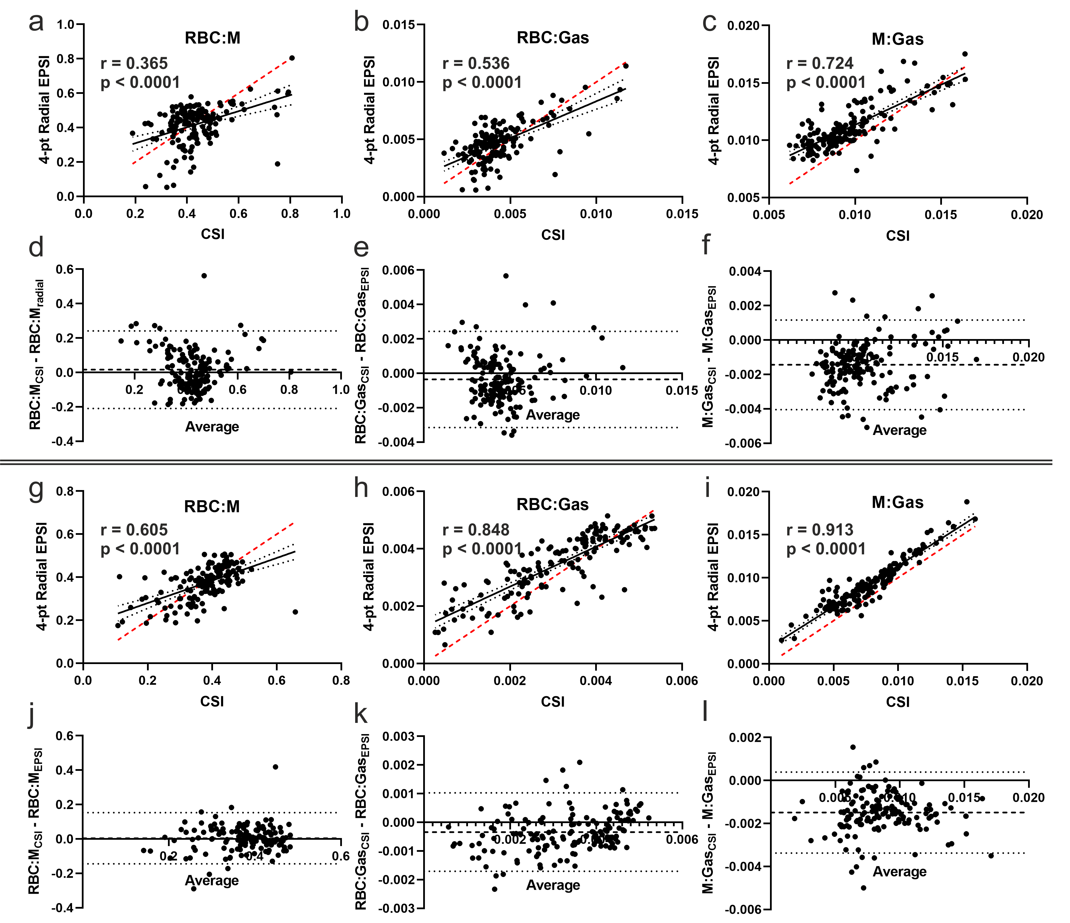
Coronal images of 129Xe gas and dissolved-phase signals from one volunteer are displayed in Figure 2, showing similar regional distributions between the two methods. The corresponding ratio maps show a mostly homogeneous distribution of 129Xe gas exchange metrics with mean RBC:M, RBC:Gas and M:Gas values of [0.43, 0.42 ], [0.0043, 0.0047] and [0.010, 0.011], respectively [CSI, EPSI], in agreement with the values expected for healthy volunteers (14). Figure 4 shows highly significant (p<0.0001) moderate to good pixel-wise correlations with Spearman r within [0.37-0.72] and biases of 0.016 (RBC:M), -0.00036 (RBC:Gas) and -0.004 (M:Gas). To compare the two methods on a dataset displaying a broader range of values and well-known regional postural related heterogeneity, the second volunteer was imaged axially. The gravitational dependence of the gas ventilation and the tissue density when imaged supine is clearly visible in the images and maps of Figure 3. The correlations improved (r within [0.61-0.91]), the bias for RBC:M remained negligible (0.004) while RBC:Gas and M:Gas were still lower for CSI than 4-pt EPSI (biases of -0.00034 and -0.0015).Discussion
CSI and 4-pt radial EPSI dissolved 129Xe imaging results were in good agreement with highly significant regional correlations (p<0.0001) between the gas exchange metrics and 95% confidence intervals of [-0.14, 0.15], [-0.0017, 0.0010], [-0.0034, 0.0004] for the RBC:M, RBC:Gas and M:Gas ratios, respectively, in the axial experiment. The biases for RBC:Gas and M:Gas could be explained by the limited spectral resolution of the CSI data to accurately measure the gas peak spectral linewidth. In future work, time-domain Voigt lineshape fitting will be implemented to circumvent spectral resolution limitations. Lower correlations were obtained for RBC:M, likely because there is no gravitational dependence or spatial inhomogeneity expected in healthy lungs for this parameter. Further validation in subjects with regionally heterogeneous lung disease / gas transfer limitation is needed.Conclusion
The results support the validity of our dissolved 129Xe 4-pt radial EPSI approach and that the chemical shift separation performed in k-space produces quantitative and reliable 129Xe gas exchange metrics.Acknowledgements
This work was supported by MRC grant MR/M008894/1 and by the National Institute for Health and Care Research (NIHR) Sheffield Biomedical Research Centre (NIHR203321). The views expressed are those of the author(s) and not necessarily those of the NIHR or the Department of Health and Social Care.References
1. Collier GJ, Schulte RF, Rao M, Norquay G, Ball J, Wild JM. Imaging gas-exchange lung function and brain tissue uptake of hyperpolarized (129) Xe using sampling density-weighted MRSI. Magn Reson Med 2023;89(6):2217-2226.
2. Kaushik SS, Robertson SH, Freeman MS, He M, Kelly KT, Roos JE, Rackley CR, Foster WM, McAdams HP, Driehuys B. Single-breath clinical imaging of hyperpolarized (129)Xe in the airspaces, barrier, and red blood cells using an interleaved 3D radial 1-point Dixon acquisition. Magnetic resonance in medicine 2016;75(4):1434-1443.
3. Qing K, Ruppert K, Jiang Y, Mata JF, Miller GW, Shim YM, Wang C, Ruset IC, Hersman FW, Altes TA, Mugler JP, 3rd. Regional mapping of gas uptake by blood and tissue in the human lung using hyperpolarized xenon-129 MRI. J Magn Reson Imaging 2014;39(2):346-359.
4. Collier GJ, Eaden JA, Hughes PJC, Bianchi SM, Stewart NJ, Weatherley ND, Norquay G, Schulte RF, Wild JM. Dissolved (129) Xe lung MRI with four-echo 3D radial spectroscopic imaging: Quantification of regional gas transfer in idiopathic pulmonary fibrosis. Magn Reson Med 2021;85(5):2622-2633.
5. Kammerman J, Hahn AD, Cadman RV, Malkus A, Mummy D, Fain SB. Transverse relaxation rates of pulmonary dissolved-phase Hyperpolarized (129) Xe as a biomarker of lung injury in idiopathic pulmonary fibrosis. Magn Reson Med 2020;84(4):1857-1867.
6. Wang JM, Robertson SH, Wang Z, He M, Virgincar RS, Schrank GM, Smigla RM, O'Riordan TG, Sundy J, Ebner L, Rackley CR, McAdams P, Driehuys B. Using hyperpolarized (129)Xe MRI to quantify regional gas transfer in idiopathic pulmonary fibrosis. Thorax 2018;73(1):21-28.
7. Weatherley ND, Stewart NJ, Chan HF, Austin M, Smith LJ, Collier G, Rao M, Marshall H, Norquay G, Renshaw SA, Bianchi SM, Wild JM. Hyperpolarised xenon magnetic resonance spectroscopy for the longitudinal assessment of changes in gas diffusion in IPF. Thorax 2019;74(5):500-502.
8. Qing K, Tustison NJ, Mugler JP, 3rd, Mata JF, Lin Z, Zhao L, Wang D, Feng X, Shin JY, Callahan SJ, Bergman MP, Ruppert K, Altes TA, Cassani JM, Shim YM. Probing Changes in Lung Physiology in COPD Using CT, Perfusion MRI, and Hyperpolarized Xenon-129 MRI. Acad Radiol 2019;26(3):326-334.
9. Saunders LC, Collier GJ, Chan HF, Hughes PJC, Smith LJ, Watson JGR, Meiring JE, Gabriel Z, Newman T, Plowright M, Wade P, Eaden JA, Thomas S, Strickland S, Gustafsson L, Bray JB, Marshall H, Capener DA, Armstrong L, Rodgers J, Brook M, Biancardi AM, Rao MR, Norquay G, Rodgers O, Munro R, Ball JE, Stewart NJ, Lawrie A, Jenkins RG, Grist JT, Gleeson F, Schulte RF, Johnson KM, Wilson FJ, Cahn A, Swift AJ, Rajaram S, Mills GH, Watson L, Collini PJ, Lawson R, Thompson AAR, Wild JM. Longitudinal Lung Function Assessment of Patients Hospitalized With COVID-19 Using (1)H and (129)Xe Lung MRI. Chest 2023.
10. Norquay G, Collier GJ, Rao M, Stewart NJ, Wild JM. 129Xe-Rb Spin-Exchange Optical Pumping with High Photon Efficiency. Physical Review Letters 2018;121(15):153201.
11. Ruppert K, Amzajerdian F, Hamedani H, Xin Y, Loza L, Achekzai T, Duncan IF, Profka H, Siddiqui S, Pourfathi M, Sertic F, Cereda MF, Kadlecek S, Rizi RR. Assessment of flip angle-TR equivalence for standardized dissolved-phase imaging of the lung with hyperpolarized 129Xe MRI. Magn Reson Med 2018.
12. Niedbalski PJ, Hall CS, Castro M, Eddy RL, Rayment JH, Svenningsen S, Parraga G, Zanette B, Santyr GE, Thomen RP, Stewart NJ, Collier GJ, Chan HF, Wild JM, Fain SB, Miller GW, Mata JF, Mugler JP, 3rd, Driehuys B, Willmering MM, Cleveland ZI, Woods JC. Protocols for multi-site trials using hyperpolarized (129) Xe MRI for imaging of ventilation, alveolar-airspace size, and gas exchange: A position paper from the (129) Xe MRI clinical trials consortium. Magn Reson Med 2021;86(6):2966-2986.
13. Kaushik SS, Freeman MS, Yoon SW, Liljeroth MG, Stiles JV, Roos JE, Foster W, Rackley CR, McAdams HP, Driehuys B. Measuring diffusion limitation with a perfusion-limited gas--hyperpolarized 129Xe gas-transfer spectroscopy in patients with idiopathic pulmonary fibrosis. J Appl Physiol 2014;117(6):577-585.
14. Collier GJ, Chan HF, Stewart NJ, Norquay G, Smith LJ, Rao M, Marshall H, Schulte RF, Wild JM. Age and lung volume dependence of dissolved xenon-129 imaging parameters. Proceedings of the ISMRM 2022. Abstract 1174.
Figures