4751
New Dual-Element 13C/1H Endorectal Coil for Improving mpMR-TRUS Guided Fusion Prostate Biopsies With Hyperpolarized C-13 Molecular Imaging1Radiology and Biomedical Imaging, University of California, San Francisco, San Francisco, United Kingdom, 2Bioengineering, University of California, San Francisco, San Francisco, CA, United States, 3Radiology and Biomedical Imaging, University of California, San Francisco, San Francisco, CA, United States, 4University of California, San Francisco, San Francisco, CA, United States, 5School of Medicine, University of California, San Francisco, San Francisco, CA, United States, 6Radiology and Biomedical Imaging, University of California, San Francisco, CA, United States
Synopsis
Keywords: Prostate, Hyperpolarized MR (Non-Gas), TRUS guided biosy
Motivation: To address the issues with the original dual-element 13C/1H endorectal coil (ERC).
Goal(s): We designed and fabricated a novel dual-element 13C/1H ERC, validated with bench testing and then evaluated in 3T MRI studies.
Approach: Phantoms were imaged using the new ERC and the original ERC. Acquired data was then analyzed and respective SNR compared.
Results: The new ERC demonstrated substantially improved 13C and 1H MRI sensitivity on bench electronic testing. In addition, the new ERC demonstrated substantial improvement in signal-to-noise ratio, and since the coil has successfully been used in hyperpolarized 13C-pyruvate, co-polarized 13C-urea+pyruvate multiparametric MRI patient studies for guiding prostate cancer biopsy.
Impact: Hyperpolarized 13C MRI, RF Coils, Metabolic imaging, Molecular imaging, Endorectal coil
Introduction
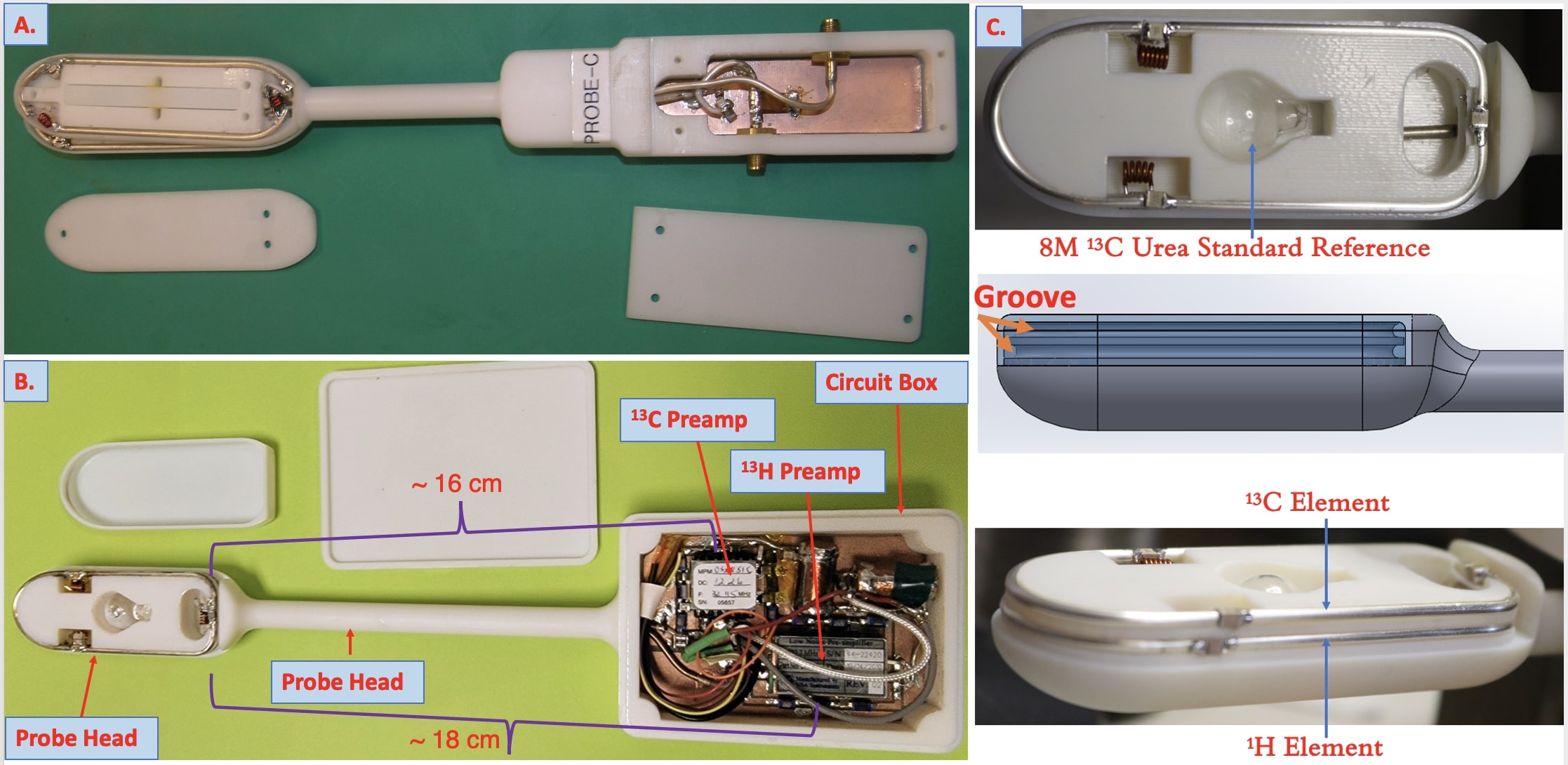
RF coils are critical components of a MRI system by which the MRI signal is excited and received.1 Thus, informed specification, design and construction, evaluation, and application of properly selected RF coils are critical to a safe and successful MRI scan.1 Endorectal coil (ERC) is inserted in the rectum like an ultrasound probe to enable improved prostate MRI. Although ERCs with two or more channels have been developed,2 they usually are only for proton imaging. In the case of hyperpolarized (HP) metabolic imaging, we focus on common C-13 metabolites that are approved for patient studies. The original 13C/1H ERC created 15 years ago has been used in hundreds of prostate cancer patient exams (Figures 1A) but is limited by suboptimal geometry with the probe head being too large and the neck too short, residual RF coupling during transmit, limited direct disinfection in chemical bath, transmission cables did not have built-in RF blocks, and preamps located ~154cm away from the probe in T/R box. To address the above-mentioned issues, the project was designed to construct a new dual-element 13C/ 1H ERC that, compared to prior designs, would provide substantially improved sensitivity, rf isolation, and reliability with improved electronics for hyperpolarized C-13 prostate MRI for guiding biopsies.Methods
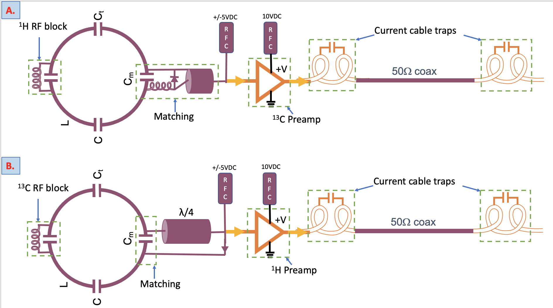
The housing was designed with nurse and technologist input to be contoured to anatomy, with tolerability and size more similar to transrectal ultrasound (TRUS) probes, using computer design software (SolidWorks 2020) and commercially 3D printed by Stratasys with a ultem1010 resin(Figure 1B). The probe is designed to have a shorter head (1.0in wide X 3in long) to increase reduce patient discomfort and longer neck for ease of maneuver. The preamps were placed 16-18cms from the probe head in the circuit box (Figure 1B). The 1H and 13C elements were made using silver plated copper wire and fit in the groves on the probe head housing (Figure 1C). To obtain the highest sensitivity C-13 element is placed on top to be closer to the prostate as 13C is less sensitive than 1H (Figure 1C). The proton element is detuned using the semi-rigid coaxial line connection to the pre-amplifier. The diode is located near the pre-amplifier and serves as pre-amplifier protection and active element detuning. For 13C a normal trap circuit was built at the taping capacitor with a diode to ground to actively detune the element during transmit. An 8 molar solution of doped 13C-urea in a NMR-compatible 1cc spherical glass phantom was constructed and placed at the center of the probe head for standard reference in 13C imaging. Miniature parallel resonant (1/16 ”) circuits was constructed for passively blocking 1H frequency in the 13C prostate element and 13C frequency in the 1H prostate element (Figure 2). The circuits of both elements are then tuned & matched. Keysight/Agilent E5071C ENA Vector Network Analyzer was used to test the components and the coil. Qunloaded for 1H = 301, Qloaded for 1H = 44, Qunloaded for 13C = 172, Qloaded for 13C = 55, resulting Q-factors 3.4 for the 13C and 6.9 for 1H. The new ERC has satisfied the body noise dominance requirement.3 The respective Q factors for the respective elements of the original coil are 1.5 and 2.8. Phantom imaging was performed using 8mm isotropic EPI imaging with a 2s temporal resolution, FOV= 24X24cm, slice thickness=10mm, matrix=16X16 on 3T GE MRI.Results and Discussion
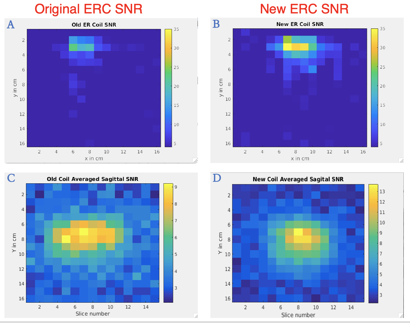
The new ERC showed twice the Q factor value of the original ERC. In addition, on phantom testing the new ERC has shown an improved SNR by about 60% in axial and 80% in the sagittal data compared to the original ERC as shown in Figure 3.Conclusion
We designed and constructed a novel dual-element 13C/1H ERC contoured to the anatomy similar to clinical TRUS probes that are used for prostate biopsies, but enables HP 13C metabolic prostate MR imaging for guiding biopsies with higher anatomic and MR molecular image quality.Acknowledgements
No acknowledgement found.References
1. Vaunghan, J. T & Griffiths, J. R, RF Coils for MRI (JohnWiley and Sons Ltd.,2012)
2. Mo Z, Zhang X, Liang H, Chen Q, Tie C, Xiao W, Cao Q, Liu C, Zou C, Wan L, Zhang X, Li Y. A Novel Three-Channel Endorectal Coil for Prostate Magnetic Resonance Imaging at 3T. IEEE Trans Biomed Eng. 2023 Jun 15;PP. doi: 10.1109/TBME.2023.3286488. Epub ahead of print. PMID: 37318962.
3. Roemer PB, Edelstein WA, Hayes CE, et al. The NMR phased array. Magn Reson Med. 1990; 16:192–225. [PubMed: 2266841]
Figures