4741
3D vector MR elastography (MRE) assessed tissue heterogeneity increases with chronic liver disease progression1Radiology, Mayo Clinic, Rochester, MN, United States, 2Gastroenterology and Hepatology, Mayo Clinic, Rochester, MN, United States
Synopsis
Keywords: Liver, Elastography, heterogeneity, MASLD
Motivation: Hepatic fibrosis, despite traditionally viewed as a diffuse occurrence, has been shown to vary spatially with disease progression.
Goal(s): MR elastography (MRE) measures mechanical properties and their spatial variations can potentially provide holistic insights into tissue inhomogeneity.
Approach: Twenty-five rats based a metabolic dysfunction associated steatotic liver disease (MASLD) model monthly underwent MRE to investigate tissue inhomogeneity during cirrhosis development.
Results: Results demonstrated increases in both microscopic and macroscopic inhomogeneities with disease progression. In cirrhotic livers, while mean MRE measurements correlated subtly with histological fibrosis and portal pressure, tissue heterogeneity presented stronger associations. Liver tissue heterogeneity is a complementary predictor of disease progression.
Impact: Assessing liver tissue heterogeneity via 3D vector MR
elastography could enhance disease progression monitoring in chronic liver
diseases and potentially predict clinically significant outcomes, offering a
more comprehensive diagnostic approach than traditional mean liver stiffness
and loss modulus measurements alone.
Introduction
Hepatic tissue heterogeneity is a recognized phenomenon during fibrosis development, despite fibrosis being traditionally considered a diffuse disease. Biopsy studies have revealed histologic disparities in different liver regions 1. MRE is a well-established tool for hepatic fibrosis, with the mean liver stiffness measurement typically utilized in clinical management 2. In conjunction with the overall mean measurements, spatial variation of tissue mechanical properties might provide a more complete assessments of tissue response during disease progression. Given this context, our study aims to bridge the knowledge gap by linking tissue heterogeneity with disease progression, especially in the widespread MASLD. We hope this connection can provide a more nuanced tool for monitoring treatment responses, especially in scenarios where mean measurements fall short.Methods
Our study involved 25 rats: twenty rats received a choline-deficient high-fat diet (CDHFD), leading to progressive MASLD, while five age-matched control rats had a normal chow diet. Monthly 3D vector MRE examinations were conducted on all animals, assessing liver mechanical properties from baseline to endpoint under CDHFD or normal diet conditions. At 12 weeks (CDHFD=10, Control=5) and 16 weeks (CDHFD=10), rats were sacrificed for portal pressure measurement and tissue harvesting, respectively. Histologic analyses were performed manually by an experienced pathologist and digitally using HALO software (version 3.6). Sirius red-stained liver specimens were quantified by subdividing each slide into 2 x 2 mm² segments for different lobes to measure fibrosis area ratio.We used Coefficient of Variation (CV) and standard deviation (STD) to assess the dispersion of mechanical properties and histologic fibrosis ratio distribution, indicating tissue heterogeneity. Group differences were compared using Mann–Whitney U tests, while Pearson correlation was employed to evaluate relationships between tissue mechanics, histologic heterogeneity, and portal pressure, considering correlations ≥0.7, 0.6, and 0.5 as strong, moderate, and weak, respectively.
Results
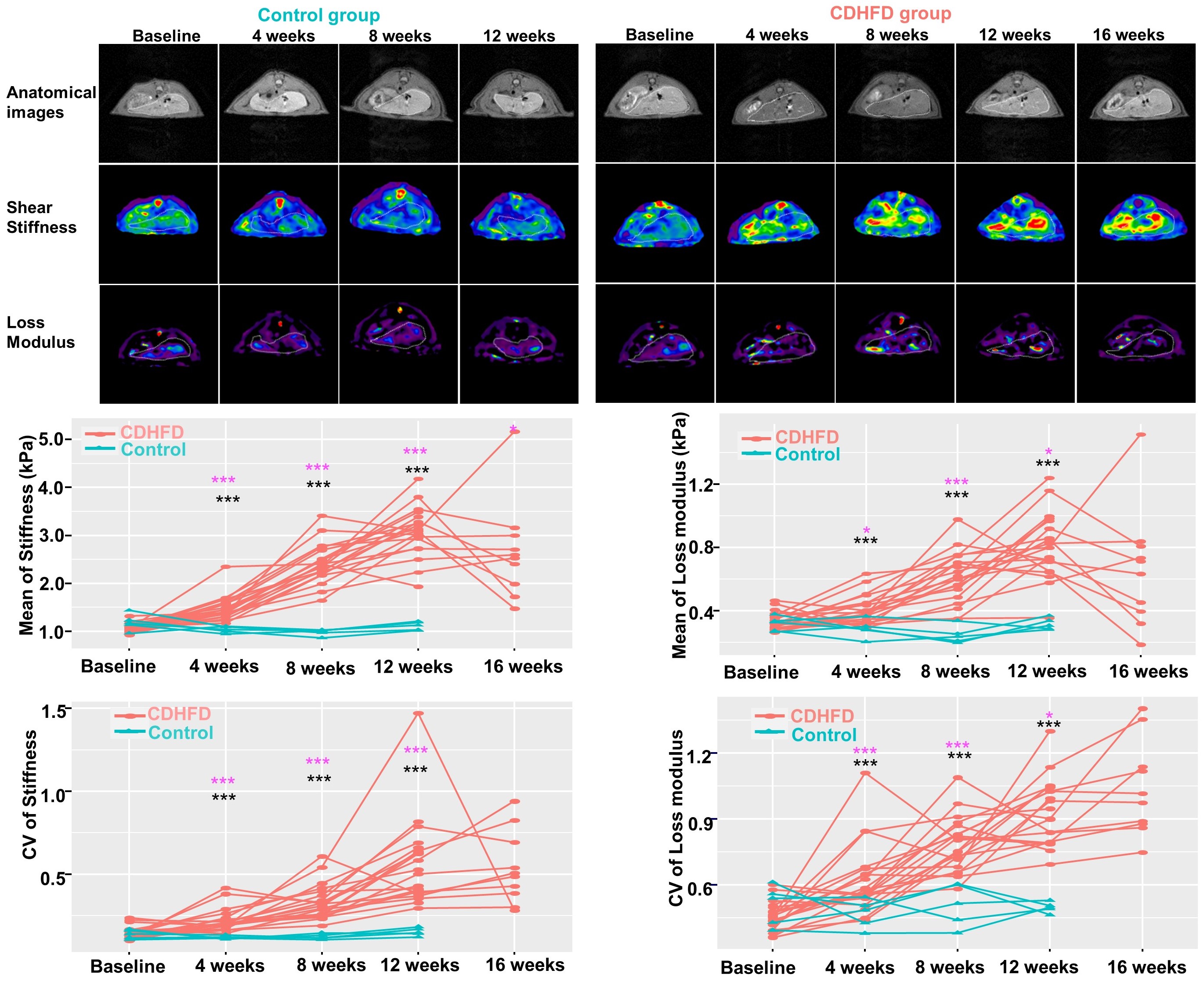
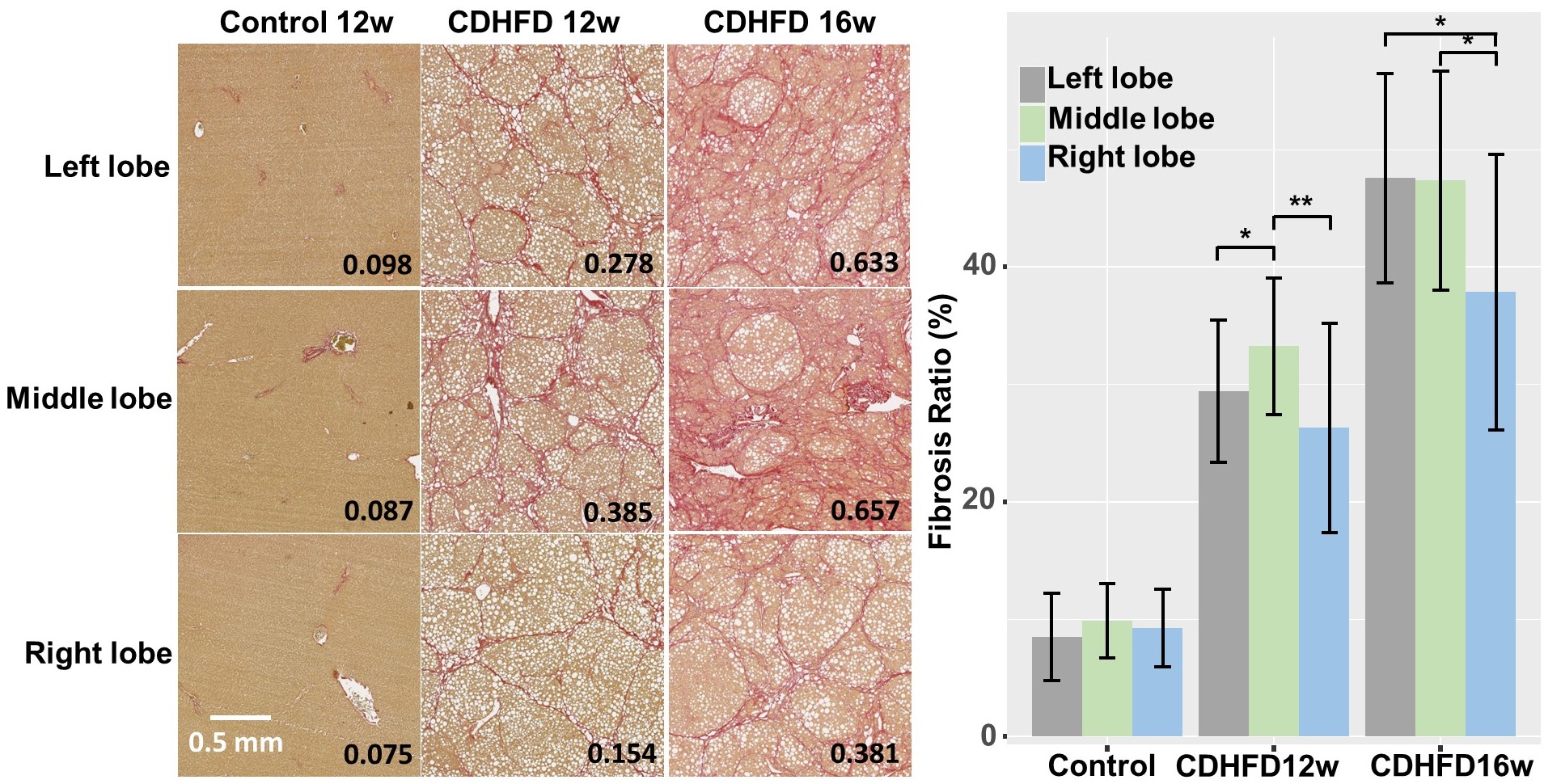
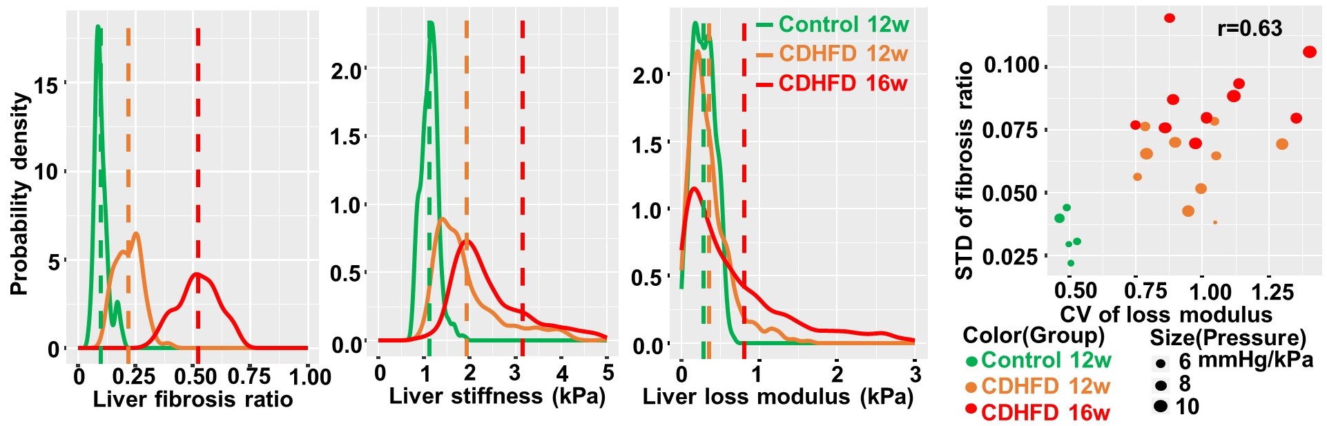
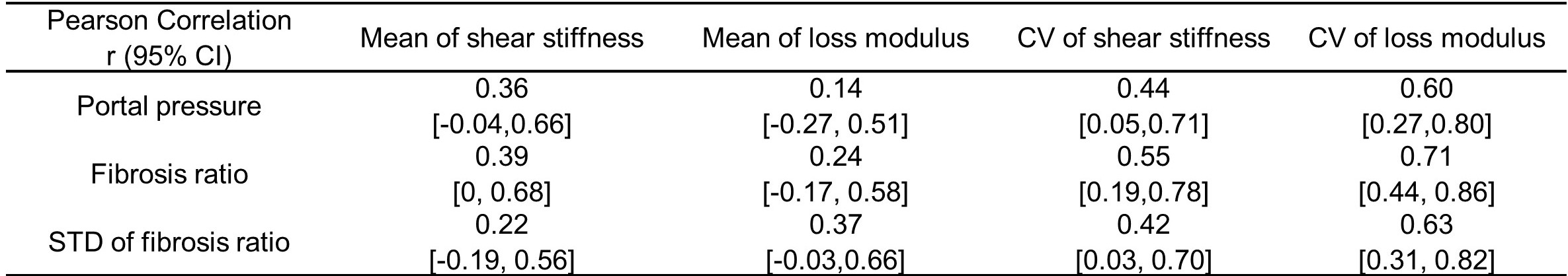
In Figure 1, the CV for shear stiffness in the CDHFD group increased from 0.12 at baseline to 0.54 at 16 weeks, while the CV for loss modulus rose from 0.50 to 1.14 over the same time points. All CDHFD mice developed cirrhosis and portal hypertension at the time of sacrifice. In the control group, both shear stiffness and loss modulus remained relatively stable. The statistical results indicate that liver inhomogeneity, as assessed in the maps of shear stiffness and loss modulus, significantly increased with the progression of fibrosis in this MASLD rat model.Figure 2 demonstrated differences in the fibrosis ratio across the left, middle, and right lobes of the liver in CDHFD rats, which evolved as the disease advanced. Figure 3 displayed changing histograms for fibrosis ratio, shear stiffness, and loss modulus, indicating varying shapes and differentiation ability with disease progression. Table 1 presented Pearson correlations and 95% confidence intervals between pathophysiological and MRE measurements. In cirrhotic livers, while mean measurements of mechanical properties showed limited associations with portal pressure and fibrosis ratio, the inhomogeneity exhibited a strong/moderate and positive correlation with them.
Discussion
In chronic liver diseases, the fibrogenic wound-healing response leads to alterations in liver stiffness. Over time, both the mean and CV generally show a marked increase in each individual animal with MASLD disease progression. Even in the absence of longitudinal histologic evidence, it can be inferred that tissue heterogeneity is amplified as MASLD progresses. This rise in tissue heterogeneity may make biopsy results unreliable in severe fibrosis 3. During the end-stage of liver disease, loss modulus is linked to fluid-associated inflammation and portal hypertension 4. As a result, CV of loss modulus demonstrates a stronger correlation with portal pressure than stiffness in our study.In cirrhosis, complex mechanisms like microvascular thrombosis and localized necroinflammation can reduce mean stiffness while increasing tissue inhomogeneity, as confirmed by our preclinical findings 5. Cirrhosis raises the risk of liver complications, such as hepatocellular carcinoma (HCC), which often exhibits pronounced heterogeneity, characterized by dense malignant cells, increased transcriptional diversity, and reduced spatial continuity 6. The inherent heterogeneity in hepatic parenchyma, characterized by variations in tissue mechanics and structural integrity, can foreshadow pathologic changes. This complexity, when accentuated, may create an environment conducive to the onset and progression of conditions like HCC, underscoring the importance of studying such heterogeneities in predicting significant clinical outcomes in cirrhosis at a macroscopic level. Thus, tissue heterogeneity, when assessed via 3D vector MR elastography, could be a promising prognostic indicator for predicting cirrhosis-associated complications.
Conclusion
Tissue mechanical heterogeneity has a positive correlation with disease progression based on 3D MRE biomarkers in the rat model. Spatial variation of tissue mechanical properties has the potential to complement mean measurements for comprehensively revealing tissue response during disease progression.Acknowledgements
No acknowledgement found.References
1.Ratziu V, Charlotte F, Heurtier A, et al. Sampling variability of liver biopsy in nonalcoholic fatty liver disease. Gastroenterology. 2005; 128(7):1898-1906.
2.Lim JK, Flamm SL, Singh S, et al. American gastroenterological association institute guideline on the role of elastography in the evaluation of liver fibrosis. Gastroenterology. 2017;152(6): 1536-1543.
3.Kawamura N, Imajo K, Kalutkiewicz KJ, et al. Influence of liver stiffness heterogeneity on staging fibrosis in patients with nonalcoholic fatty liver disease. Hepatology 2022;76(1):186-195.
4.Li J, Allen AM, Shah VH, et al. Longitudinal changes in MR elastography-based biomarkers in obese patients treated with bariatric surgery. Clin Gastroenterol Hepatol 2023;21(1):220-222.
5.Anaparthy R, Talwalkar JA, Yin M, et al. Liver stiffness measurement by magnetic resonance elastography is not associated with developing hepatocellular carcinoma in subjects with compensated cirrhosis. Aliment Pharmacol Ther. 2011;34(1):83-91.
6.Zhang Q, Lou Y, Yang J, et al. Integrated multiomic analysis reveals comprehensive tumour heterogeneity and novel immunophenotypic classification in hepatocellular carcinomas. Gut. 2019;68(11):2019-2031.
Figures