4735
Diagnostic value of GRASP in the differentiation of breast malignancy1The Fourth Affiliated Hospital of Hebei Medical University, Shijiazhuang, China, 2MR Research Collaboration, Siemens Healthineers Ltd, Beijing, China
Synopsis
Keywords: Perfusion, Contrast Mechanisms, breast tumor; dynamic contrast-enhanced magnetic resonance imaging (DCE-MRI)
Motivation: Breast MRI plays a vital role in early breast cancer screening, staging, and surgical guidance. The core technology used in breast MRI is DCE-MRI.
Goal(s): This study aims to assess the effectiveness of using the GRASP sequence in distinguishing highly malignant breast lesions.
Approach: We utilized the GRASP sequence, which offers high temporal resolution, to compare and analyze the microvascular characteristics of benign and malignant breast lesions.
Results: We found that Kep was significantly higher in malignant lesions compared to benign lesions, suggesting a potential relationship between the abundant blood supply and high wall permeability observed in malignancies.
Impact: The GRASP dynamic enhancement technique provides both morphological and hemodynamic characteristics of breast lesions, aiding in the differentiation of high-grade malignancies.
Introduction
Tumorigenesis, development, and metastasis are closely associated with angiogenesis, which is the formation of new blood vessels. Tumors are characterized by a significant increase in the number of blood vessels, which can lead to disturbed blood flow in the microcirculation. In contrast, benign tumors have relatively mature vascular morphology and good permeability, while malignant tumors have lower permeability compared to normal breast tissue. DCE-MRI of the breast provides valuable information about the microvascular features of benign and malignant lesions. In benign lesions, the microvessel walls remain intact, whereas malignant tumor tissue exhibits the development of numerous tiny blood vessels with incomplete walls. Nutrients from the bloodstream can penetrate the vessel walls and nourish the surrounding tumor cells, supporting their invasive growth. DCE-MRI based on the physiological model of "neoangiogenesis" offers morphological and hemodynamic features of the breast lesion1. The temporal resolution of the imaging technique is a crucial factor influencing the results of quantitative DCE-MRI analysis. GRASP-based dynamic enhanced scans can acquire quantitative parameters with high temporal resolution, thereby improving the accuracy of breast lesion diagnosis. Therefore, this study seeks to explore the diagnostic value of GRASP dynamic enhanced scanning in differentiating highly malignant breast lesions.Method
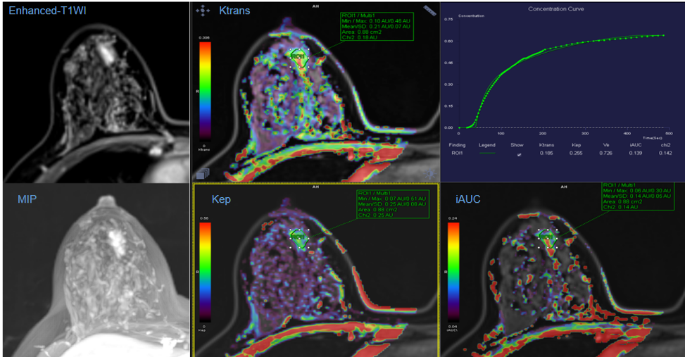
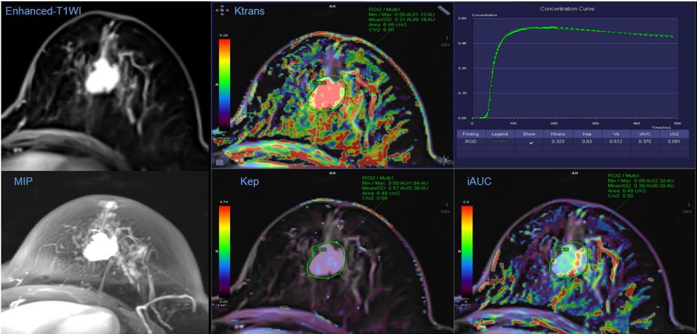
This study recruited a total of 14 patients. Based on their pathological results and clinical grade, 6 cases were classified as benign or low-grade malignant breast cancer, while 8 cases were classified as high-grade malignant breast cancer. All patients underwent breast MRI using a 3T MR scanner (MAGNETOM Vida, Siemens Healthcare, Erlangen, Germany). Before and after the administration of contrast media (0.2 mmol/kg, Gadovist, Bayer) at a rate of 3mL/s, followed by a 10-mL saline chaser, ultrafast dynamic contrast-enhanced (UF DCE)-MRI was acquired using a GRASP sequence with free breathing, with a total acquisition time of 8 minutes and 25 seconds. The scanning parameters were as follows: time resolution = 3s, field of view = 380 mm x 380 mm, matrix = 380 x 320, voxel size = 1.2 mm x 1.2 mm x 2.5 mm.For the analysis of the acquired data, dynamic enhanced post-processing analysis was performed using the Tissue 4D toolbox on a workstation (Syngo.via VB20A, Siemens Healthcare). This analysis enabled the extraction of pharmacokinetic parameters, specifically Ktrans, Kep, and Ve. These parameters were subsequently compared between highly malignant and benign tumors using an independent sample t-test.
Results
As shown in Table 1, the Kep indicator of the benign group was 0.27 ±0.19min-1, while the Kep indicator of the highly malignant group was 0.53 ±0.21min-1. The overall difference between the two groups was found to be statistically significant (P = 0.037). However, no statistically significant differences were observed for the other parameters (P> 0.05).Discussion
The Kep value reflects the reflux of the contrast agent from the extracellular space to the vascular lumen. A higher Kep value indicates a worse degree of vascular endothelium differentiation, higher vascular wall permeability, increased local blood perfusion of the tumor, and a higher degree of malignancy. The present study discovered a significantly increased Kep value in the highly malignant group, suggesting that the return of the contrast agent to the vascular lumen was significantly faster in this group compared to the benign group. This finding is related to the abundant blood supply and high wall permeability of malignant tumors. Ktrans reflects the absolute parameters of vascular perfusion speed and Ve reflects the vascular extracellular space of the whole voxel volume ratio. Previous studies have reported that Ktrans is an important indicator for distinguishing between benign and malignant breast tumors, while the value of Ve in this differentiation is controversial 2,3. In our study, the overall difference in the Ktrans index did not reach statistical significance. This may be due to the limited number of cases and suggests the need for further analysis with a larger sample size in the future.Conclusion
Our findings show that in DCE-MRI based on the GRASP technique, the Kep value was higher in highly malignant breast tumors compared to benign tumors. Therefore, Kep has potential as a reference value for differentiating between benign and malignant breast tumors and could become a common diagnostic method for breast tumors.Acknowledgements
No acknowledgement found.References
1.Cho N. Breast Cancer Radiogenomics: Association of Enhancement Pattern at DCE MRI with Deregulation of mTOR Pathway. Radiology. 2020 Aug;296(2):288-289.
2.Sun TT, Liu WH, Zhang YQ,et al. Diagnostic value of quantitative pharmacokinetic parameters and relative quantitative pharmacokinetic parameters in breast lesions with dynamic contrast-enhanced MRI. Zhonghua Yi Xue Za Zhi. 2017 Aug 1;97(29):2266-2270. Chinese.
3.Amarnath J, Sangeeta T, Mehta SB. Role of quantitative pharmacokinetic parameter (transfer constant: K(trans)) in the characterization of breast lesions on MRI. Indian J Radiol Imaging. 2013 Jan;23(1):19-25.
Figures
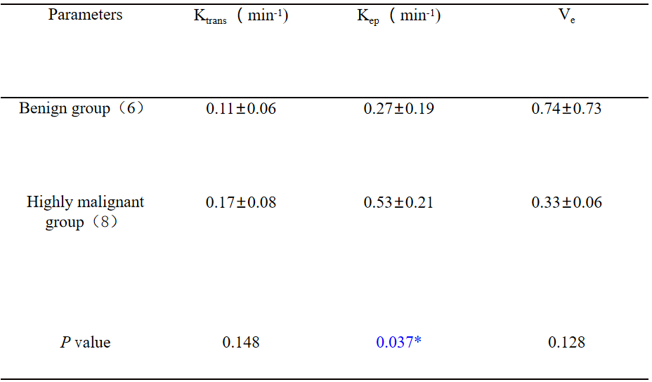
Table1 Comparison of pharmacokinetic parameters between benign and malignant breast lesions.
Note:Values are presented as mean ± standard deviation."*" indicates that the P value is less than 0.05, and the difference is considered statistically significant .