4725
Monitoring Extracellular Oxygenation Modulation with myo-inositol tripyrophosphate using EPRI1MD Anderson Cancer Center, Houston, TX, United States, 2The University of Texas Health Science Center at Houston Graduate School of Biomedical Sciences, Houston, TX, United States
Synopsis
Keywords: Biomarkers, Cancer, ITPP, EPRI, Immunotherapy, Radiosensitizer, Breast Cancer
Motivation: We showed previously that multispectral optoacoustic tomography (MSOT) measures oxygen saturation (%sO2) of intravascular hemoglobin which can indirectly evaluate the effect of a radiosensitizer, inositol tripyrophosphate (ITPP), on tumor oxygenation. Electron paramagnetic resonance imaging (EPRI) could serve as a more direct biomarker by measuring the extravascular, extracellular oxygen pressure (pO2) in pre-clinical tumor models.
Goal(s): Validate prior MSOT results showing decrease in hemoglobin saturation with ITPP.
Approach: Use EPRI to observe extracellular oxygenation before and after treatment with ITPP compared with vehicle control.
Results: Tumors treated with ITPP demonstrated lower extracellular oxygenation compared to pretreatment levels (p = 0.003).
Impact: An imaging biomarker to determine how much a radiosensitizer improves tumor pO2 would dramatically impact clinical care by potentially improving response to radiotherapy and immunotherapy. EPRI could evaluate the effect of a radiosensitizer allowing for more personalized treatment approaches.
Introduction
Extracellular oxygenation is an important player in cancer treatment. Increased oxygenation improves response to radiation therapy. These radiosensitizers could also improve response to immunotherapy because extracellular hypoxia has been known to cause immunotherapy resistance.1 Hypoxia imaging techniques such as electron paramagnetic resonance imaging (EPRI) to measure extracellular pO2 can be used as an imaging biomarker to evaluate tumor oxygenation. Inositol tripyrophosphate (ITPP) is an allosteric modifier of hemoglobin causing a rightward shift of the hemoglobin oxygen dissociation curve.2,3 Prior work by our group has shown that ITPP followed 3h later by immune checkpoint blockade (αPD-1, αCTLA-4) caused both tumor regression and increase in survival.4 Multispectral optoacoustic tomogrophy (MSOT) measurement of hemoglobin saturation of 4T1 tumors treated with ITPP showed decrease in hemoglobin saturation 3h after ITPP injection.4 We hypothesized this was due to increased oxygen offloading into the tumor microenvironment, and using EPRI we could observe a corresponding increase in extravascular, extracellular oxygen. Our goals for this study were two-fold: 1) to establish an EPRI protocol to monitor a change in tumor oxygenation after ITPP treatment, and 2) to confirm an increase in extracellular oxygenation with ITPP treatment to further validate our observations measuring hemoglobin saturation with MSOT after treatment with ITPP.Methods
We implanted 4T1 tumors in BALB/c mice and allowed them to grow until they reached a diameter of 5 mm in an identical fashion to our prior MSOT studies. EPRI imaging was performed as follows: a computed tomography (CT) scan was acquired for anatomical reference which was segmented and registered along with an echo spin echo (ESE) experiment on the JIVA-25 EPRI instrument (o2M Technologies, Inc.). We evaluated oxygenation within the tumor by performing an inversion recovery echo spin echo (IRESE) experiment within a JIVA-25 instrument 27 minutes after intraperitoneal injection with OX071. We performed EPRI as a baseline and then 3h after either 36.5 mg ITPP or PBS i.p. injection. Average oxygenation was obtained by averaging pO2 values for each voxel within the tumor. The change in oxygenation was obtained by subtracting oxygenation at baseline from the average oxygenation after treatment. Baseline oxygenation was compared with post-treatment oxygenation using a paired two-tailed Student‘s t-test. ITPP treatment versus PBS control was compared using an unpaired two-tailed Student’s t-test for both average oxygenation and the change in oxygenation.Results
Tumor treated with ITPP demonstrated an average oxygenation of 37.6 Torr prior to treatment and 30.4 Torr post treatment (p = 0.003). Tumors treated with PBS demonstrated an average oxygenation of 33.6 torr prior to treatment and 38.7 Torr post treatment. The difference between the pre and post treatment groups for PBS control were not significant. Differences between the ITPP treatment group and vehicle control were not statistically significant before (p = 0.144) or after treatment (p = 0.064). The average change in oxygenation after ITPP treatment was –10.2 Torr. The average change in oxygenation after PBS control was +5.1 Torr. The change in oxygenation after ITPP treatment was statistically significantly different from the change in oxygenation after PBS control (p = 0.01).Discussion
Our prior work using MSOT to evaluate the effect of ITPP on tumor oxygenation demonstrated a decrease in hemoglobin saturation 3h after ITPP administration.4 We expected an increase in the extracellular oxygenation as we hypothesized the decrease in hemoglobin saturation was due to oxygen offloading into the extracellular space. We observed the opposite of this. Other groups have reported an increase in pO2 in response to ITPP, however, none of these other reported results have used our 3h timescale.2,3 Overall, EPRI is an excellent noninvasive technique for elucidating the time course of pO2 in response to ITPP.Conclusion
For 4T1 orthotopic tumors, there is a decrease in extracellular oxygenation 3h post-ITPP treatment. The mechanism behind this change is unclear and merits further consideration. It is possible physiologic compensation is present resulting in a temporarily lowered oxygenation or another effect of ITPP dominates at this timescale such as vascular normalization. Regardless, this study emphasizes the importance of timing to ensure the most effective response to treatment.Acknowledgements
The research has been funded in part by R01CA231513, CPRIT (RP220270), and CCTS TL1. We would also like to thank the CCSG-funded Small Animal Imaging (SAIF) for their expertise and assistance in image acquisition.References
1. Kopecka J, Salaroglio IC, Perez-Ruiz E, et al. Hypoxia as a driver of resistance to immunotherapy. Drug Resist Updat 2021; 59: 100787. DOI: 10.1016/j.drup.2021.100787.
2. Krzykawaska-Serda M, Szczygiel D, Gawel S, et al. Oxygen therapeutic window induced by myo-inositol tripyrophosphate (ITPP) – Local pO2 study in murine tumors.
3. Tran L, Cao-Pham T, Jordan B, et al. Impact of myo-inositol tripyrophosphate (ITPP) on tumour oxygenation and response to irradiation in rodent tumour models. J Cell Mol Med 2019; 23(3): 1908-1916. DOI: 10.1111/jcmm.14092.
4. Chin R, Liang X, Li T, et al. Potentiation of the immune checkpoint blockade response by tumor acidosis and hypxoia modulation is predictable using molecular imaging. J Immunother Cancer 2022; 10(Suppl 2):A1-A1603. DOI:10.1136/jitc-2022-SITC2022.0004.
Figures
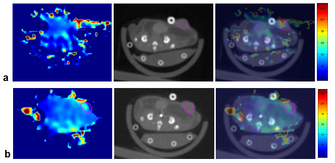
Figure 1. Representative oxygen map from EPRI, CT image and CT with overlay. Tumor ROI is circled in pink. Range of partial pressures restricted to –5 torr to 128 torr. A) Baseline imaging. Average pO2 for this sample was 46.8 Torr. Images are from left to right pO2 map, CT anatomical image, and pO2 map overlaid on anatomical CT. B) Imaging 3 hours after treatment with ITPP. Average pO2 for this sample was 38.9 Torr. Image order is same as in part A.
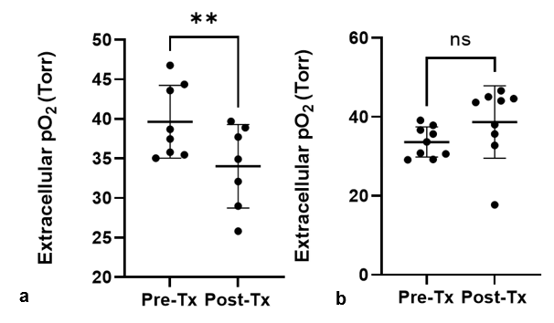
Figure 2. Average extracellular oxygen partial pressure within tumors treated with ITPP and PBS. A) Oxygenation 24h prior to ITPP administration (Pre-Tx) and 3h post-ITPP administration (Post-Tx). Oxygenation decreased significantly **p = 0.003. B) Oxygenation 24h prior to PBS administration (Pre-Tx) and 3h post-ITPP administration (Post-Tx). Oxygenation did not change significantly p = 0.259.
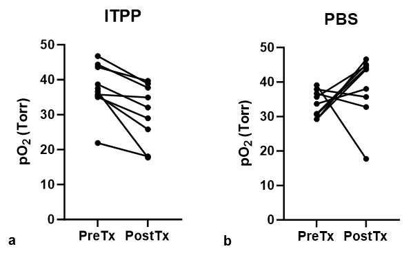
Figure 3. Mouse-wise comparison of extracellular partial pressure. A) Average pO2 of mice imaged 24h before ITPP injection and 3h post injection. In all mice, pO2 decreased. B) Average pO2 of mice imaged 24h before PBS injection and 3h post injection. Change in pO2 was variable.