4705
Prediction of myelin stainings using 7T MRI and deep learning1Department of Medical Engineering, Carinthia University of Applied Sciences, Klagenfurt, Austria, 2Division of Neuropathology and Neurochemistry, Department of Neurology, Medical University of Vienna, Vienna, Austria
Synopsis
Keywords: Diagnosis/Prediction, Machine Learning/Artificial Intelligence, Brain Histological Prediction
Motivation: Histological examination of the brain provides precise analyses of brain tissue, but requires ex vivo tissue samples. In vivo MRI offers an alternative but still has limitations.
Goal(s): Predict histologic images from in vivo MR images.
Approach: Utilize deep learning generative models to create brain histological images from multi contrast MR images.
Results: The appropriate combination of MRI contrasts can generate myelin histology images.
Impact: Generating myelin histological images significantly impacts brain tissue property research by providing ex vivo information. This adaptable technique extends its applicability to various tissue properties, providing broader insights into histology and beyond.
Introduction
Histological properties of brain tissue, like myelin and iron, are important biomarkers for neurodegenerative diseases. Quantitative susceptibility mapping (QSM) and R2* mapping of magnetic resonance imaging (MRI) has been found to be effective in assessing iron levels but are limited in their ability to assess myelin. In vivo MRI aims to connect cellular-level microstructure to macroscopic brain properties. However, our understanding of the exact relationship between these tissue properties (e.g., myelin and iron distribution) and MRI images is limited, and furthermore, these parameters are currently accessible only through ex vivo histology. This concept leads to the utilization of deep learning, which has the potential to predict histology from MRI data. This offers an opportunity to enhance our understanding of brain development, function, and diseases.Methods
Data acquisition:In this study, which was approved by the ethics committee of the Medical University of Vienna (EK Nr. 1727/2014), a human cadaver head from an 88-year-old female was imaged using a 7 T Siemens MAGNETOM MRI system.
T1 weighted images were acquired using an MP2RAGE sequence and to create SWI, QSM and R2* data a single- (TE = 12 ms, TR = 25 ms, voxel size = 0.23 mm x 0.23 mm x 0.62 mm) and a multi-echo gradient echo scan (TE1/TE2/TE3/TE4 = 4 ms/9 ms/15 ms/22 ms, TR = 26 ms, voxel size = 0.43 mm x 0.43 mm x 0.65 mm) was used.
After MRI, the brain was removed and prepared for histological staining. Myelin staining was performed using the Luxol fast blue with periodic acid Schiff method (Vincent et al. 2016).
Image registration:
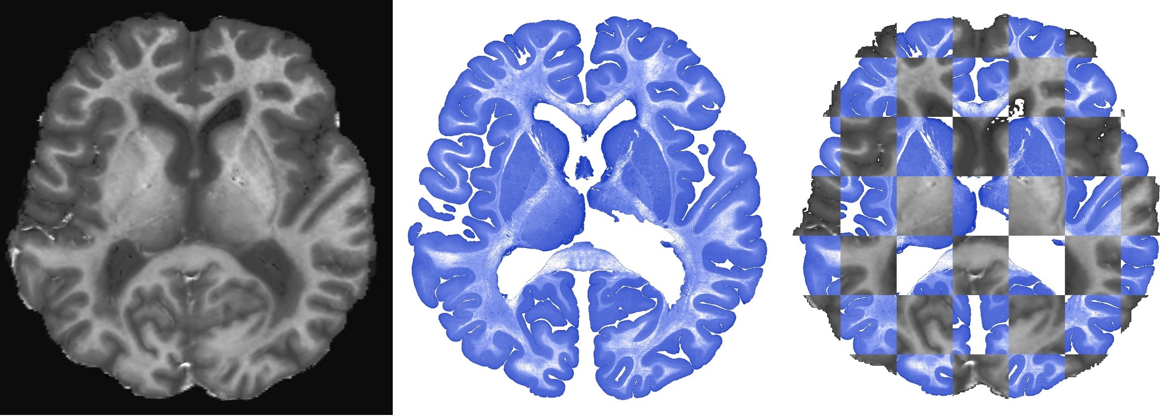
MRI to myelin registration was performed using the T1 images. Prior to registration histogram matching was performed to increase registration accuracy. Registration between myelin stainings and T1 images was performed using a combination of manual landmarks and non-linear registration using the minc-toolbox (Vincent et al. 2016). All MRI contrasts were transformed into the histology space using the T1 to myelin transform (Figure 1).
Prediction of histological features:
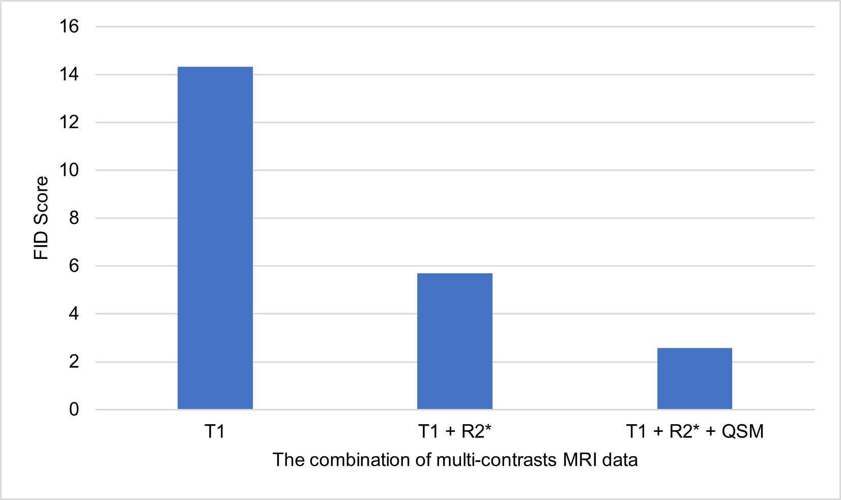
A Self-Attention Generative Adversarial Network (SAGAN) featuring nine residual blocks for both encoder and decoder was trained using combinations of the registered MR images (T1, QSM and R2*; T1 and R2*; T1) and the myelin stainings (132 slices). Due to the large image size (4158x3933 pixels per slice), we partitioned these images into smaller patches with 256x256 pixel prior to network training. The network was trained with a batch size of 128, 100 epochs and a learning rate of 0.0002. In the validation process, the perceptual loss (Johnson et al. 2016; Kim und Lee 2021) was introduced alongside the conventional L1 loss and adversarial loss. The Fréchet Inception Distance (FID score) was selected to evaluate the quality of the generated images (Heusel et al. 2017).
Results
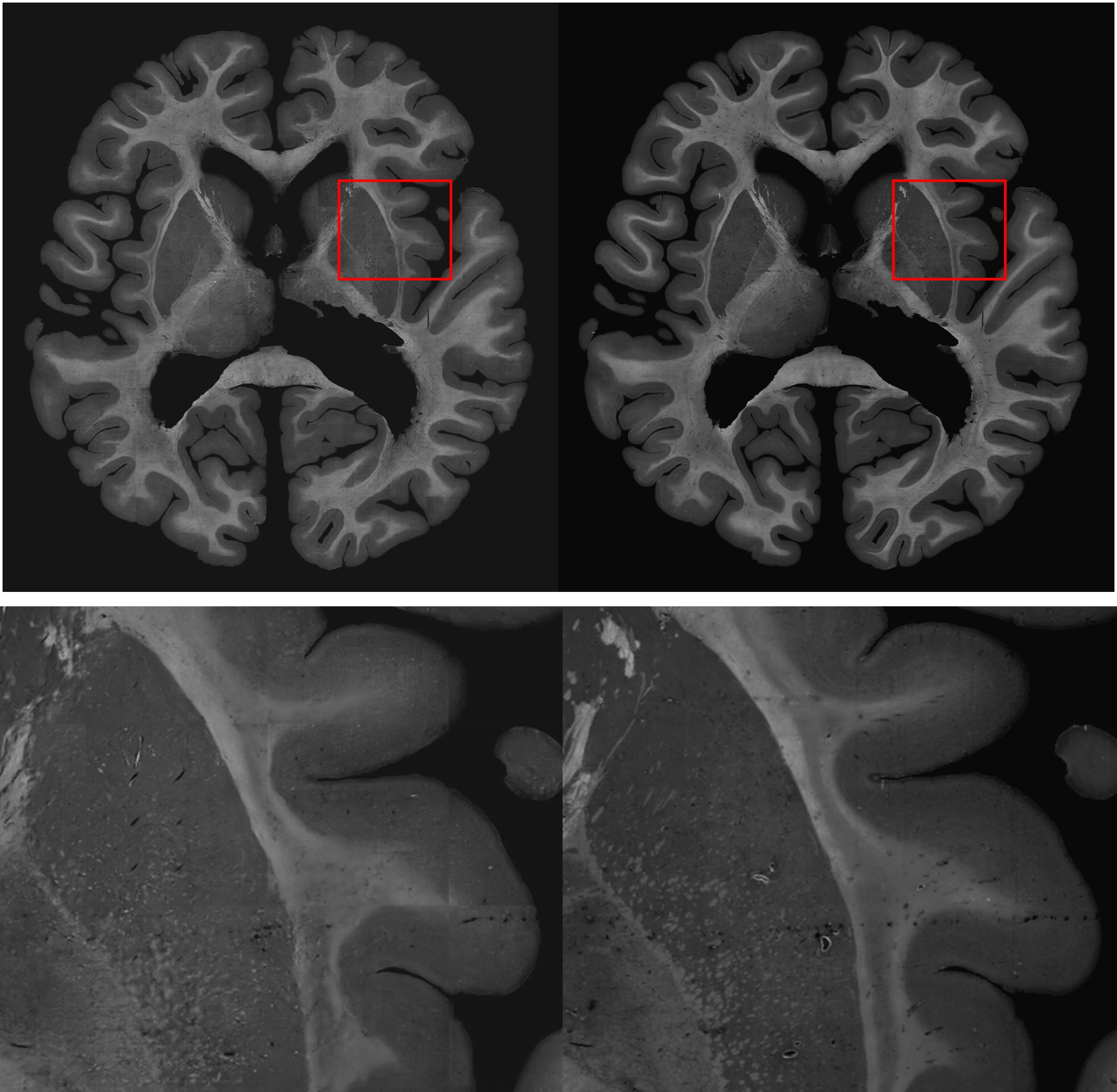
A SAGAN was successfully used to generate 2D images representing myelin stainings from MRI data (Figure 2). For predicting myelin, the best FID score (2.57) was achieved by combining T1, QSM, and R2* data (Figure 3). This highlights the value of using multiple MRI contrasts for training and prediction. Predictions of individual patches raised concerns about the impact of edges, leading to structural discontinuities (Figure 2). Addressing these issues necessitates the exploration of techniques for improvement.Conclusions
The generative model has effectively demonstrated its ability to produce myelin histology images from multi-sequence MRI data. This represents a significant advancement to generate ex vivo histology data from in vivo MRI data. Nevertheless, there exist opportunities for further refinement in image quality, such as increasing the quantity of training samples or fine-tuning the network's architecture and parameters.Acknowledgements
This research was funded in whole, or in part, by the Austrian Science Fund (FWF): [DFH 50-B].
References
Heusel, Martin; Ramsauer, Hubert; Unterthiner, Thomas; Nessler, Bernhard; Hochreiter, Sepp (2017): GANs Trained by a Two Time-Scale Update Rule Converge to a Local Nash Equilibrium. DOI: 10.48550/arxiv.1706.08500.
Johnson, Justin; Alahi, Alexandre; Fei-Fei, Li (2016): Perceptual Losses for Real-Time Style Transfer and Super-Resolution.
Kim, J.; Lee, C. (2021): Reliable Perceptual Loss Computation for GAN-Based Super-Resolution With Edge Texture Metric. In: IEEE Access 9, S. 120127–120137. DOI: 10.1109/ACCESS.2021.3108394.
Vincent, Robert D.; Neelin, Peter; Khalili-Mahani, Najmeh; Janke, Andrew L.; Fonov, Vladimir S.; Robbins, Steven M. et al. (2016): MINC 2.0: A Flexible Format for Multi-Modal Images. In: Frontiers in neuroinformatics 10, S. 35. DOI: 10.3389/fninf.2016.00035Figures