4701
Data-Driven MRS Signal Decomposition Using Wavelet Analysis1Department of Electrical Engineering, Eindhoven University of Technology, Eindhoven, Netherlands, 2Department of Biomedical Engineering, Eindhoven University of Technology, Eindhoven, Netherlands, 3High Field Research Group, Center for Image Sciences, University Medical Center Utrecht, Utrecht, Netherlands
Synopsis
Keywords: Analysis/Processing, Spectroscopy, Wavelet Analysis, Signal Decomposition, Proton MRS
Motivation: Magnetic resonance spectroscopy (MRS) is currently limited by noise, low spatial resolution, and artifacts that compromise the accuracy of metabolite quantification.
Goal(s): This work aims to enhance MRS signal quality without compromising signal integrity, employing wavelet analysis for robust signal decomposition.
Approach: A novel method utilizing wavelet analysis and a U-Net architecture creates masks that segment scalograms, effectively isolating individual metabolites in MRS signals.
Results: The method has shown in simulations the ability to distinctly separate and characterize metabolite signals, offering a promising direction for refining MRS data analysis.
Impact: Provides a data-driven method for MRS signal decomposition based on wavelet analysis that shows success in extracting metabolite and baseline information. It holds the potential for accurate characterization of nuisance signals, which could lead to improved MRS fitting.
Introduction
The potential of magnetic resonance spectroscopy (MRS) is hindered by intrinsically low signal-to-noise ratio (SNR) and limited spatial resolution. Moreover, spectra are often contaminated with residual water signals, lipid contributions, and macromolecular baselines that are difficult to model accurately, thereby obstructing precise quantification 1, 2. Wavelet analysis has shown promise in characterizing spectral components of MRS signals, specifically in disentangling overlapping signal components, including the challenging baseline contributions from macromolecules 3, 4. A recent data-driven method applied a continuous wavelet transform (CWT) to identify and characterize the spectral wavelets representing signals and noise in MRS spectra. Employing a support vector machine, this approach classified these features to selectively remove noise components while preserving valuable metabolite peaks 5.This work presents a novel method for decomposing MRS signals in the frequency domain through wavelet analysis, utilizing a U-Net architecture to generate masks that segment the signals' CWT. This approach effectively separates the signals into their individual metabolite components without compromising their structure. Results on simulated MRS data demonstrate that this technique can successfully learn appropriate weighting masks to decompose metabolites across a wide range of MRS signals with different amplitudes, frequency and phase variations, broadenings, and baselines. This offers a promising direction for the decomposition, noise reduction, and artifact segmentation of MRS signals.
Methods
The signal model for the simulation of the MRS spectra takes the following form: $$X(f) = \sum_{m=1}^M X_m(f) = e^{i(\phi_0 + \phi_1 f)} \sum_{m=1}^M a_m \text{FFT}\{s_m(t) e^{-(i\epsilon + \gamma + \sigma^2 t)t}\} + B(f), $$where $$$\gamma$$$ and $$$\sigma$$$ account for Lorentzian and Gaussian broadening, $$$\phi_0$$$ and $$$\phi_1$$$ are zero and first order phases, and $$$\epsilon$$$ represents frequency shifts. The metabolite basisset $$$\{s_m\}_{m=1}^M$$$ is taken from the fitting challenge data6 (21 metabolites and a macromolecule spectrum), $$$a_m$$$ are the concentrations, and the baseline $$$B(f)$$$ is represented by a second-order polynomial. To enhance data variability each batch is created during training by randomly drawing the model parameters from uniform distributions, along with adding complex Gaussian noise to the spectra (peak SNR 5-15 dB).The principle of wavelet analysis is to represent a signal as a superposition of wavelets, which are generated from a single mother wavelet by scaling (dilating or compressing) and translating (shifting). The forward CWT is defined as $$W(s,\delta) = \frac{1}{\sqrt{s}} \int X(f) \, \Psi^*\left(\frac{f-\delta}{s}\right) \, df, $$where $$$s$$$ is the scale factor, $$$\delta$$$ is the translation parameter, and $$$\Psi(f)$$$ is the mother wavelet (with denoting the complex conjugate). This transformation results in a two-dimensional representation of the signal, known as scalogram $$$W(s,\delta)$$$. The inverse CWT (ICWT) is obtained by integrating over all possible scales and translations of the wavelet coefficients, allowing reconstruction of the spectra. Figure 1 motivates the use of wavelet-based decomposition. It displays a sample spectrum alongside its corresponding scalogram and a selection of basis spectra, highlighting the enhanced separability achieved through the use of the CWT.
The proposed decomposition method is outlined in Figure 2. The real and imaginary parts of the MRS spectra (0.5-4.5 ppm) are converted to scalograms, which are then fed into a U-Net architecture. The network applies convolutions and pooling to downsample and increase feature representation, followed by upsampling and feature merging. It produces masks for the signal's real and imaginary components and applies them to the scalograms to isolate metabolite-specific information, which can be converted back to spectra using the ICWT. The network is trained with the mean squared error (MSE) of these spectra and the ground truths $$$X_m$$$ (both normalized). By incorporating a differentiable CWT and ICWT into the training pipeline, the U-Net weights are updated with backpropagation through the entire pipeline of Figure 2.
Results & Discussion
Figure 3 and Figure 4 show the real and imaginary component channels, respectively, of a randomly selected test sample, showcasing the actual part of the spectrum alongside its scalogram. This is followed by the masks created by the U-Net and the separate scalograms obtained post-segmentation. The spectra resulting from the ICWT of the segmented scalograms are then displayed, alongside the actual ground truth spectra of the individual metabolites for comparison. The masks manage to capture the fundamental attributes, including correct broadening, phase and frequency alignment, and fine nuances of each signal for both real and imaginary parts.Conclusion
Using a U-Net architecture to generate masks that segment scalograms, the method successfully separates individual metabolite components within complex MRS signals. Results from simulated data validate the method's precision in capturing key spectral features and subtle details, demonstrating its capacity to improve metabolite quantification by precisely identifying individual signals and characterizing noise and artifacts.Acknowledgements
This work was (partially) funded by Spectralligence (EUREKA IA Call, ITEA4 project 20209).References
1. Cristina Cudalbu, Kevin L. Behar, Pallab K. Bhattacharyya, et al. “Contribution of macromolecules to brain 1H MR spectra: Experts' consensus recommendations.” NMR in Biomedicine, 34 (2021).
2. Aimamorn Suvichakorn, Hélène Ratiney, Sophie Cavassila, and Jean-Pierre Antoine, “Wavelet-based Techniques in MRS,” Signal Processing. InTech (2010).
3. Hacene Serrai, Lotfi Senhadji, Jacques D. de Certaines, and Jean-Louis Coatrieux. “Time-domain quanification of amplitude, chemical shift, apparent relaxation time T2, and phase by wavelet-transform analysis. Application to biomedical magnetic resonance spectroscopy.” Journal of magnetic resonance, 124 1 (1997): 20-34.
4. Aimamorn Suvichakorn, Hélène Ratiney, Adriana Bucur, Sophie Cavassila, and Jean-Pierre Antoine, “Analyzing Magnetic Resonance Spectroscopic Signals with Macromolecular Contamination by the Morlet Wavelet.” ECIFMBE (2008).
5. Bing Ji, Zahra Hosseini, Liya Wang, Lei Zhou, Xinhua Tu, and Hui Mao. “Spectral Wavelet‐feature Analysis and Classification Assisted Denoising for enhancing magnetic resonance spectroscopy.” NMR in Biomedicine, 34 (2021).
6. Małgorzata Marjańska, Dinesh K. Deelchand, and Roland Kreis. “Results and interpretation of a fitting challenge for MR spectroscopy set up by the MRS study group of ISMRM.” Magnetic Resonance in Medicine, 87 (2021): 11 - 32.
Figures
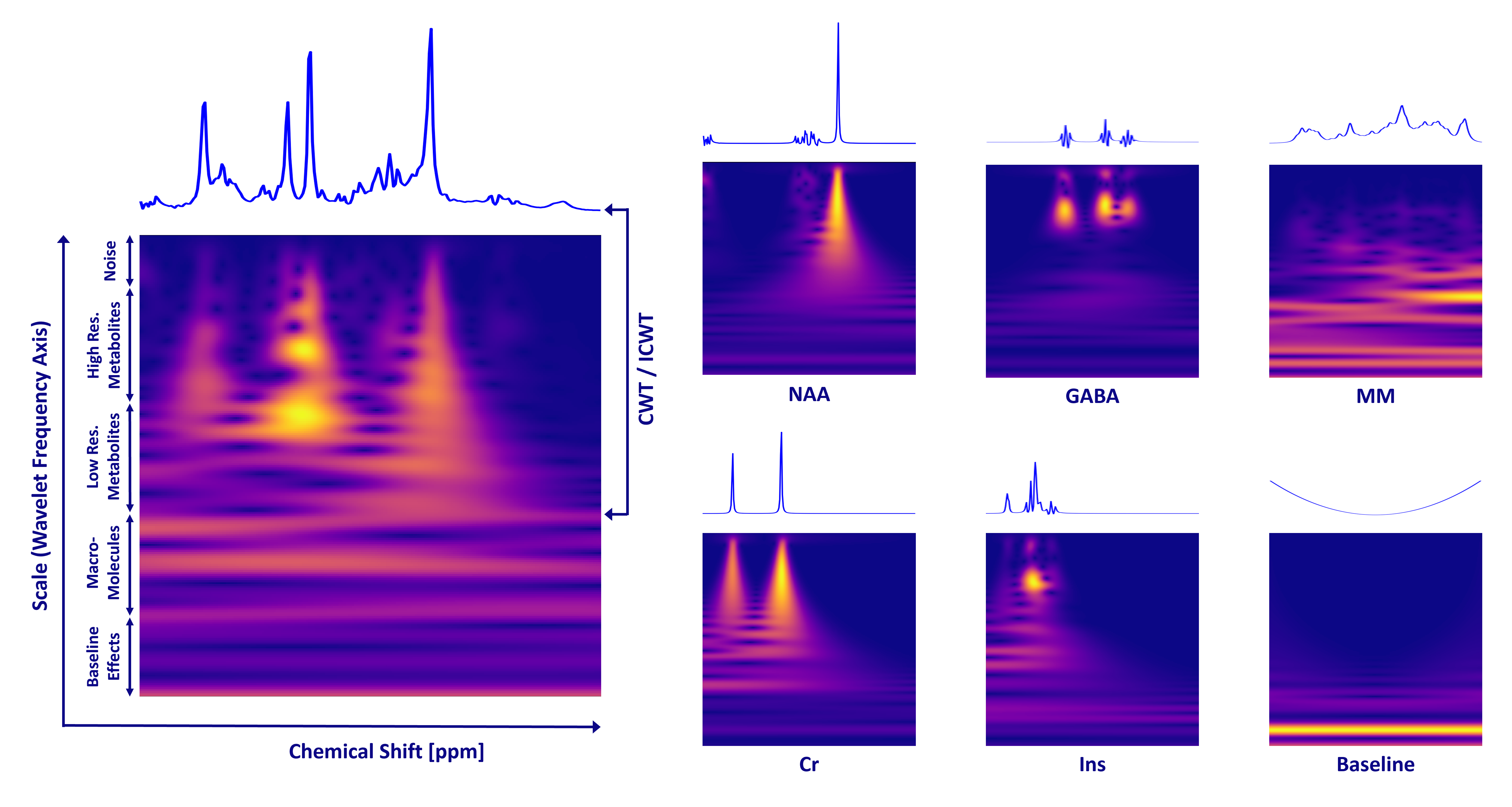
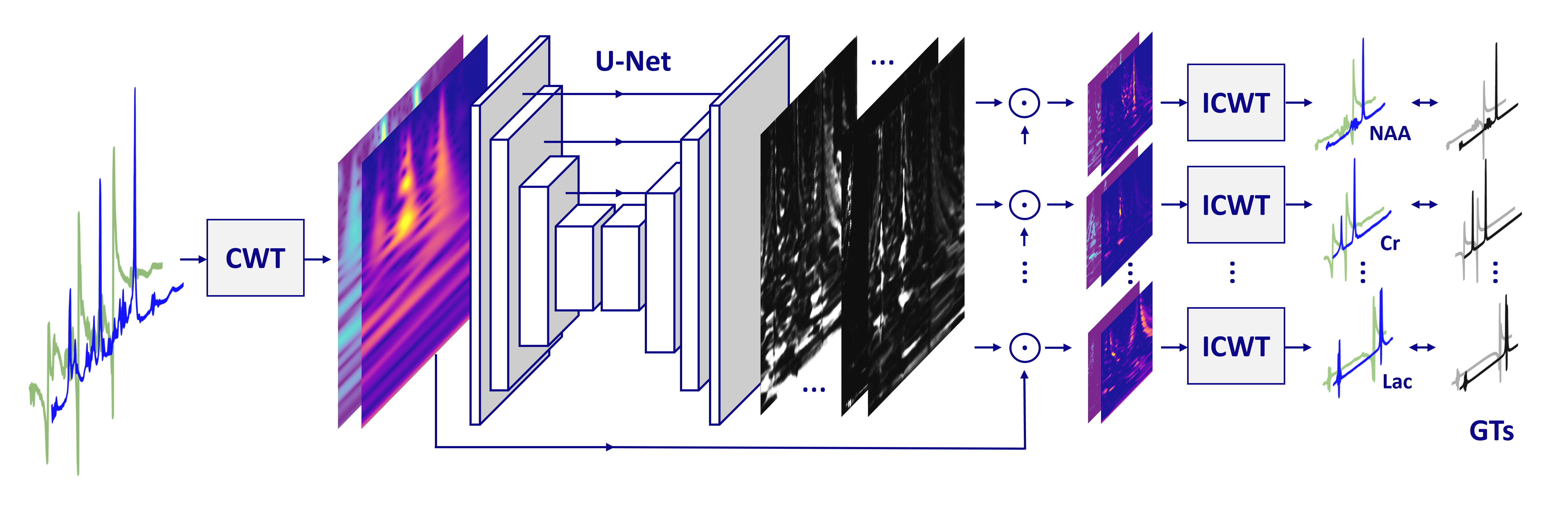
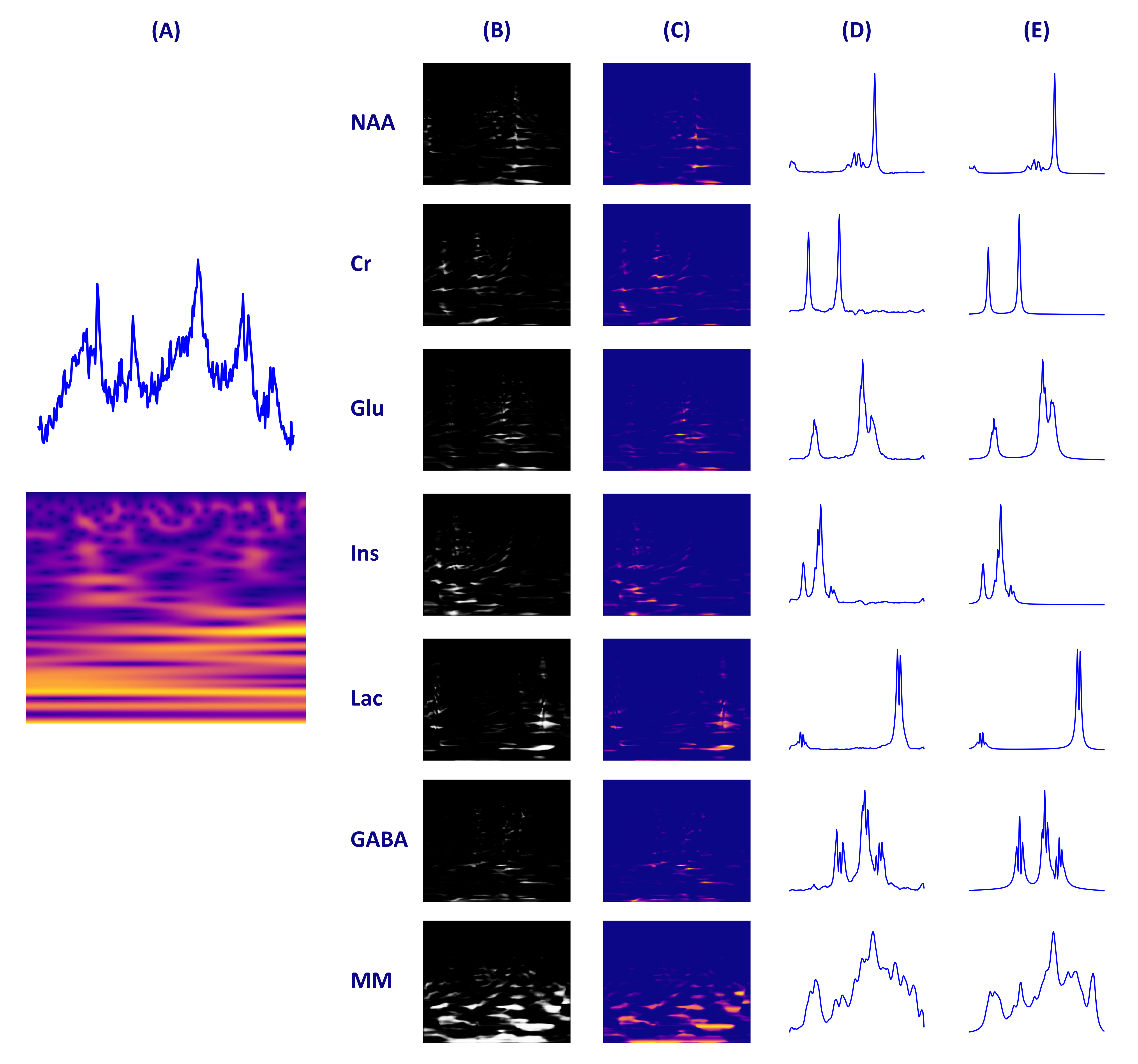
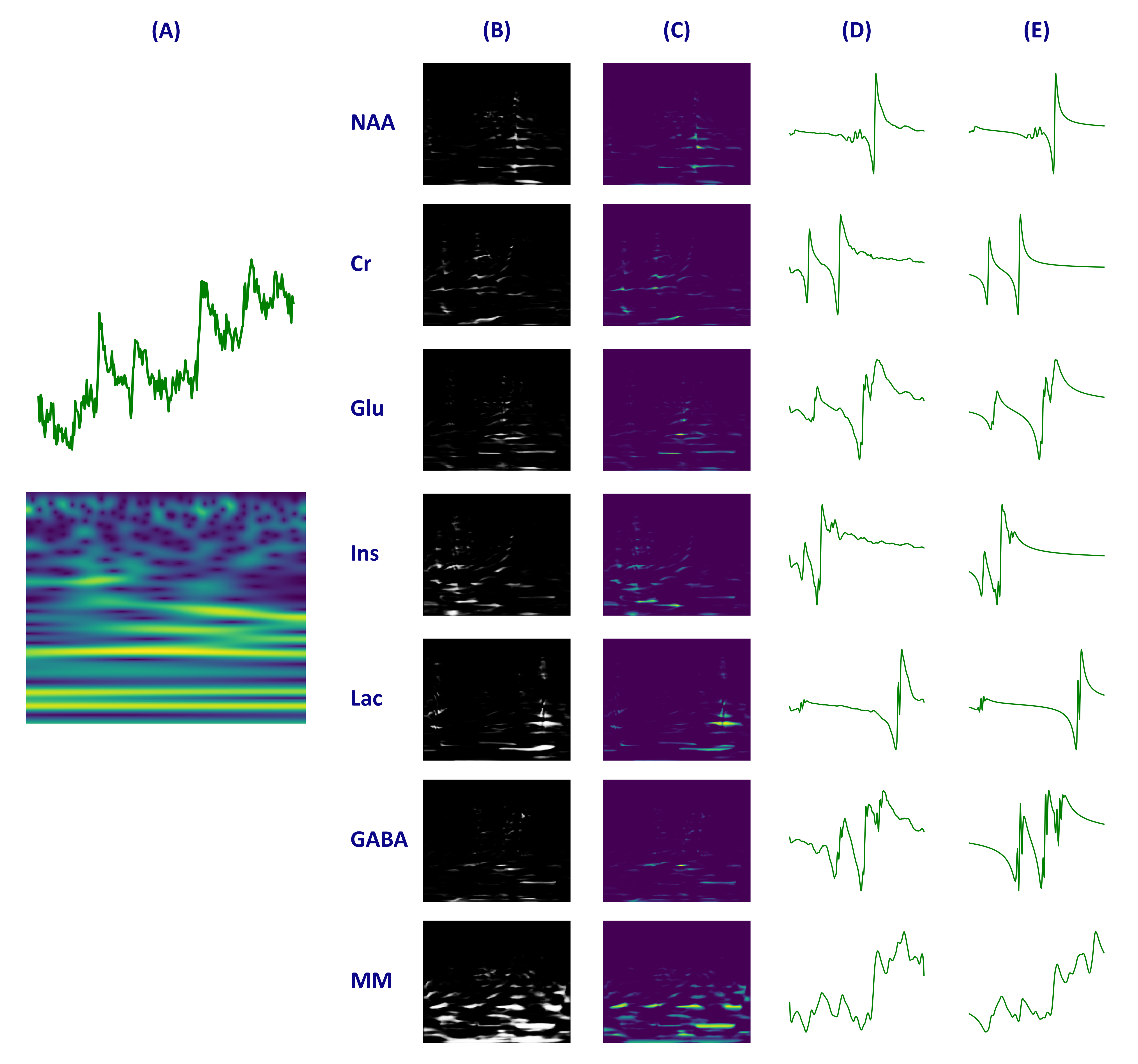
Figure 4. Depicts the imaginary component of (A) a test example spectrum chosen at random and its associated scalogram, (B) the masks produced by the U-Net, (C) the separate scalograms obtained after segmentation, (D) the spectra derived from applying the inverse continuous wavelet transform (ICWT) to the segmented scalograms, and (E) the actual ground truth (GT) spectra of the metabolites that make up the spectrum.