4699
Intraoperative Precision Sampling of Tumor Microenvironments based on 7T MRSI: A pipeline development1Medical University of Vienna, Vienna, Austria, 2University of Vienna, Vienna, Austria
Synopsis
Keywords: Spectroscopy, Contrast Mechanisms, Spectroscopy, Neuro Tumors (Pre-Treatment), Surgery
Motivation: Molecular and pathological diagnosis requires fresh tissues. It is important to achieve precise sampling and preservation of the tumor is for maintaining the tumor integrity.
MRSI can accurately identify tumor hotspots and can be used for obtaining high quality samples.
Goal(s): Develop MRSI pipeline for precise intraoperative tumor sampling.
Approach: We processed 7T MRSI metabolic ratio maps and identified the tumor hotspots. We then correlated them with their quantitatively analyzed metabolic profiles.
Results: We were able to achieve high quality tumor samples based on our 7T MRSI maps.
Impact: Due to high resolution 7T metabolic ratio maps we could identify and define tumor hotspots which resulted in precise tumor sampling. We were also able to preserve the tissue specimens and obtain high quality results.
Introduction
Magnetic resonance spectroscopic imaging (MRSI) is capable of imaging neurochemical compounds in the human brain [4]. 7T MRSI has a higher signal-to-noise (SNR) ratio and is beneficial due to better resolution, coverage and identifying the molecules of interest [5]. While publications on 7T MRSI show great promise for neurochemical mapping of the healthy and diseased brain, validation with gold standards such as mass spectrometry remains challenging due to the difficulty to acquire precise tissue samples from the human brain in vivo.Gliomas represent a complex and heterogeneous group of brain tumors, for which the WHO’s 2021 update on the classification of tumors of the central nervous system introduced integrative diagnoses incorporating molecular parameters alongside histology, setting a new standard for glioma characterization [1] currently only determined from biopsy or resection. We previously demonstrated high-resolution 7T 3D-MRSI in high grade glioma (HGG) patients, resolving metabolic heterogeneities within tumors [2].Our high-resolution maps could be used to obtain samples from specific intratumoral hotspots and molecular pathology and analytical chemistry could validate these findings. Therefore, the purpose of this study is to develop a pipeline that spans MRSI acquisition, transfer to neuronavigation, surgical sampling, and tissue analysis in order to correlate our MRSI findings with clinical and analytical gold standards[3].Methods
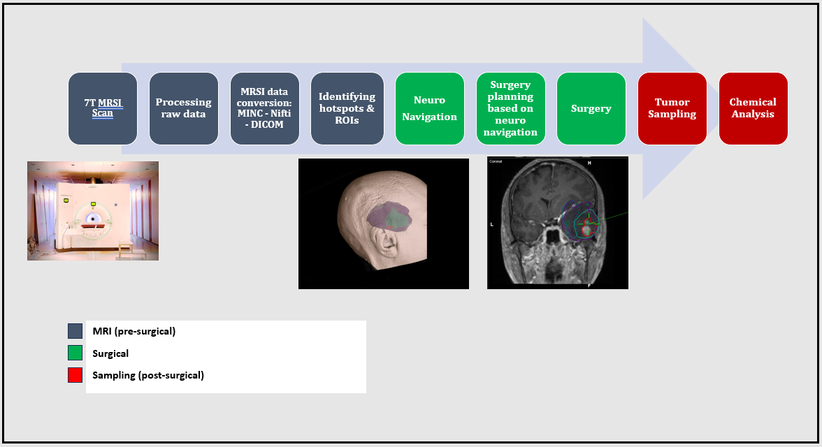
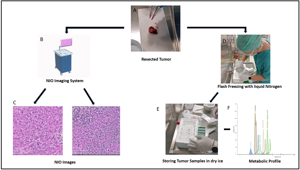
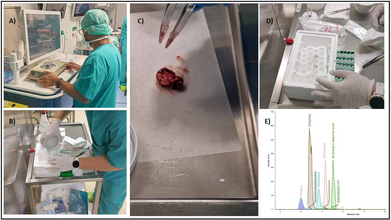
For this study we used a 7T scanner (Siemens Healthineers, Magnetom/Magnetom plus after an upgrade) and a 1Tx/32Rx head coil (Nova Medical,). We acquired MRSI data with 3.4 mm isotropic resolution and a matrix size of 64×64×39 in 15 min [6] and processed it using in-house scripts and LCModel quantification. Based on our previous studies’ results, selected metabolites N-acetylaspartate (NAA), choline (Cho), glutamine (Gln), glycine (Gly), and myo-inositol (Ins), to process the ratio maps tCho/tNAA, Gln/tNAA, Gly/tNAA and Ins/tNAA for transfer to neuronavigation.To this end, our MINC outputs were converted to NiFTI and finally to DICOM using the KARAWUN software [8]. These DICOM ratio maps together with morphological reference data were transferred to our neuronavigation planning server (Elements, Brainlab). Metabolic hotspot ROIs were then contoured by neurosurgeons and saved for sampling.During surgery, samples were taken from the ROIs with 1-2 mm precision and separated into three parts: One for histopathology/molecular pathology, one for intraoperative Raman spectroscopy (NIO system, Invenio Imaging) [7], and one for analytical chemistry. This last one was weighed and then flash-frozen using liquid nitrogen. Mass spectrometry created quantitative metabolic profiles of the tumors using mass spectrometry. Fig.1 depicts the steps of our pipeline. Overall, for every sampling location, NIO-derived pathologic profiles, molecular pathology, and spectrometry were available for validation [9].Results
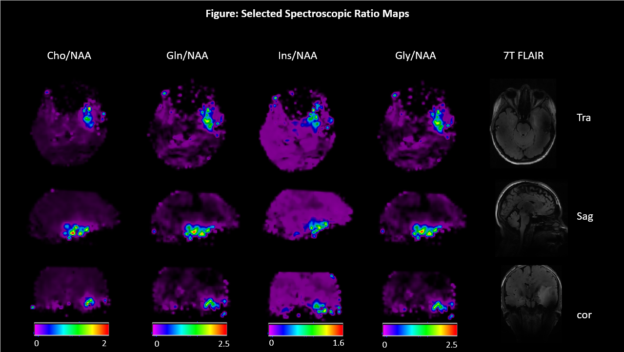
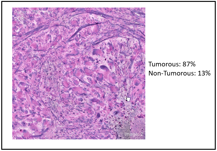
Prior to the first patient application, all steps except resective sampling were individually and successfully tested using existing data from previous studies. At time of submission, we have applied this pipeline in one patient (age 29, sex: F, HGG suspected). This first patient’s metabolic ratio maps (Fig.2.) show the intratumoral increases of Cho, Gln, Ins and Gly ratios to tNAA,. Four samples were obtained during surgery based on MRSI data. The tumor samples were intraoperatively scanned with the NIO device which identified a majority of tumorous tissue in all four samples (Fig.3.). Preservation for further chemical analysis was successful (Fig.4. D and E), mass spectrometric data was still processed at the time of writing, but preliminary results (Fig 5E) confirms at least the presence of choline and glutamine.Preliminary chemical and NIO results show that we were able to successfully sample tumor hotspots based on 7T MRSI maps.Discussion and Conclusion
We successfully demonstrated that even highly post-processed data as our 7T MRSI method can be transferred back into clinical neuronavigation systems. Our first, exploratory, patient shows that while a fully study and detailed evaluation is still required, our intended workflow is feasible in generating hotspot samples that are usable for validation by other methodologies.To address this main limitation, more patient scans and complete molecular-pathologic and mass-spectrometric reports will be necessary.We are confident that by doing so, we will validate our MRSI method using these gold standards, strengthening the case for 7T MRSI as a future diagnostic tool. If we can establish correlations of metabolic patterns to specific tumor microenvironments, future research could investigate its use to improve neurosurgical planning, especially for a better maximal safe resection.Acknowledgements
This study was supported by the Austrian Science Fund (FWF) project KLI 1089. The financial support by the Austrian Federal Ministry for Digital and Economic Affairs, the National Foundation for Research, Technology and Development and the Christian Doppler Research Association is gratefully acknowledged.References
- Louis, D. (2021, August 2). The 2021 WHO Classification of Tumors of the Central Nervous System: a summary. PubMed. Retrieved November 3, 2023, from https://pubmed.ncbi.nlm.nih.gov/34185076/
- Hangel, G. (2020, September 15). High-resolution metabolic imaging of high-grade gliomas using 7T-CRT-FID-MRSI. PubMed. Retrieved November 3, 2023, from https://pubmed.ncbi.nlm.nih.gov/32977210/
- Hangel, G. (2022, April 26). 7T HR FID-MRSI Compared to Amino Acid PET: Glutamine and Glycine as Promising Biomarkers in Brain Tumors. PubMed. Retrieved November 4, 2023, from https://pubmed.ncbi.nlm.nih.gov/35565293/
- Moser, E. (2012). 7-T MR--from research to clinical applications? PubMed. Retrieved November 5, 2023, from https://pubmed.ncbi.nlm.nih.gov/22102481/
- Bogner, W. (2011, December 22). High-resolution mapping of human brain metabolites by free induction decay (1)H MRSI at 7 T. PubMed. Retrieved November 5, 2023, from https://pubmed.ncbi.nlm.nih.gov/22190245/
- Hingerl, L. (2020). Clinical High-Resolution 3D-MR Spectroscopic Imaging of the Human Brain at 7 T. PubMed. Retrieved November 7, 2023, from https://pubmed.ncbi.nlm.nih.gov/31855587/I
- Invenio Imaging Receives CE Mark to Detect Cancer at the Time of Surgery using Artificial Intelligence - UCSF Rosenman Institute. (2022, May 26). Rosenman Institute. Retrieved November 7, 2023, from https://rosenmaninstitute.org/blog/invenio-imaging-receives-ce-mark-to-detect-cancer-at-the-time-of-surgery-using-artificial-intelligence/
- Beare, R. (2022, September 7). Karawun: a software package for assisting evaluation of advances in multimodal imaging for neurosurgical planning and intraoperative neuronavigation. NCBI. Retrieved November 8, 2023, from https://ncbi.nlm.nih.gov/pmc/articles/PMC9883338/
- Kampa, J. (2019, March 9). Glioblastoma multiforme: Metabolic differences to peritumoral tissue and IDH-mutated gliomas revealed by mass spectrometry imaging. Retrieved November 8, 2023, from https://onlinelibrary.wiley.com/doi/full/10.1111/neup.12671
Figures