4698
Improved Low Angular Resolution Diffusion Parametric Maps Using Deep Learning1Department of Radiologic Technology, Faculty of Associated Medical Sciences, Chiang Mai University, Chaing Mai, Thailand, 2Department of Psychology, University of Totonto Mississauga, Mississauga, ON, Canada
Synopsis
Keywords: Analysis/Processing, Neuro, Brain, White Matter
Motivation: The DTI technique is used to analyze and evaluate the white matter microstructure; however, the acquisition time is too long for clinical practice and large-scale research.
Goal(s): To reduce acquisition time by improving the low angular resolution diffusion parametric maps.
Approach: The deep-learning framework was used to improve the low angular resolution diffusion parametric maps and image quality measured with PSNR, and NRMSE.
Results: Our deep-learning framework improves low angular resolution diffusion parametric maps by effectively acquiring fiber information in FA map and enhancing overall image quality with increased PSNR and decreased NRMSE.
Impact: The reduced acquisition time and improved quality of the low angular resolution diffusion parametric maps obtained with our deep-learning framework may benefit to clinicians and researchers who study in white matter microstructure in routine clinical practice and large-scale research.
INTRODUCTION
Diffusion parametric maps, including fractional anisotropy (FA), mean diffusivity (MD), axial diffusivity (AD), and radial diffusivity (RD), have emerged as a promising biomarker for detecting changes in white matter (WM) microstructures among transgender people who experience adverse effects following hormone therapy1–4. To produce high-quality diffusion parametric maps, a large number of diffusion-encoding directions are often required, resulting in prolong acquisition times which makes it highly susceptible to motion artifact. In this work, we implement a deep-learning framework for improving low angular diffusion parametric maps, in which the network was trained to predict high angular diffusion parametric maps that aim to minimize acquisition time while preserving high-quality results.METHODS
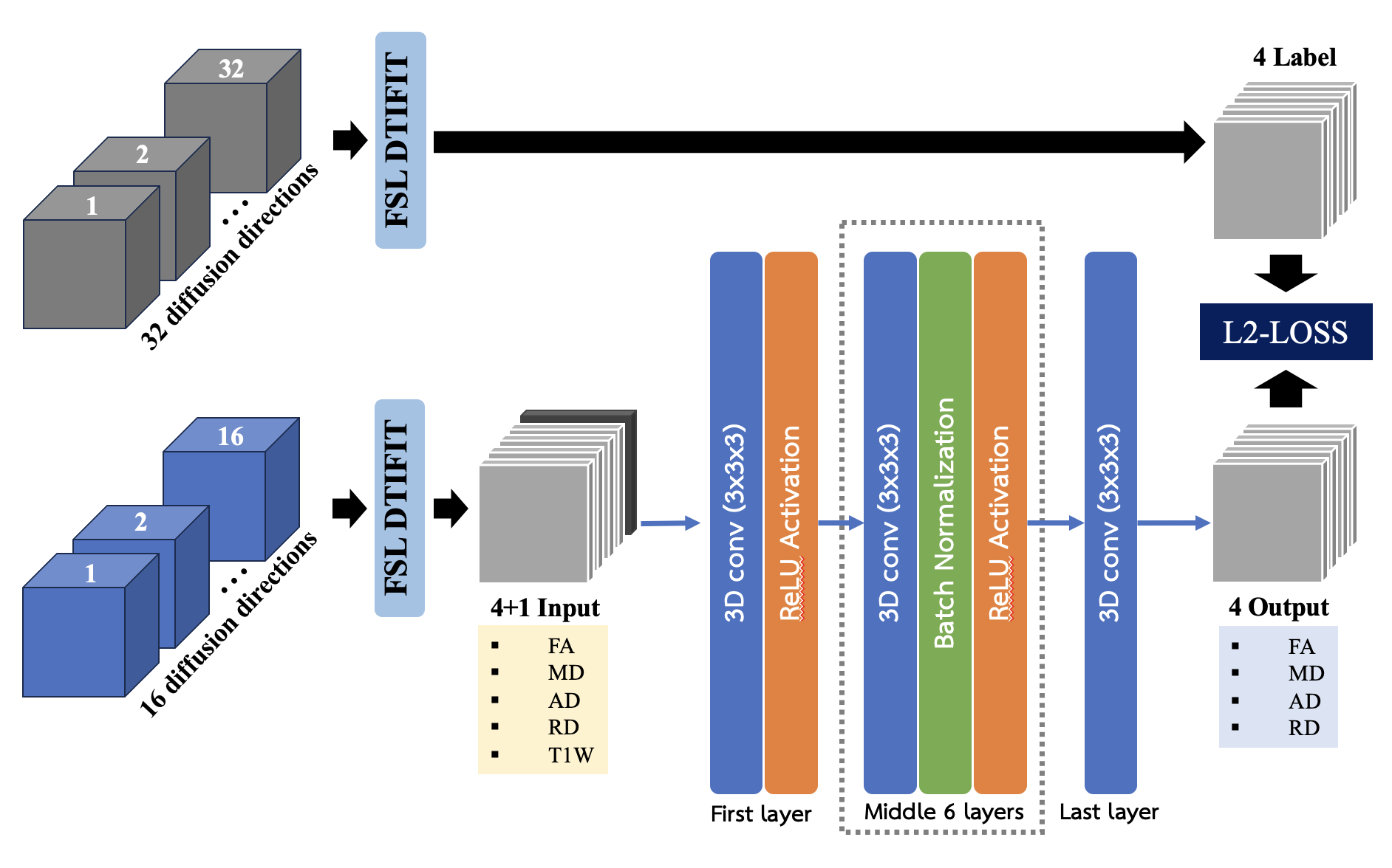
Data acquisition and Processing: A total of 130 volunteers were recruited, including 37 men (M), 35 women (F), 30 gay men (G), and 28 transgender women (TG). The mean ages of each group were 27.95 ± 4.86, 26.78±5.01, 26.8±5.82, and 24.19±4.43 years old, respectively. The study was approved by a local institutional review board. All participants were HIV-negative and passed MRI safety screening. They underwent 1.5T MRI (Ingenia; Philips, Best, the Netherlands) using the following parameters: FOV = 160x160 mm, TR/TE = 4680/85 ms, Matrix 225x225, Thickness = 2.5 mm, 76 slices, 32 diffusion-directions with b = 1000 s/mm2, phase encoding AP and PA, acquisition time 15 minutes. These data sets were artificially subsampled by a factor of two—only 16 diffusion directions were selected from the original data sets. All 260 data sets were processed to obtain diffusion parametric maps (FA, MD, AD, RD) using the FSL DTIFIT toolbox5. 100 pairs of high (32 diffusion directions) and low (16 diffusion directions) angular diffusion parametric maps were used to train the network. 20 and 10 of those maps were used for validation and testing, respectively. Anatomical 3D T1w-FFE was used as additional input data. These data were registered to mean DWI before incorporation into the network.Deep Learning Implementation: The network used in this study is illustrated in Fig.1. It was implemented in Tensorflow6 and trained by minimizing the normalized-root-mean-squared-error (NRMSE) loss between the predicted and ground truth using the Adam optimizer7 with a learning rate of 1×10⁻⁴ and batch size of 2, running on NVIDIA Geforce RTX-3090 with 24 GB GPU. To evaluate the network performance, mean and standard deviation of peak signal-to-noise ratio (PSNR) and normalized root mean square error (NRMSE) were reported.
RESULTS
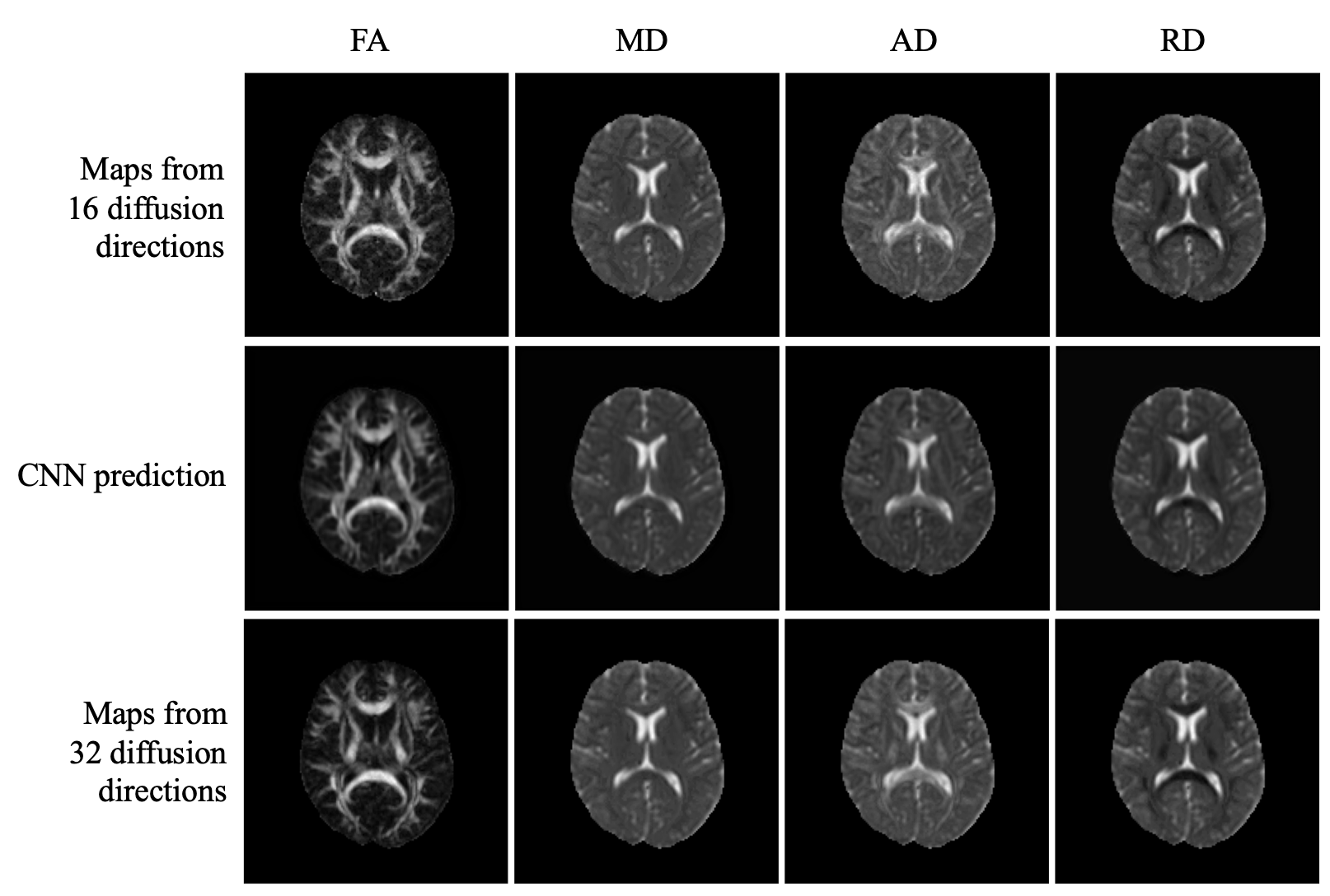
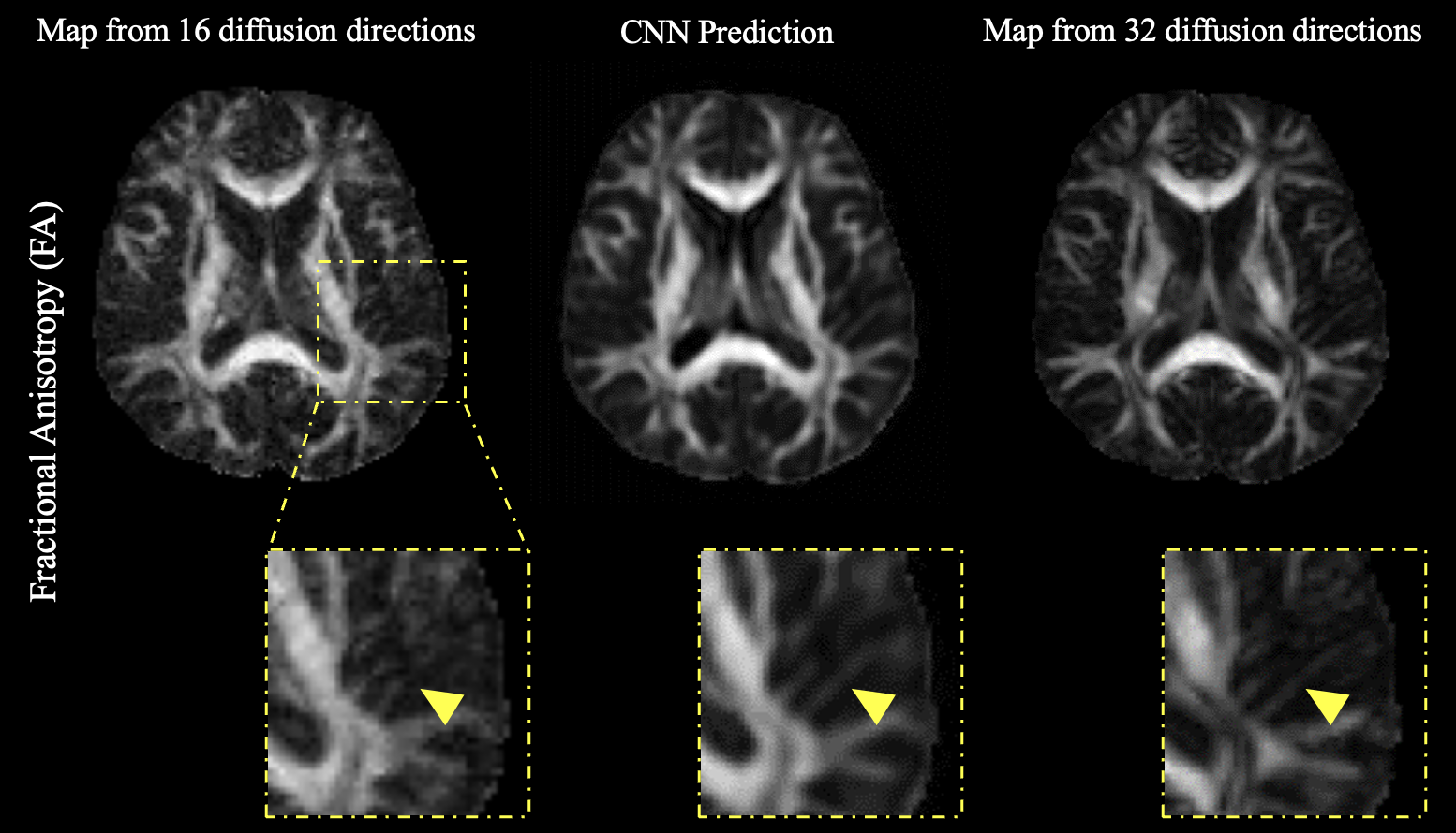
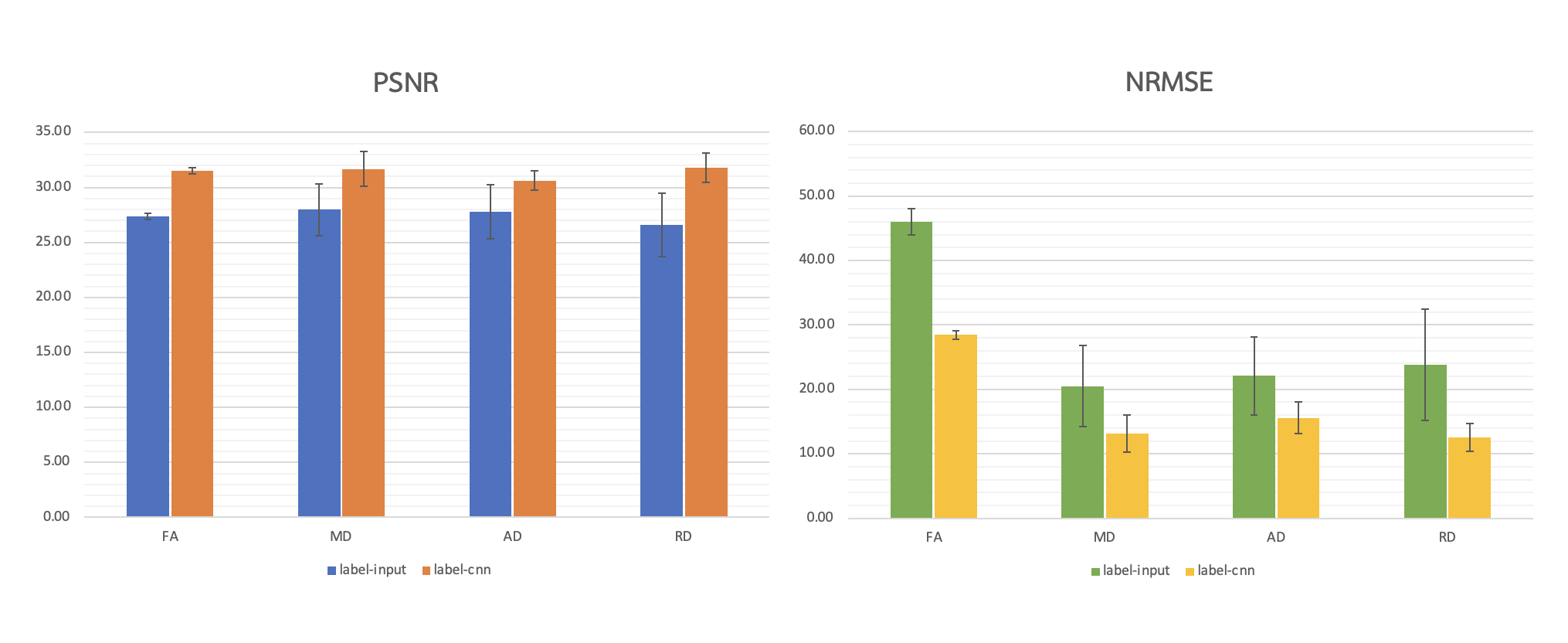
In Fig.2, FA, MD, AD, and RD maps computed from 16 diffusion direction data exhibit noise. Deep learning techniques effectively suppress this noise, resulting in smoother maps compared to those obtained from 32 diffusion direction data. Fig.3 shows the FA map in the temporoparietal region (enclosed by the yellow dashed box). Deep learning techniques effectively remove the noise in FA maps derived from 16 diffusion direction data, producing results visually comparable to those obtained from 32 diffusion direction data. Fig.4 presents the performance evaluation results. Deep learning techniques significantly enhance diffusion parametric maps, as evidenced by the 15.2%, 13.2%, 10.3%, and 19.6% increases in PSNR values for FA, MD, AD, and RD, respectively. Additionally, the NRMSE values for FA, MD, AD, and RD decrease by 38.2%, 36.1%, 29.5%, and 47.4%, respectively.DISCUSSION
This study employed a 3D deep-learning technique to enhance the quality of low angular resolution diffusion parametric maps, resulting in a notable reduction in NRMSE values. Nevertheless, certain challenges warrant further investigation. Firstly, expanding the training dataset could generalize and improve the network's performance. Secondly, the network was implemented with only eight layers due to computational limitations. Employing a larger network with more trainable parameters may yield better performance. Finally, the network's effectiveness should be validated through clinical applications, such as the study of white matter microstructure in transgender individuals.Acknowledgements
No acknowledgement found.References
1. O’Leary KB, Garcia-Altieri MA. The Impact of Gender-affirming Hormone Therapy on Anatomic Structures of the Brain among Transgender Individuals. J Psychiatr Pract. 2022;28(4):328-334. doi:10.1097/PRA.0000000000000633
2. Kranz GS, Hahn A, Kaufmann U, et al. White matter microstructure in transsexuals and controls investigated by diffusion tensor imaging. Journal of Neuroscience. 2014;34(46):15466-15475. doi:10.1523/JNEUROSCI.2488-14.2014
3. Van Hemmen J, Saris IMJ, Cohen-Kettenis PT, Veltman DJ, Pouwels PJW, Bakker J. Sex Differences in White Matter Microstructure in the Human Brain Predominantly Reflect Differences in Sex Hormone Exposure. Cerebral Cortex. 2017;27(5):2994-3001. doi:10.1093/cercor/bhw156
4. Thurston L, Rodkong A, Skorska M, et al. Sex/gender, sexual orientation, and gender-affirming hormones are associated with white matter microstructure in a Thai sample. Psychoneuroendocrinology. 2023;153:106193. doi:10.1016/J.PSYNEUEN.2023.106193
5. FDT - FslWiki. Accessed November 1, 2023. https://fsl.fmrib.ox.ac.uk/fsl/fslwiki/FDT
6. TensorFlow. Accessed November 1, 2023. https://www.tensorflow.org/?hl=th
7. Adam. Accessed November 1, 2023. https://keras.io/api/optimizers/adam/
Figures