4697
Deep Learning-Driven Generation of Diffusion-Weighted Imaging for Acute Ischemic Stroke from Non-Contrast Computed Tomography1Shenzhen Institute of Advanced Technology, Chinese Academy of Sciences, Shenzhen, China, 2Longgang Central Hospital of Shenzhen, Shenzhen, China
Synopsis
Keywords: AI/ML Image Reconstruction, Brain
Motivation: Noncontrast computed tomography(NCCT), commonly used for its rapidity in acute ischemic stroke(AIS) diagnosis, often fails to identify early ischemic changes, whereas MRI, despite its superior sensitivity , may introduce critical delays due to its lengthier image acquisition time.
Goal(s): This study aims to investigate the feasibility of converting NCCT images of AIS patients to diffusion-weighted (DW) images using deep learning techniques.
Approach: The proposed method utilizes an enhanced CycleGAN model to generate synthetic DW images from CT scans.
Results: The synthetic DW images generated by our network achieved good performance with a PSNR of 28.30, an SSIM of 0.843, and an NMSE of 0.293.
Impact: This work might be translated to clinical settings to help physicians make clinical decisions for patients by providing with high-quality MR images in emergency situations.
Introduction
Recent advancements in artificial intelligence (AI), particularly deep learning, are emerging as potential game-changers in this landscape[1]. They show promise not only in terms of AIS enhancing diagnostic precision but also in accelerating the diagnosisdiagnostic process[2-6]. Deep learning methods have already demonstrated their utility in diverse medical image translation tasks, such as synthetic CT image generation from MR images for radiation dose calculations and Ki image generation from static positron emission tomography (PET) for enhancing the specificity of cancer detection[7]. A subset of this research has started to focus on stroke identification, employing AI to achieve improved vascular imaging and DW image segmentation[8-15]. However, scant research has explored the prospective utility of end-to-end image translation from NCCT to DW images for AIS diagnosis purposes. To address this gap, we introduce a customized cyclic generative adversarial network (CycleGAN) model that is uniquely augmented by incorporating lesion presence information as prior information into the input and bolstered by the addition of the perceptual loss.Materials and methods
Data Source:CT images and DW images of the brain from May 2013 to April 2022 were queried on the picture archiving and communication system (PACS) of Longgang Central Hospital of Shenzhen. A total of 104 subjects aged 37–89 years who presented within 6 hours after the witnessed onset of stroke symptoms and who underwent pretreatment CT and 48-hour posttreatment MRI of the brain were enrolled.
Method Implements:
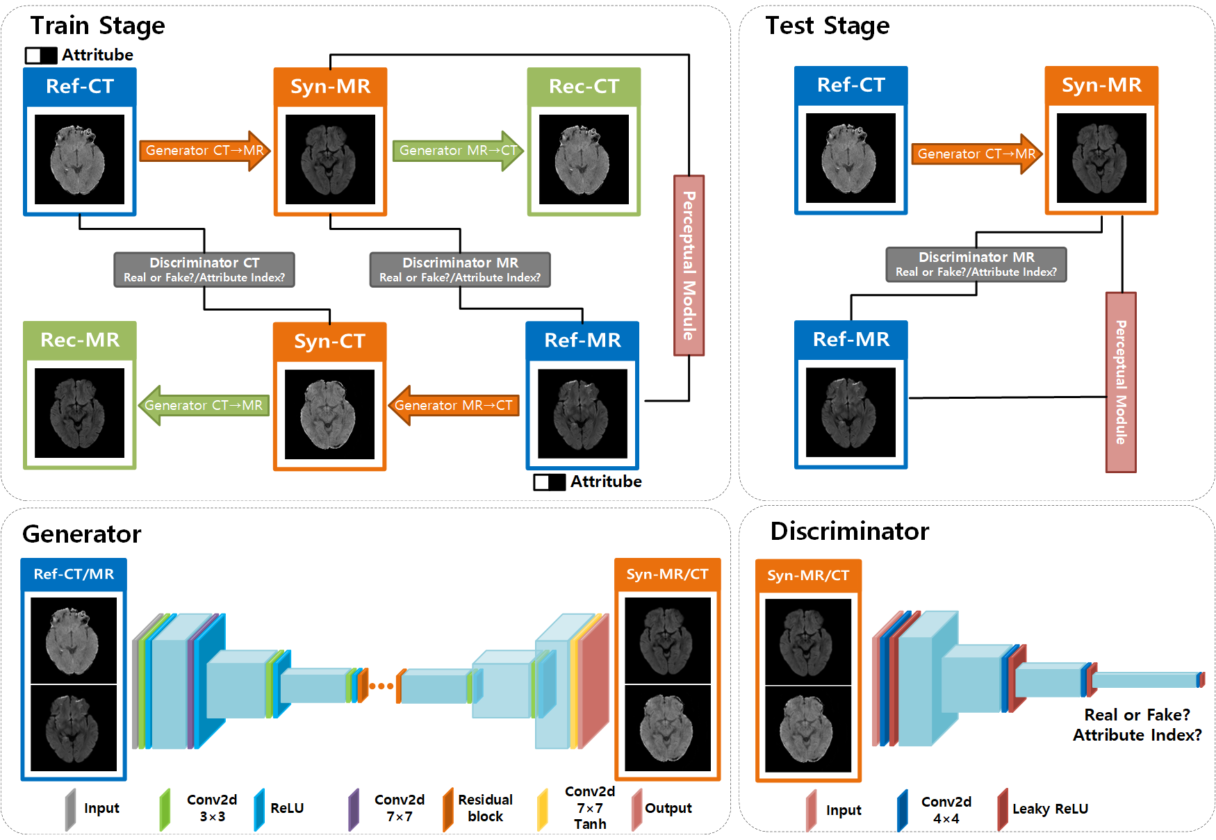
The overall structure is shown in Figure 1, our method employs a cyclic generative adversarial network (CycleGAN) to learn the mapping between CT scans and MR images, generating synthetic MR images from CT scans. We enhance the CycleGAN by introducing a new attribute to both discriminators, acknowledging that stroke lesions are more pronounced in MR images than in CT scans, where they are subtler yet detectable. Complementing this, we integrate a perceptual module, pivotal to our enhancement strategy. Rooted in the Visual Geometry Group 16 (VGG16) architecture, this module is crucial for extracting high-level features from both synthetic and reference MR images, thereby refining the transformation process and enhancing the overall accuracy of the generated images.
Data analysis:
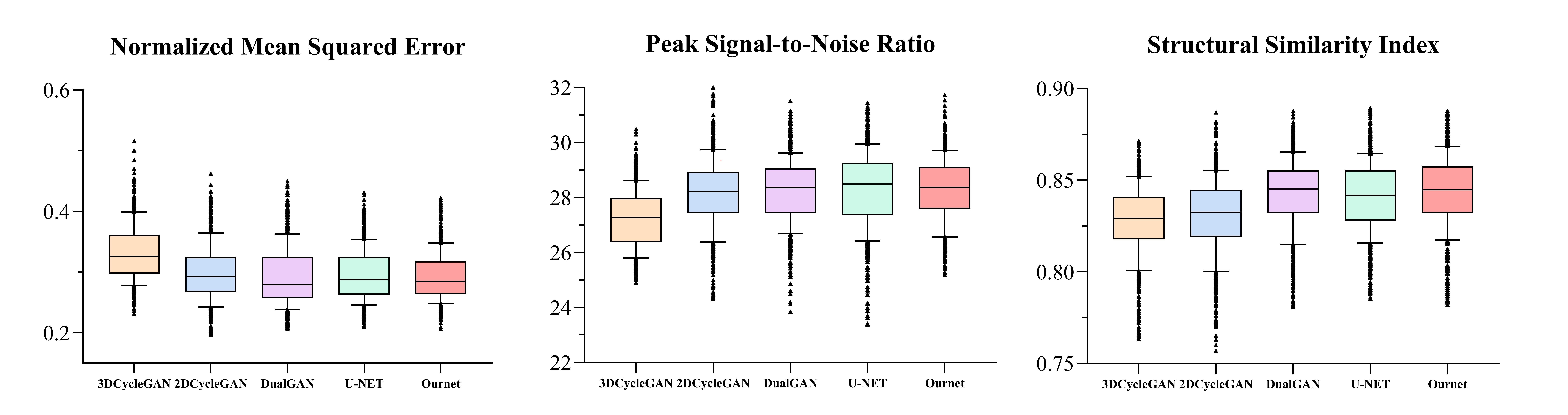
To evaluate the performance of our model, we adopt several metrics, including Peak Signal-to-Noise Ratio (PSNR), Structural Similarity Index Measure (SSIM), and Normalized Mean Squared Errorthe peak signal-to-noise ratio (PSNR), structural similarity index measure (SSIM), and normalized mean squared error (NMSE). These metrics offer a comprehensive assessment of our model's performance, encompassing aspects of image fidelity, structural preservation, and overall error respectively. Moreover, we juxtapose our model with several established algorithms, including DualGAN, U-Net, 2D CycleGAN, and 3D CycleGAN., in the field to gain a relative perspective of its performance. These models were chosen due to their prominence and proven efficacy in image-to-image translation tasks, and their comparison with our proposed model provides a valuable context.
Results
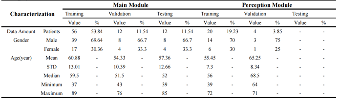
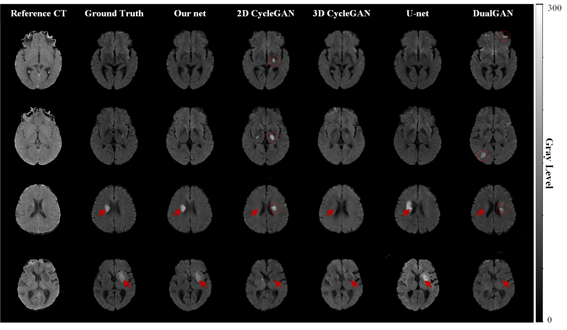
The dataset was split into two parts for training the perceptual module and the main module. Table 1 shows how patient data waswere distributed between these modules. Fig. 2 compares representative DW images from our algorithm with those from other algorithms, alongside actual DW images and input CT images for reference. Our network and U-Net notably excel in highlighting high signal intensity areas characteristic of stroke lesions, ain contrast to the less accurate lesion depiction by 2D CycleGAN, 3D CycleGAN, and DualGAN, where signal intensities in lesion areas inversely mimic those in real DW images. Fig. 3 quantitatively assesses the precision of our synthetic DW images against actual DWI images and those from other networks. Our method demonstrates superior performance, achieving a PSNR of 28.30, an SSIM of 0.843, and an NMSE of 0.293, highlighting its effectiveness in accurate image generation.Discussion and conclusion
In this research, we present a model based on CycleGAN to convert NCCT images into diffusion-weighted MR images for AIS patients. This method trains a CycleGAN to map between CT and MR images, specifically tailored for stroke diagnosis. Utilizing this model in emergency scenarios allows for the transformation of CT images into high-quality MR images, which are inherently more sensitive in detecting AIS. Crucially, this approach aims to diminish the time typically neededrequired to acquire MR images, thereby accelerating treatment initiation and potentially enhancing patient outcomes.Acknowledgements
This work was supported by the National Natural Science Foundation of China (82372038 and 62101540), the Shenzhen Excellent Technological Innovation Talent Training Project of China (RCJC20200714114436080), the Key Laboratory for Magnetic Resonance and Multimodality Imaging of Guangdong Province (2023B1212060052) and the Shenzhen Science and Technology Program (JCYJ20220818101804009 and RCBS20210706092218043).References
[1] Kamal, H., V. Lopez, and S.A. Sheth, Machine learning in acute ischemic stroke neuroimaging. Frontiers in neurology, 2018. 9: p. 945.
[2] Zhu, J.-Y., et al. Unpaired image-to-image translation using cycle-consistent adversarial networks. in Proceedings of the IEEE international conference on computer vision. 2017.
[3] Welander, P., S. Karlsson, and A. Eklund, Generative adversarial networks for image-to-image translation on multii-contrast mr images-a comparison of cyclegan and unit. arXiv preprint arXiv:1806.07777, 2018.
[4] Zhou, L., et al., Supervised learning with cyclegan for low-dose FDG PET image denoising. Medical image analysis, 2020. 65: p. 101770.
[5] Sun, B., et al., Double U-Net CycleGAN for 3D MR to CT image synthesis. International Journal of Computer Assisted Radiology and Surgery, 2023. 18(1): p. 149-156.
[6] Sun, H., et al., Synthesis of pseudo-CT images from pelvic MRI images based on an MD-CycleGAN model for radiotherapy. Physics in Medicine & Biology, 2022. 67(3): p. 035006.
[7] Wang, H., et al., Deep learning–based dynamic PET parametric K i image generation from lung static PET. European Radiology, 2023. 33(4): p. 2676-2685.
[8] Yahav-Dovrat, A., et al., Evaluation of artificial intelligence–powered identification of large-vessel occlusions in a comprehensive stroke center. American Journal of Neuroradiology, 2021. 42(2): p. 247-254.
[9] Sheth, S.A., et al., Machine learning–enabled automated determination of acute ischemic core from computed tomography angiography. Stroke, 2019. 50(11): p. 3093-3100.
[10] Amukotuwa, S.A., et al., Automated detection of intracranial large vessel occlusions on computed tomography angiography: a single center experience. Stroke, 2019. 50(10): p. 2790-2798.
[11] Chatterjee, A., N.R. Somayaji, and I.M. Kabakis, Abstract WMP16: artificial intelligence detection of cerebrovascular large vessel occlusion-nine month, 650 patient evaluation of the diagnostic accuracy and performance of the Viz. ai LVO algorithm. Stroke, 2019. 50(Suppl_1): p. AWMP16-AWMP16.
[12] Jabal, M.S., et al., Interpretable machine learning modeling for ischemic stroke outcome prediction. Frontiers in neurology, 2022. 13: p. 884693.
[13] Shaham, U. and R.R. Lederman, Learning by coincidence: Siamese networks and common variable learning. Pattern Recognition, 2018. 74: p. 52-63.
[14] Tomita, N., et al., Automatic postpost-stroke lesion segmentation on MR images using 3D residual convolutional neural network. NeuroImage: clinical, 2020. 27: p. 102276.
[15] Maier, O., et al., Correction: Classifiers for ischemic stroke lesion segmentation: A comparison study. PLoS One, 2016. 11(2): p. e0149828.
Figures