4696
High quality diffusion images from accelerated acquisition on an MR-Linac by using Deep learning.1Joint Department of Physics, The Institute of Cancer Research and The Royal Marsden NHS Foundation Trust, London, United Kingdom, 2Mathematics, St John’s College, University of Oxford, Oxford, United Kingdom, 3The Royal Marsden Hospital, London, UK; The Institute of Cancer Research, London, United Kingdom, 4Netherlands Cancer Institute, Amsterdam, Netherlands
Synopsis
Keywords: AI/ML Image Reconstruction, Diffusion/other diffusion imaging techniques, Geometric distortion, ADC variability, Deformable Registration
Motivation: Trade-offs between acquisition time and precision of the apparent diffusion coefficient (ADC) hinder the adoption of diffusion-weighted (DW) MRI for biologically-adaptive MR-guided radiotherapy.
Goal(s): To obtain high quality DW images and precise ADC maps using deep learning while shortening acquisition times on an MR-Linac.
Approach: We trained U-net models to generate high quality DW images and ADC maps from only one average per b-value. Four models were trained using either trace-weighted or single DW direction images and with or without registration to the b0 image.
Results: Trained models effectively generated high-quality images from subsampled data. Registration reduced ADC variability.
Impact: Using deep learning we obtained high quality diffusion-weighted MRI from subsampled MR-Linac data. Shortened acquisitions and increased precision of the apparent diffusion coefficient facilitate integration into adaptive MR-guided radiotherapy workflows that use diffusion-weighted MRI for treatment response assessment and prediction.
Introduction
The apparent diffusion coefficient (ADC) is a potential biomarker for radiotherapy response assessment and could aid in daily treatment planning, particularly on hybrid MR-Linacs1. However, diffusion-weighted imaging (DWI) with EPI readout is susceptible to distortions from field inhomogeneity and eddy-currents. On the Unity MR-Linac system, ADC measurements over 7 cm from the isocentre may be compromised, especially with in-plane DW gradients2. Radio-translucent coils further reduce signal-to-noise ratio, potentially increasing bias and variability. We propose employing the quickDWI3 deep learning algorithm, utilizing a U-net architecture to enhance subsampled noisy DWI data. Prior work4 utilized DL algorithms to denoise less averaged high-resolution diffusion images. Additionally, recent research5 estimates ADC maps from accelerated diffusion data using a combination of CNNs and vision transformers.Our goal is to employ deformable image registration to mitigate distortions, enabling the use of accelerated DWI in treatment planning and reducing eddy current-related ADC biases.
Methods
We collected data from 10 prostate patients in the HERMES trial (NCT04595019), each involving 2-4 scans at various treatment stages on a 1.5T Unity MR-Linac (Elekta AB, Stockholm). Model training utilized data from 7 patients (22 scans), with one each reserved for validation (3 scans) and testing (2 scans); one patient was excluded due to implant-related artifacts. The acquired images covered 15 slices (224 rows/columns) with b-values (and averages) of 0(6), 30(6), 150(6), 500 (14) s/mm² and 3 DW directions.
All diffusion-weighted images underwent 2D deformable B-Spline registration6 to align with the corresponding b0 image, utilizing Elastix7 with a rigidity penalty term for local deformation control. Four distinct models were trained: All directions (AD), single direction (SD), all direction registered (AD+R), and single direction registered (SD+R).
Fig. 2A depicts the shared input and output dimensions across all models, with 12 input channels, including additional inputs from adjacent slices. A software patch in the reconstruction pipeline facilitated the extraction of diffusion images from individual averages and directions for training. For all models, individual images along each direction were arithmetically averaged, and geometrically averaged along directions for AD and AD+R to form the ground truths. Training loss was mean absolute error (MAE with a learning rate of 1e-4, batch size 30, spanning 50 epochs.
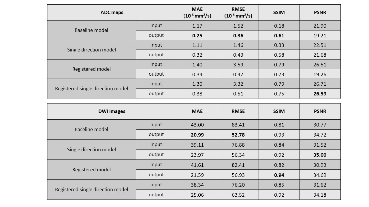
We assessed mean absolute error (MAE), root mean square error (RMSE), structural similarity index (SSIM), and peak signal-to-noise ratio (PSNR) for both DWI and ADC images. Additionally, we compared ADC distributions in a rectangular region of interest (ROI) within the prostate test dataset between ground truth, model inputs, and model outputs.
Results and Discussion
DW images showed distortion mainly along the phase encode direction (AP), highlighted in Fig. 1A, which varied with DW direction. Lower b-values showed less distortion. Registration to the b0 image reduced distortion, indicated by dashed red lines (Fig. 1B).Training and validation losses converged to ~0.2 for non-registered models but converged to ~0.5 for registered models (Fig. 2B). This disparity may be due to the model struggling to generalize to the large amount of freedom in deformable image registration.
All models reduced MAE and RMSE for ADC in the sub-sampled diffusion data (Fig. 3). The best model for each metric is highlighted. SSIM for DW images improved compared to inputs across all models, but for ADC maps, SSIM decreased for registered models (AD+R and SD+R). This aligns with the loss curve findings, suggesting registered models may not have generalized the registrations, or that the input resembled the 'noisy' ground truth, impeding noise learning.
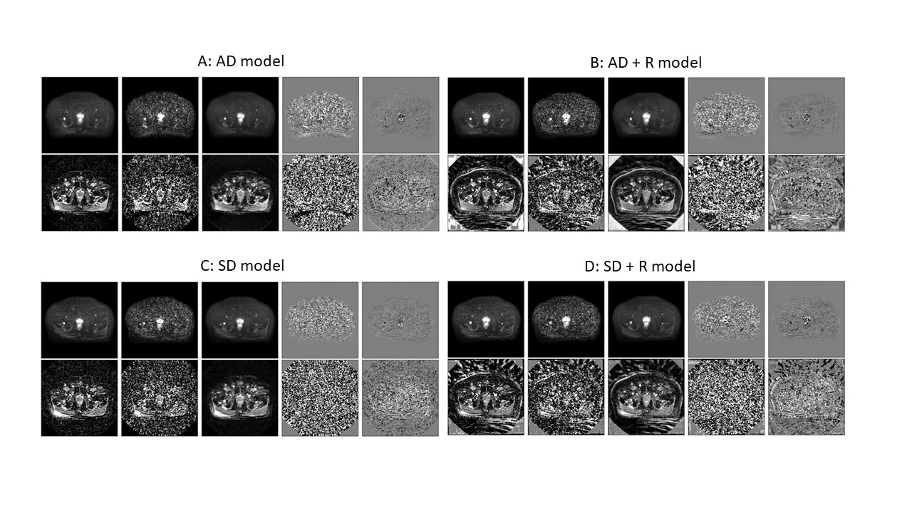
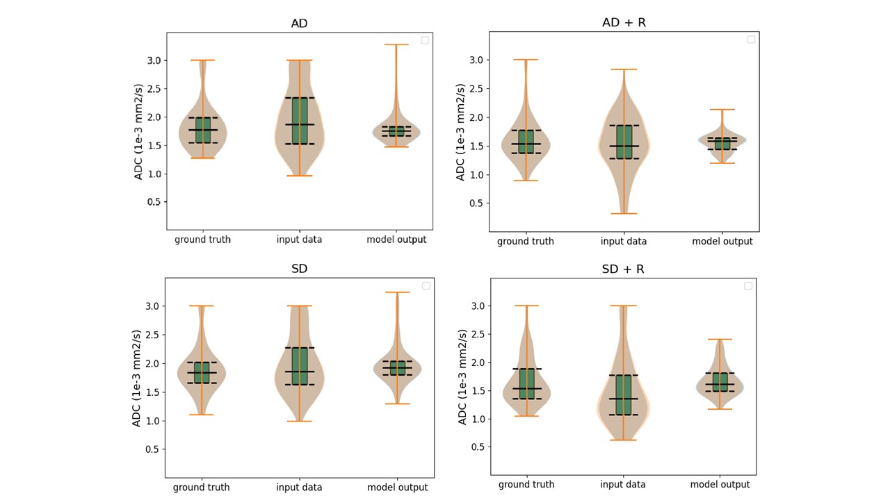
Sample ground truth, raw input, model output and respective difference images for each model are displayed in Fig. 4. The SD model output [Fig. 4C] showed direction-specific signal pile-up due to diffusion gradient-related distortions, reduced in the AD model, possibly due to ground truth averaging (Fig. 4A). In registered ADC maps [Fig. 4B,D], ground truth images are noisy, suggesting registration may need refinement. Predicted output, particularly in all-directions models, was less noisy, potentially explaining the decrease in SSIM for registered models [Fig. 3]. This is supported by the predicted ADC values having less variability in the registered models (AD + R and SD + R) [Fig. 5]. The single direction models (SD and SD +R) tend to induce a positive bias. Predicted images from all models improved over noisy data [Fig. 3]. However, obtaining evaluations from radiation oncologists will be crucial, particularly given the variation in ground truth for different models. Additionally, exploring the possibility of training models with data from alternative diffusion directions could provide valuable insights.
Conclusion
DL models can generate high-quality images from subsampled diffusion data, enabling DWI in biologically adaptive MR-guided radiotherapy workflows.Acknowledgements
We acknowledge Rosie Goodburn, Magali Nuixe, Joan Chick and Bjoern Eiben for valuable discussions.
This work was supported by Cancer Research UK programme grant (C33589/A28284).
The Institute of Cancer Research and The Royal Marsden NHS Foundation Trust are members of the Elekta MR-Linac Research Consortium.
We acknowledge research support from Elekta and Dave Higgins (Philips MR) for providing MR source code, research licences, and support
References
1. van Houdt PJ, Saeed H, Thorwarth D, Fuller CD, Hall WA, McDonald BA, et al. Integration of quantitative imaging biomarkers in clinical trials for MR-guided radiotherapy: Conceptual guidance for multicentre studies from the MR-Linac Consortium Imaging Biomarker Working Group. Eur J Cancer. 2021;153:64-71.
2. Kooreman ES, van Houdt PJ, Keesman R, Pos FJ, van Pelt VWJ, Nowee ME, et al. ADC measurements on the Unity MR-linac - A recommendation on behalf of the Elekta Unity MR-linac consortium. Radiother Oncol. 2020;153:106-13.
3. Zormpas-Petridis K, Tunariu N, Curcean A, Messiou C, Curcean S, Collins DJ, et al. Accelerating Whole-Body Diffusion-weighted MRI with Deep Learning-based Denoising Image Filters. Radiol Artif Intell. 2021;3(5):e200279.
4. Kawamura M, Tamada D, Funayama S, Kromrey ML, Ichikawa S, Onishi H, et al. Accelerated Acquisition of High-resolution Diffusion-weighted Imaging of the Brain with a Multi-shot Echo-planar Sequence: Deep-learning-based Denoising. Magn Reson Med Sci. 2021;20(1):99-105.
5. Li Y, Joaquim MR, Pickup S, Song HK, Zhou R, Fan Y. Learning ADC maps from accelerated radial k-space diffusion-weighted MRI in mice using a deep CNN-transformer model. Magn Reson Med. 2023.
6. Rueckert D, Sonoda LI, Hayes C, Hill DL, Leach MO, Hawkes DJ. Nonrigid registration using free-form deformations: application to breast MR images. IEEE Trans Med Imaging. 1999;18(8):712-21.
7. Klein S, Staring M, Murphy K, Viergever MA, Pluim JP. elastix: a toolbox for intensity-based medical image registration. IEEE Trans Med Imaging. 2010;29(1):196-205.
Figures
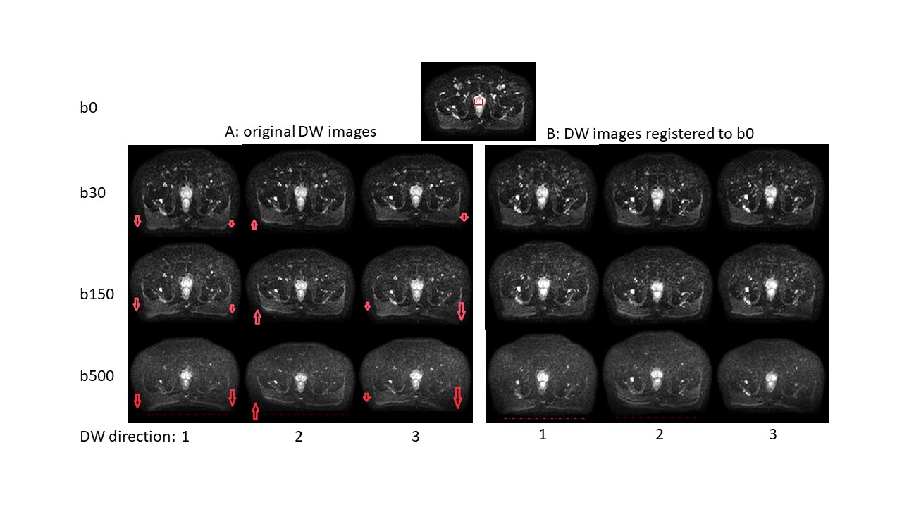
Figure1: Example diffusion-weighted images, serving as inputs for the models without (A) and with registration (B) to the b0 image to account for eddy-current-related image distortions. Distortions appear primarily along the phase encoding direction (AP), are more pronounced at higher b values (arrows) and reduced after registration. Distortions differ for diffusion-encoding directions (1-3) .Dashed red lines serve as visual aide.
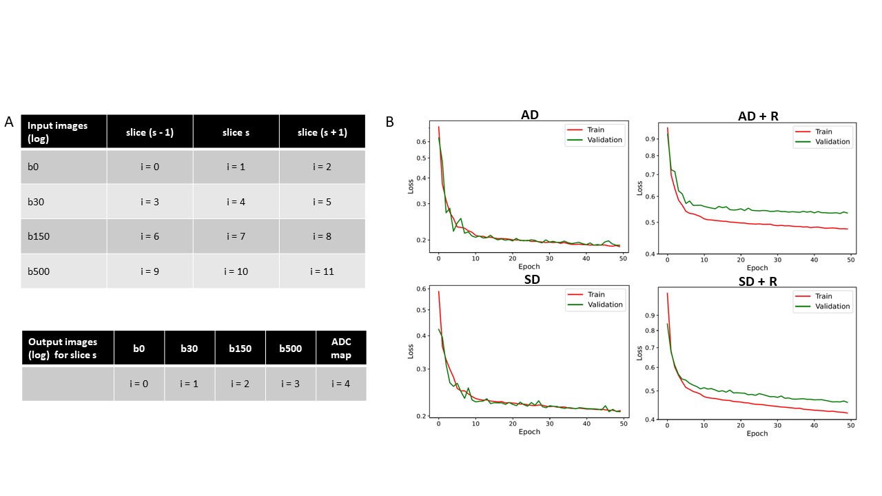
Figure2: (A) Model input includes adjacent slices (s-1, s, s+1), each 224x224 pixels. This forms 12 channels with images from 4 b-values. For the first and last slices, slice s is used instead of s-1 and s+1 respectively. Output comprises 5 channels: 4 DW images and an ADC map. Logarithm of signal intensity is applied to both. (B) Training loss curves depict that non-registered models exhibit superior generalization to validation data compared to registered models.