4694
Deep Learning-based Super-Resolution Reconstruction for Brain Diffusion-weighted MRI1The First Affiliated Hospital of Dalian Medical University, Dalian, China, 2Clinical & Technical Support, Philips Healthcare, Beijing, China
Synopsis
Keywords: AI/ML Image Reconstruction, Neuro, Super-Resolution Reconstruction
Motivation: A deep learning-constrained algorithm has been integrated into MRI data acquisition and image reconstruction processes, encompassing compressed sensing, image denoising, and resolution upscaling techniques. Nonetheless, limited prospective studies are available that evaluate the application of this algorithm for brain diffusion-weighted imaging.
Goal(s): The primary objective of this study was to compare the recently developed deep learning-constrained algorithm with conventional compressed sensing reconstruction.
Approach: This study comprehensively assessed images, both qualitatively and quantitatively, employing rigorous methodologies and analytical tools.
Results: The results demonstrated that the newly developed deep learning-constrained algorithm significantly enhanced image sharpness while maintaining signal-to-noise ratio, thus advantaging clinical diagnosis.
Impact: Deep learning-constrained super-resolution reconstruction leads to a significant increase in image sharpness of brain DWI, which holds potential to improve clinical diagnosis of diseases, such as stroke and tumors.
Introduction
Conventional brain diffusion weighted imaging (DWI), relying on the echo planar imaging (EPI) acquisition, exhibits limited spatial resolution compared to other routine neuroimages due to its heightened sensitivity to B0 inhomogeneities and its high requirement on the hardware of magnetic field gradients. Recent developed deep-learning (DL) based super-resolution networks offer potential to improve image resolution1,2; however, the application of such methods for brain DWI has been scarcely evaluated in prospective studies. The primary objective of this study was to evaluate the performance of DL-based super-resolution reconstruction on randomly under-sampled brain DWI, with comparison to the conventional compressed sensing reconstruction.Method
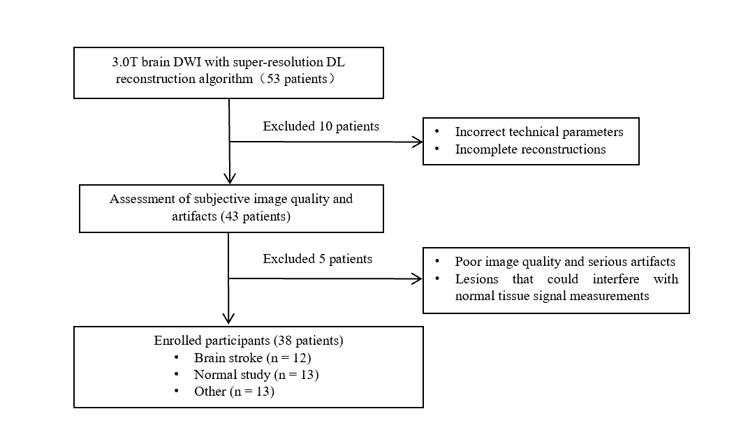
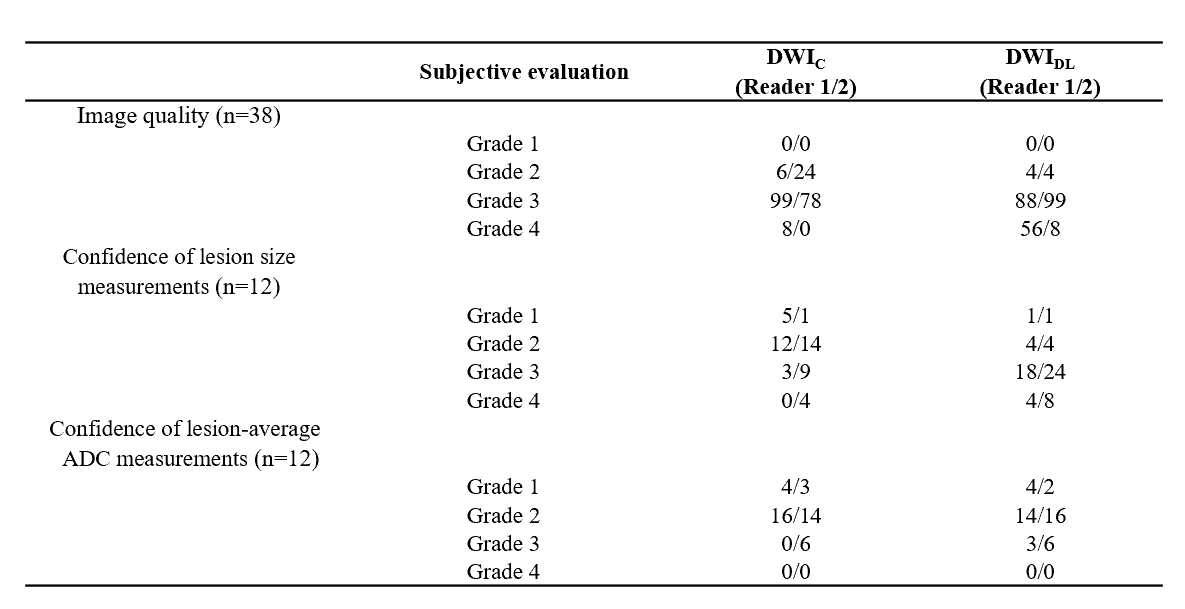
In this prospective study, 38 participants were examined using a 3.0T MRI system (Ingenia CX, Philips Healthcare, Best, the Netherlands). The study was approved by the local IRB, and informed consent was obtained from all subjects. The study workflow is shown in Figure 1. MRI scans, including T2-weighted imaging, T1-weighted imaging, fluid attenuated inversion recovery imaging, and DWI, were acquired using a 32-channel head coil. DWI was acquired based on single-shot EPI with two b values (0 and 1000 s/mm2) with a randomly under-sampling factor of 2 in the phase encoding direction and a 24-second acquisition time. The DWI images were reconstructed with two schemes: (a) DL-based super-resolution reconstruction (DWIDL); (b) conventional compressed sensing reconstruction (DWIC). The DL super-resolution algorithm (Precise-Image-Net), provided by Philips Healthcare, as an extension of the Adaptive-CS-Net algorithm, was trained on 6 million image pairs (originally high-resolution and secondary downscaled images) for removal of ringing artifacts and upscaling of image resolution1,3. Quantitative analysis was conducted by calculating the signal-to-noise ratio (signal intensity in white matter and grey matter divided by the signal standard deviation of cerebrospinal fluid) with measurement of averaged signal intensity values inside an equal-sized region of interest (25 mm2) in corpus callosum, caudate nucleus, and cerebrospinal fluid. Additionally, the edge rise distance (ERD) was determined as a quantitative measure of image sharpness using ImageJ software (https://imagej.nih.gov/ij/)4,5. Two radiologists independently reviewed the images in a random order. The readers assessed the maximum dimension and lesion-average ADC, reported their measurement confidence, and evaluated the quality of each image using a 4-point scale6 quantitative measurements and qualitative scores between DWIDL and DWIC groups of images were compared using the Wilcoxon signed-rank test.Result
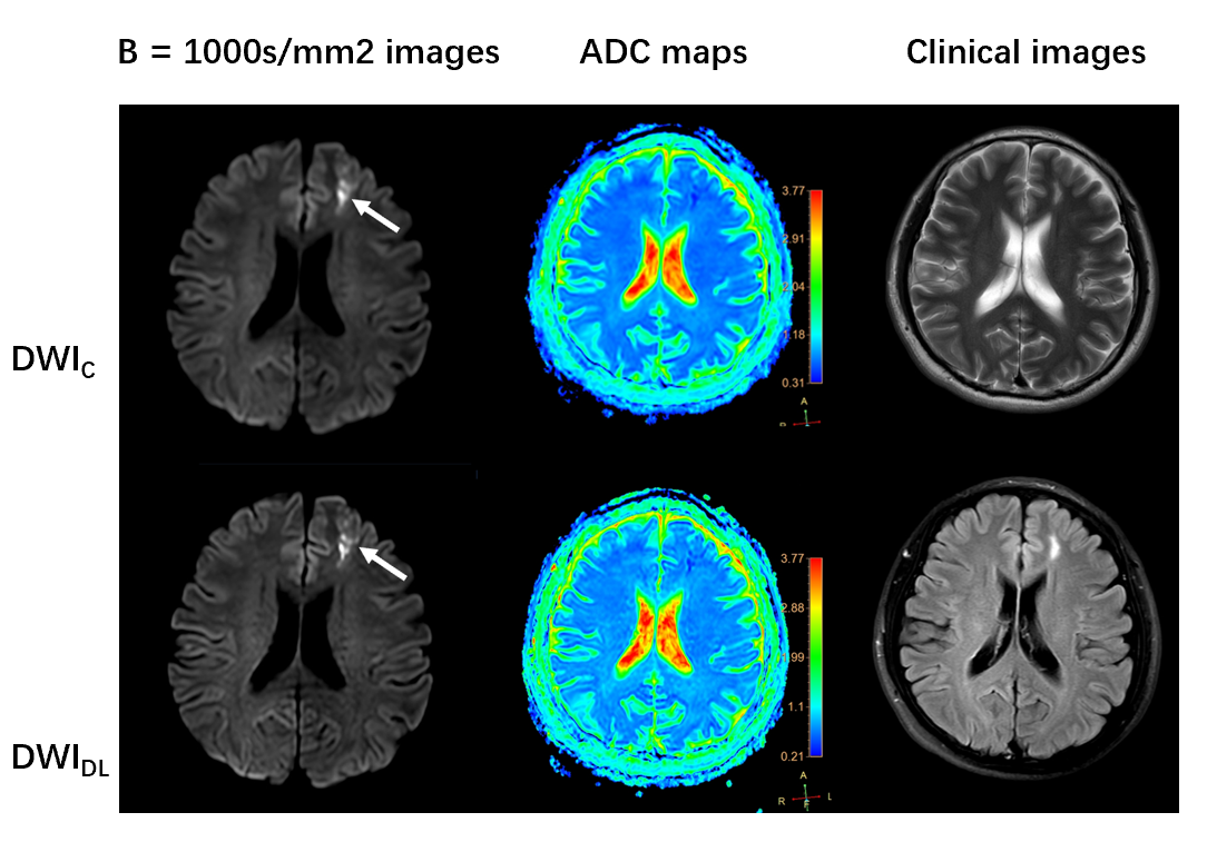
No significant differences were observed between DWIDL and DWIC images in terms of SNR, but with DWIDL images exhibiting significantly reduced ERD (P < 0.001) (Figure 2). Table 1 shows DWIDL received higher scores of image quality and confidence in the measurement of lesion size than DWIC (P < 0.001). Figures 3 displays representative DWI (b = 1000 s/mm2) images and ADC maps obtained from DWIDL and DWIC reconstructions, respectively.Conclusion
DL-constrained super-resolution reconstruction significantly enhances the image sharpness of brain DWI with comparison to conventional reconstruction. Given that this technique is straightforward and does not necessitate additional acquisition time, it shows promise for robust high-resolution diffusion imaging, and great potential for improved diagnosis of diseases, such as stroke and tumors.Acknowledgements
NoneReferences
1. Leon M. Bischoff, Johannes M. Peeters, Leonie Weinhold, et al. Deep Learning Super-Resolution Reconstruction for Fast and Motion-Robust T2-weighted Prostate MRI. Radiology.2023;308:3.
2. Li Y, Sixou B, Peyrin F. A review of the deep learning methods for medical images super resolution problems. IRBM 2021;42(2):120–133.
3. Pezzotti N, Yousefi S, Elmahdy MS, et al.An Adaptive Intelligence Algorithm for Undersampled Knee MRI Reconstruction. IEEE Access.2020;8:204825-204838.
4. Kim M, Lee SM, Park C, et al.Deep learning-enhanced parallel imaging and simultaneous multislice acceleration reconstruction in knee MRI. Invest Radiol.2022;57:826–833.
5. Wang, Q., Zhao, W., Xing, X. et al. Feasibility of AI-assisted compressed sensing protocols in knee MR imaging: a prospective multi-reader study. Eur Radiol (2023).
6. Jung JY, Yoon YC, Kim HR, Choe BK, Wang JH, Jung JY. Knee derangements: comparison of isotropic 3D fast spin-echo, isotropic 3D balanced fast field-echo, and conventional 2D fast spin-echo MR imaging. Radiology 2013; 268:802–813.
Figures