4693
Image Super-Resolution Using Deep Convolutional Networks Improve the Image quality of Compressed Sensing MRI for Pancreatic DWI.1radiology department, west China hospital of Sichuan university, chengdu, China, 2Clinical Science, Philips Health Care,Chengdu,China, chengdu, China, 3radiology Department, west China hospital of Sichuan university, chengdu, China
Synopsis
Keywords: AI/ML Image Reconstruction, Pancreas, super-resolution convolutional neural network, high-resolution diffusion weighted imaging
Motivation: Image resolution achieved with compressed sensing was inferior compared to that obtained with sense technique.Current state of the art in Super-Resolution enables enhanced image resolution at a finer level of detail.
Goal(s): Objective is to enhance visualization of anatomical details in high-resolution pancreatic DWI by leveraging SR.
Approach: In our study, we employed integrating super-resolution convolutional neural network-compressed sensing (SR-CS) algorithm and integrating artiffcial intelligence-compressed sensing (AI-CS) algorithm for the reconstruction of pancreatic HR-DWI raw data.
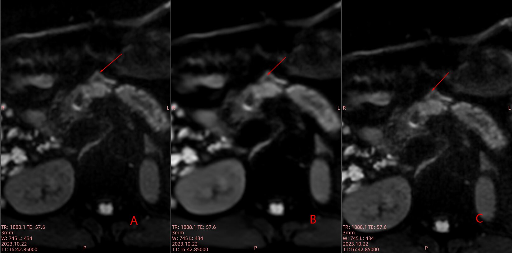
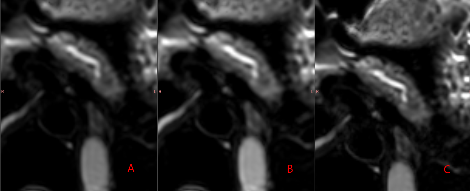
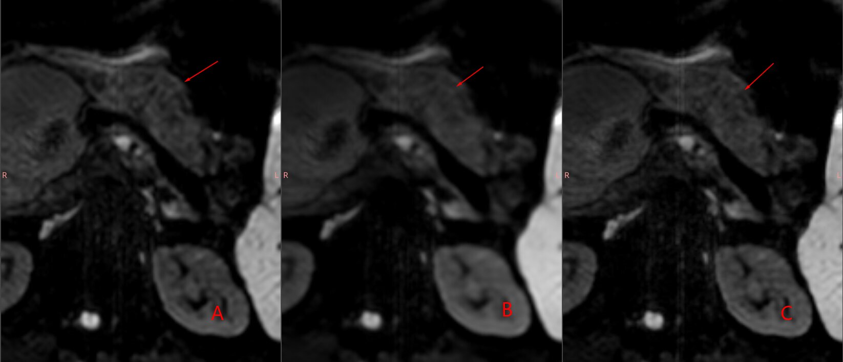
Results: Images with SR-CS generally exhibit superior performance compared to traditional images in terms of tumor border delineation and reduction of background noise from peritoneum and spine.
Impact: Current utilization of AI is extensive, while application of SR in medical images remains rare. Utilization of SR allows for execution of MRI within a concise timeframe, while simultaneously considering the aspect of resolution.
Synopsis
The high-resolution diffusion weighted imaging (HR-DWI) sequence holds significant value in the differential diagnosis of pancreatic masses. In our study, we employed integrating super-resolution convolutional neural network-compressed sensing (SR-CS) algorithm and integrating artiffcial intelligence-compressed sensing (AI-CS) algorithm for the reconstruction of pancreatic HR-DWI raw data. Images with SR-CS generally exhibit superior performance compared to traditional images in terms of tumor border delineation and reduction of background noise from the peritoneum and spine.Introduction
There were motion artifacts observed in high-resolution DWI despite the use of respiratory gating for long acquisition time. In the past five years, Compressed Sensing (CS) technique has been widely employed to reduce the acquisition time of various pulse sequences in magnetic resonance imaging (MRI). Unlike other digestive organs, only high-resolution images can reveal the microanatomy of the pancreas. The image resolution achieved with CS technique was inferior compared to that obtained with sense technique.The application of super-resolution convolutional neural networks can enhance the fine resolution of images.The problems in CS are possiblely being addressed through the application of SR technology.Methods
In total, 25 patients who underwent pancreatic HR-DWI at 3.0T, reconstructed with AI-CS algorithm, SR-CS algorithm and CS algorithm, were prospectively enrolled.All pancreatic examinations were conducted on a 3.0-T system (Ingenia Elition, Philips Healthcare). Patients were scanned in the supine position using a 16-element phased-array coil. Two radiologists independently evaluated overall background suppression, image quality, artifacts and visualization of the pancreas using a five-point scale. Signal-to-noise ratio (SNR) of the head, neck and tail of the pancreas, as well as contrast-to-noise (CNR) of the pancreas and renal cortex were measured. The Friedman test was conducted to compare the three algorithms.Results
1.SR-CS and AI-CS were signiffcantly better than CS in terms of overall background suppression, image quality and pancreas tissue delineation (P <0.001).2.For pancreatic morphology in b-value 50 and lesion conspicuity in b-value 800, SR-CS exhibited superior visualization of intricate structural details compared to both CS and AI-CS.
3.The three SNR of SR-CS and AI-CS was signiffcantly higher for CS. There was no signiffcant difference in CNR among SR-CS,SR-CS and CS.
Discussion
Our study identified two main findings: SR-CS can enhance image resolution specifically at b-value 50; on b-value 800 with SR technique, normal pancreas microanatomy appears more blurred compared to conventional CS images, but tumor borders are sharper on SR-CS images. Although AI-CS significantly reduces noise and improves Signal-to-Noise Ratio (SNR), it also smooths out tiny textures and blurs contour details of the pancreas microanatomy. At b-value 50, while SR-CS assisted images exhibit slightly-increased noise levels, they are able to display anatomical structures that appear blurred or even disappear in traditional CS images due to volume effects caused by high-signal water content. AI-CS's manifestation at both b-values 50 and 800 remains consistent with similar outcomes. SR-CS serves as a supplement solely of AI-CS for enhancing resolution in high-contrast tissues but may result in loss of details for low-signal ones.The presence of a low signal on b-value 800 generally indicates the presence of unhindered diffusion of water molecules or a minimal water content in tissues.This particular manifestation of SR in high b values is more appropriate for illustrating the infiltration of tumors , albeit it may impact the visualization of the boundaries of certain inflammatory masses.Conclusion
The utilization of SR-CS can effectively mitigate noise and improve microanatomical identification of pancreatic CS DWI images.Acknowledgements
No acknowledgement found.References
1.Yang F, Pan X, Zhu K, Xiao Y, Yue X, Peng P, Zhang X, Huang J, Chen J, Yuan Y, Sun J. Accelerated 3D high-resolution T2-weighted breast MRI with deep learning constrained compressed sensing, comparison with conventional T2-weighted sequence on 3.0 T. Eur J Radiol. 2022 Nov;156:110562. doi: 10.1016/j.ejrad.2022.110562. Epub 2022 Oct 17. PMID: 36270194.
2.Dong C, Loy CC, He K, Tang X. Image Super-Resolution Using Deep Convolutional Networks. IEEE Trans Pattern Anal Mach Intell. 2016 Feb;38(2):295-307. doi: 10.1109/TPAMI.2015.2439281. PMID: 26761735.
3.Wu X, Deng L, Li W, Peng P, Yue X, Tang L, Pu Q, Ming Y, Zhang X, Huang X, Chen Y, Huang J, Sun J. Deep Learning-Based Acceleration of Compressed Sensing for Noncontrast-Enhanced Coronary Magnetic Resonance Angiography in Patients With Suspected Coronary Artery Disease. J Magn Reson Imaging. 2023 Nov;58(5):1521-1530. doi: 10.1002/jmri.28653. Epub 2023 Feb 27. PMID: 36847756.
4.Harder FN, Jung E, McTavish S, Van AT, Weiss K, Ziegelmayer S, Gawlitza J, Gouder P, Kamal O, Makowski MR, Lohöfer FK, Karampinos DC, Braren RF. High-Resolution, High b-Value Computed Diffusion-Weighted Imaging Improves Detection of Pancreatic Ductal Adenocarcinoma. Cancers (Basel). 2022 Jan 18;14(3):470. doi: 10.3390/cancers14030470. PMID: 35158737; PMCID: PMC8833466.
5.Wu X, Tang L, Li W, He S, Yue X, Peng P, Wu T, Zhang X, Wu Z, He Y, Chen Y, Huang J, Sun J. Feasibility of accelerated non-contrast-enhanced whole-heart bSSFP coronary MR angiography by deep learning-constrained compressed sensing. Eur Radiol. 2023 Nov;33(11):8180-8190. doi: 10.1007/s00330-023-09740-8. Epub 2023 May 20. PMID: 37209126.
6.Fattahi R, Balci NC, Perman WH, Hsueh EC, Alkaade S, Havlioglu N, Burton FR. Pancreatic diffusion-weighted imaging (DWI): comparison between mass-forming focal pancreatitis (FP), pancreatic cancer (PC), and normal pancreas. J Magn Reson Imaging. 2009 Feb;29(2):350-6. doi: 10.1002/jmri.21651. PMID: 19161187.
7.Jang KM, Kim SH, Min JH, Lee SJ, Kang TW, Lim S, Choi D. Value of diffusion-weighted MRI for differentiating malignant from benign intraductal papillary mucinous neoplasms of the pancreas. AJR Am J Roentgenol. 2014 Nov;203(5):992-1000. doi: 10.2214/AJR.13.11980. PMID: 25341136.
8.Schima W, Böhm G, Rösch CS, Klaus A, Függer R, Kopf H. Mass-forming pancreatitis versus pancreatic ductal adenocarcinoma: CT and MR imaging for differentiation. Cancer Imaging. 2020 Jul 23;20(1):52. doi: 10.1186/s40644-020-00324-z. PMID: 32703312; PMCID: PMC7376657.
Figures