4685
Comprehensive raw data collection using a Philips Research Imaging Development Environment (PRIDE) tool1Philips Healthcare, Rochester, MN, United States, 2Department of Radiology, Mayo Clinic, Rochester, MN, United States, 3Russell H. Morgan Department of Radiology and Radiological Science, The Johns Hopkins University School of Medicine, Baltimore, MD, United States, 4. M. Kirby Research Center for Functional Brain Imaging, Kennedy Krieger Institute, Baltimore, MD, United States
Synopsis
Keywords: Software Tools, Software Tools
Motivation: Automate collecting of MRI raw data from the Philips MR scanners.
Goal(s): To create a tool that will allow users to extract selected scan data in raw format and save specific scan information for later use.
Approach: Using a Philips PRIDE interface to gathers raw data and schedule a job to execute during afterhours on the Scanner Console.
Results: The simple tool allows to collect complete raw data in under 3 mins at the end of the scan and associate the data with respective DICOM information for later association or annotation.
Impact: Collection and storage of the complete acquisition raw data with associated DICOM information for potential use in the future using a flexible tool with a small footprint.
Introduction
MRI studies—both clinical and research—usually only retain the final reconstructed images (as DICOM1 data), discarding the raw data that was used during the reconstruction. Nonetheless, a library of pre-existing raw data enables the quicker creation of innovative reconstruction methods, such as those based on machine learning and deep learning, which are continually being developed. Moreover, certain artifacts can be frequently corrected far more easily in the raw data than in the reconstructed images, which requires the raw data be accessible/available. Unfortunately, the raw data storage requirements are often prohibitive even in research settings due to involved process in extracting the raw data from the scanner console and associating it with DICOM data for future comparison and analysis, especially in large-scale population studies. On Philips MR scanners, the purest raw data (labeled as LAB/RAW/SIN data format) does not contain any Protected Health Information (PHI) and so it is challenging to associate the obtained raw data with the corresponding DICOM data from a given scan of a particular subject. The existing export solutions either require significant manual steps or purchasing of costly data management software such as Flywheel2 or Agora3. For the manual method, one must manually export each individual scan and label them after that scan, which makes for a significant booking keep work for the researchers. It is evident that these approaches are not suitable for multi-site large-scale population studies such as HBCD4. The aim of this project is to develop a flexible simple software tool for exporting the MRI raw data, associate it with DICOM data (using each series specific SOP and Instance UID tags) and send it to a remote server or local network or directly store it on the console. All additional meta information necessary can be then extracted on the remote server by using DICOM queries to PACS using the Study Instance UID number and Series Instance UID tags.Methods
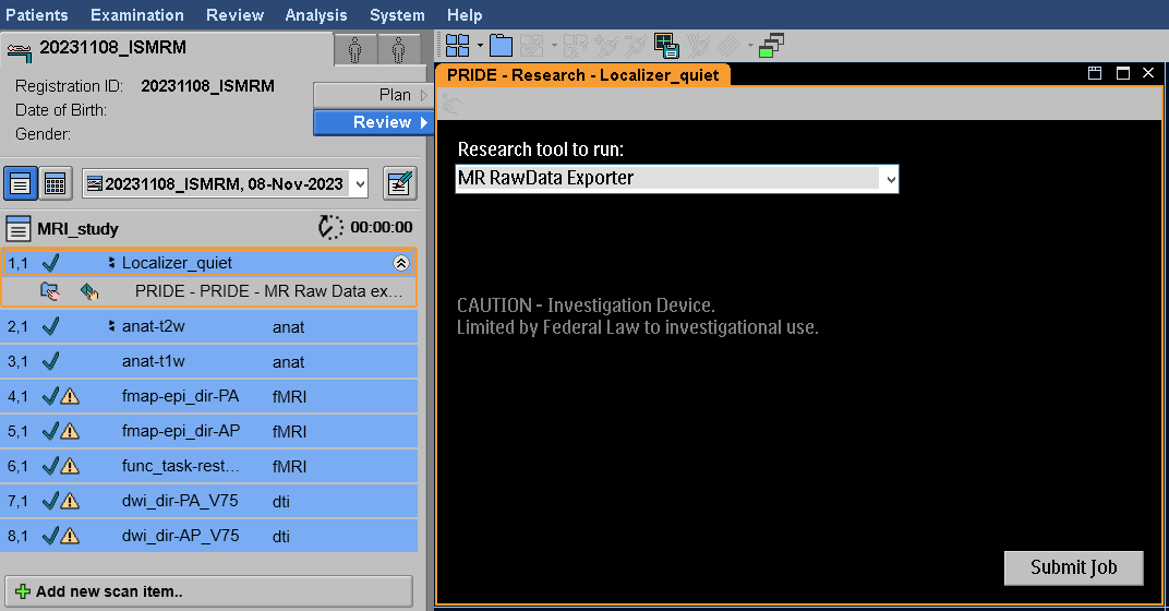
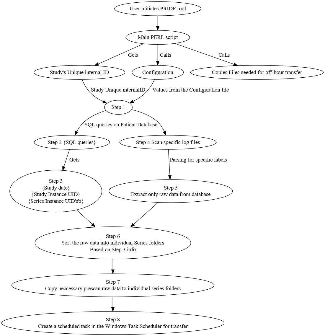
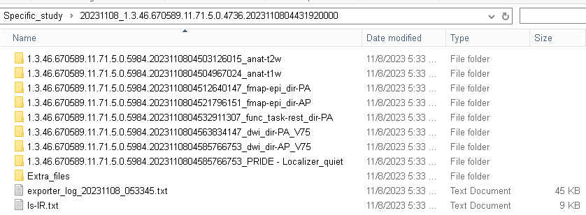
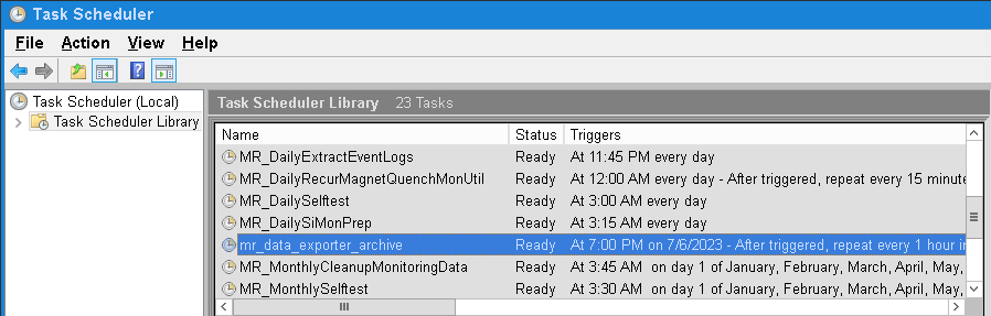
The Philips Research Imaging Development Environment (PRIDE)5,6,7 interface on the Philips MR scanners enables researchers to run processing software/tools (either for analysis or for performing administrative tasks) on the scanner console. Such a tool can be incorporated into ExamCard (sequences and examination settings file) for easy launching, as shown in Fig. 1. Once integrated into an ExamCard, the processing step will remain for any study that will use that particular ExamCard in the future. The PRIDE interface then calls a custom-built PERL script that initiates the extraction of the collected raw data in the given study while extracting the Study Instance UID tag and Series Instance UID tags from the database to associate them with the corresponding raw data. The various steps that happen in the PERL code are shown in the Fig. 2 flowchart. The study’s raw data is neatly organized into a folder structure shown in the Fig. 3 (based on the configuration settings this folder can be stored either locally or to a network location). Also, a scheduled task can be created at the time of the execution of the PRIDE tool in the Windows Task Scheduler (Fig. 4), that can perform additional steps if needed (e.g. removing identifiable information or compressing the data). This task can run in the background during off-hours of the scanner, even when user is logged out from the scanner console. Such a background task can be configured to also send the data to a remote server, such as FIONA8.Results
The PRIDE-based tool created was able to extract and properly organize the raw data for several studies, including the full protocol of the HBCD study, in under 3 minutes (a total of 25+ GB of data for each HBCD scan). The tool is currently in use at the six Philips sites as part HBCD study (under an IRB). After the initial exporting of the raw data, the files were compressed and transferred to a remote FIONA box using the background task in Windows Task Scheduler without interfering with the normal operation of the scanner. The PRIDE tool developed will work on any Philips MR scanners running on software releases R5.1.7 or higher.Discussion
We present a software tool for (Philips) raw data that can extract, organize (using the associated DICOM Series UID tags), and send the data to a local or remote server. This tool can be shared with any site using Philips MR scanners, allowing for long-term data collection. Since saving raw data for future reconstruction requires patient consent, this tool is currently shared with sites that have a research agreement with Philips and a local IRB in place.Acknowledgements
No acknowledgement found.References
1Bidgood WD Jr, Horii SC, Prior FW, Van Syckle DE. Understanding and using DICOM, the data interchange standard for biomedical imaging. J Am Med Inform Assoc. 1997;4(3):199-212.
2Flywheel (https://flywheel.io)
3Agora (https://www.gyrotools.com/gt/index.php/products/agora)
4HEALthy Brain and Child Development Study (HBCD) (https://heal.nih.gov/research/infants-and-children/healthy-brain)
5Covington K, Welch EB, Jeong HK, Landman BA. Integrating Medical Imaging Analyses through a High-throughput Bundled Resource Imaging System. Proc SPIE Int Soc Opt Eng. 2011;7967:79670E.
6Zou T, Yu H, Jiang C, et al. Differentiating the histologic grades of gliomas preoperatively using amide proton transfer-weighted (APTW) and intravoxel incoherent motion MRI. NMR Biomed. 2018;31(1):10.1002/nbm.3850.
7Soher, B. J., Deelchand, D. K., Ganji, S. (2021). Across-vendor, inline standardized spectral analysis for single voxel MRS data acquisition at 3T. Proc. Intl. Soc. Mag. Reson. Med. 29 (2021), Program number: 0727.
8FIONA https://github.com/ABCD-STUDY/FIONASITE
Figures