4684
Using digital reference objects to optimize advance reconstruction methods for abdominal DCE MRI in the presence of motion1Biomedical Engineering, University of Iowa, Iowa City, IA, United States, 2Radiology, University of Wisconsin Madison, Madison, WI, United States, 3Location AMC, Amsterdam UMC, Amsterdam, Netherlands, 4Radiology, University of Iowa, Iowa City, IA, United States
Synopsis
Keywords: Software Tools, Phantoms, digital reference object
Motivation: Concern over gadolinium contrast injections for research purposes limits in vivo testing of new imaging methods. An in-silica optimization strategy is needed when developing advanced reconstructions in the setting of free-breathing abdominal DCE MRI.
Goal(s): Demonstrate a framework for testing performance of different reconstructions.
Approach: A publicly available DRO was used to generate simulated free-breathing abdominal DCE k-space data. Data was then reconstructed using different reconstruction settings for MOCCO and SENSE.
Results: This approach allowed head-to-head comparisons of different reconstruction methods as well as comparison with the ground truth data used to generate the DRO.
Impact: The proposed testing approach allows researchers to test numerous combinations of acquisitions and reconstructions while having ground truth as a benchmark.
Introduction
High temporal and spatial resolution abdominal DCE MRI remain a challenge due to the wide range of contrast dynamics and respiratory motion. Further, quantitative DCE MRI has shown promise to improve reliability and consistency across sites and during longitudinal studies. Advanced acquisition and reconstruction strategies have made progress and show promise to further improve the spatial and temporal resolution in the presence of motion (1-7). However optimization and validation of these advanced algorithms remains a challenge. In vivo studies are the gold standard but even these suffer from shot-to-shot variability and recent concerns around gadolinium staining (8) serve further limit utilization of multiple contrast injections for research. Digital Reference Objects (DROs) provide a simulation framework for testing new methods in scenarios including DCE contrast changes (9). A recent DRO was made publicly available that includes both DCE capabilities and respiratory motion (10). In this work we present a framework for optimizing advance reconstructions in silica, in the setting of free-breathing DCE-MRI.Methods
A prime challenge was to identify a DRO with the ability to modulate contrast enhancements. It should further provide regulation of respiratory dynamics to simulate abdominal respiratory motion. A review of the published manuscripts and publicly available DROs identified the MRXCAT by Eric Schrauben (10) as a potential candidate. The open access code allowed flexibility for k-space synthesis and data for contrast enhancement with and without motion was generated for reconstruction algorithm testing. The number of respiratory states was modified to generate 10 settings of increasing respiratory motion excursion. This framework allowed for methodically testing reconstruction algorithm performance using varying degrees of respiratory motion and contrast enhancement. To demonstrate the capability of this approach, we chose to test different reconstruction parameters of the MOCCO algorithm (11) which has shown promising results in a DCE setting. Testing was performed using different combinations of principal components (PCAs) and their impact on with and without motion.Results
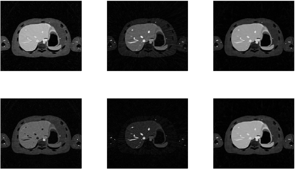
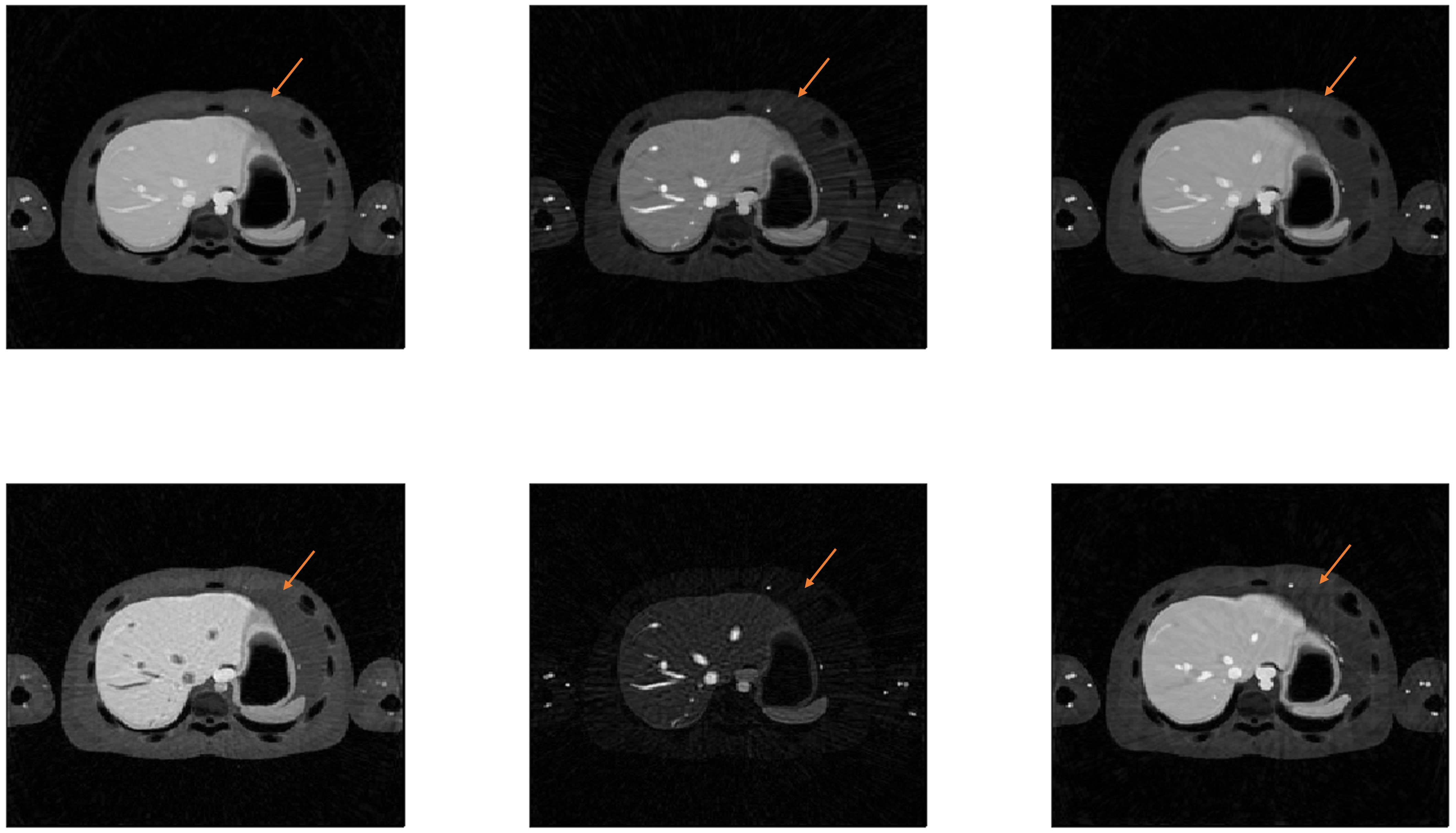
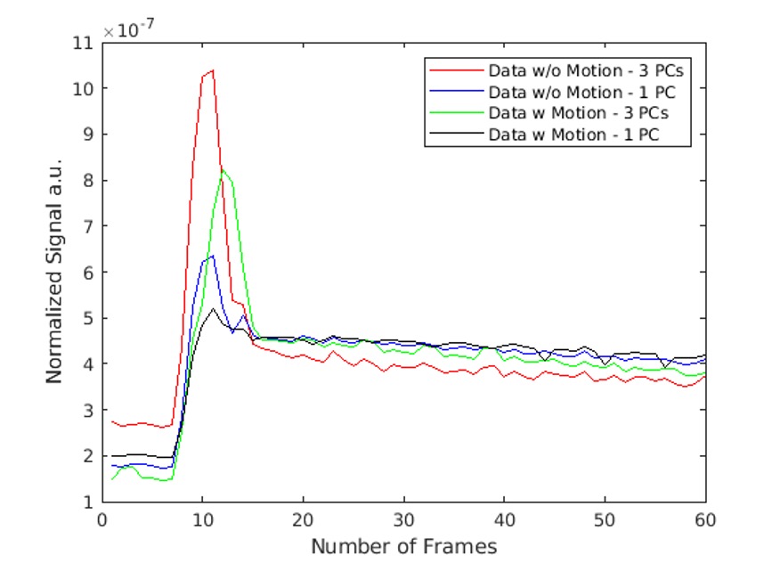
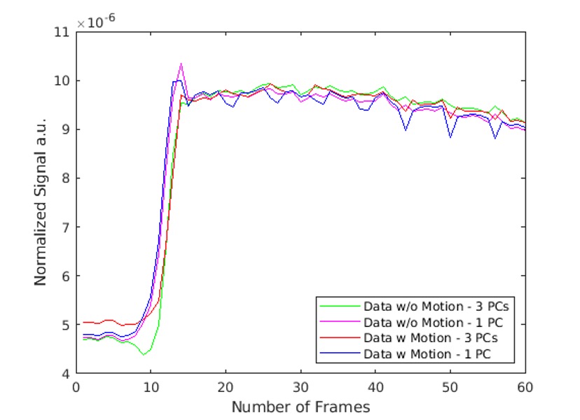
The DRO and open-source code provided a flexible platform to readily extract simulated data for the reconstruction performance testing. Example axial images from 3 unique time-frames show the ability to evaluate the overall image quality differences between MOCCO using 1 vs. 3 PCAs (Fig. 1). Further, with the introduction of motion we can see the impact on image quality using the same matched reconstruction settings although the settings have not been optimized for image quality (Fig. 2). Signal time-course plots taken from the aorta (Fig. 3) and the liver (Fig. 4) demonstrate the ability to quantitatively compare the signal changes due to combinations of motion and reconstruction settings. In this example we confirm that a single PCA is insufficient to fully represent the more complex enhancement patterns of this DRO.Discussion and Conclusion
In this abstract we have outlined a strategy for optimizing and comparing advanced image reconstruction methods for free-breathing abdominal DCE based on publicly available DROs. We demonstrate this using the DRO from Schrauben et al. to explore an example of performance trade-offs using different reconstruction settings with the MOCCO algorithm. However, the approach can be readily applied to testing in other reconstructions including the XD-GRASP implementation that is embedded into Schrauben’s DRO code. This testing approach provides an important step in the development and validation process of new reconstruction methods due to the flexibility to readily introduce and control effects of contrast changes, motion, and temporal sampling while providing a gold standard reference. Future work will aim to increase the number of motion states to simulate more continuous respiratory motion changes from imaging TR to TR.Acknowledgements
NIH grants R01CA248192 and R01CA266879. A grant from NVida to JHH. University of Iowa and University of Wisconsin-Madison receive research support from GE Healthcare.References
1. Deng Z, Pang J, Yang W, Yue Y, Sharif B, Tuli R, Li D, Fraass B, Fan Z. Four-dimensional MRI using three-dimensional radial sampling with respiratory self-gating to characterize temporal phase-resolved respiratory motion in the abdomen. Magnetic Resonance in Medicine. 2016;75(4):1574–1585. doi:10.1002/mrm.25753
2. Feng L, Grimm R, Block KT, Chandarana H, Kim S, Xu J, Axel L, Sodickson DK, Otazo R. Golden-angle radial sparse parallel MRI: Combination of compressed sensing, parallel imaging, and golden-angle radial sampling for fast and flexible dynamic volumetric MRI: iGRASP: Iterative Golden-Angle RAdial Sparse Parallel MRI. Magnetic Resonance in Medicine. 2014;72(3):707–717. doi:10.1002/mrm.24980
3. Kim SG, Feng L, Grimm R, Freed M, Block KT, Sodickson DK, Moy L, Otazo R. Influence of temporal regularization and radial undersampling factor on compressed sensing reconstruction in dynamic contrast enhanced MRI of the breast: Temporal Regularization and Radial Undersampling Effects on DCE-MRI. Journal of Magnetic Resonance Imaging. 2016;43(1):261–269. doi:10.1002/jmri.24961
4. Feng L, Axel L, Chandarana H, Block KT, Sodickson DK, Otazo R. XD-GRASP: Golden-angle radial MRI with reconstruction of extra motion-state dimensions using compressed sensing. Magnetic Resonance in Medicine. 2016;75(2):775–788. doi:https://doi.org/10.1002/mrm.25665
5. Weiss J, Taron J, Othman AE, Grimm R, Kuendel M, Martirosian P, Ruff C, Schraml C, Nikolaou K, Notohamiprodjo M. Feasibility of self-gated isotropic radial late-phase MR imaging of the liver. European Radiology. 2017;27(3):985–994. doi:10.1007/s00330-016-4433-0
6. Shahzadi I, Siddiqui MF, Aslam I, Omer H. Respiratory motion compensation using data binning in dynamic contrast enhanced golden-angle radial MRI. Magnetic Resonance Imaging. 2020;70:115–125. doi:10.1016/j.mri.2020.03.011
7. Mansour R, Thibodeau Antonacci A, Bilodeau L, Vazquez Romaguera L, Cerny M, Huet C, Gilbert G, Tang A, Kadoury S. Impact of temporal resolution and motion correction for dynamic contrast-enhanced MRI of the liver using an accelerated golden-angle radial sequence. Physics in Medicine & Biology. 2020;65(8):085004. doi:10.1088/1361-6560/ab78be
8 McDonald RJ, McDonald JS, Kallmes DF, Jentoft ME, Murray DL, Thielen KR, Williamson EE, Eckel LJ. Intracranial Gadolinium Deposition after Contrast-enhanced MR Imaging. Radiology. 2015 Jun;275(3):772-82. doi: 10.1148/radiol.15150025. Epub 2015 Mar 5. PMID: 25742194.
9 Wang PN, Velikina JV, Bancroft LCH, Samsonov AA, Kelcz F, Strigel RM, Holmes JH. The Influence of Data-Driven Compressed Sensing Reconstruction on Quantitative Pharmacokinetic Analysis in Breast DCE MRI. Tomography. 2022; 8(3):1552-1569. https://doi.org/10.3390/tomography8030128.
10 Schrauben E, 2023, Accessed Nov. 8, 2023. https://github.com/schrau24/XCAT-ERIC
11 Velikina JV, Samsonov AA. Reconstruction of dynamic image series from undersampled MRI data using data-driven model consistency condition (MOCCO). Magnetic Resonance in Medicine. 2015;74(5):1279–1290. doi:10.1002/mrm.25513
Figures