4683
Adjustment and Basic Imaging Sequences for the Open-Source MRI4ALL Console Using the PyPulseq and MaRCoS Libraries1Bernard and Irene Schwartz Center for Biomedical Imaging, Department of Radiology, New York University Grossman School of Medicine, New York University, New York, NY, United States, 2Center for Advanced Imaging Innovation and Research (CAI2R), Department of Radiology, New York University Grossman School of Medicine, New York University, New York, NY, United States, 3Athinoula A. Martinos Center for Biomedical Imaging, Department of Radiology, Massachusetts General Hospital, Harvard Medical School, Charlestown, MA, United States, 4Institute of Imaging Science, Vanderbilt University Medical Center, Nashville, TN, United States, 5Oxford Ionics Ltd, Oxford OX5 1PF, United Kingdom, 6Department of Biomedical Engineering, Yale University, New Haven, CT, United States, 7Accessible Magnetic Resonance Laboratory, Biomedical Imaging and Engineering Institute, Department of Diagnostic, Molecular and Interventional Radiology, Icahn School of Medicine at Mt. Sinai, New York, NY, United States
Synopsis
Keywords: Software Tools, Software Tools
Motivation: Open-source imaging significantly advances MR accessibility. The construction of an open-source scanner required the development of an open-source console
Goal(s): This work aims to develop acquisition tools to calibrate the system hardware, acquire signals in the form of 1D, 2D, and 3D images, and provide a visualization of the acquisition trajectories.
Approach: Using the PyPulseq and the MaRCoS libraries, we implemented the adjustment (RF, gradients, shim), and acquisition sequences (1D, 2D, 3D) as well as a plotting tool for Cartesian trajectories.
Results: The acquisition tools were run on the scanner, successfully defining calibration values, obtaining signals, and plotting trajectories.
Impact: As part of the MRI4ALL Hackathon, this work integrates and advances a toolset for acquisition support, compatible with vendor-agnostic libraries like PyPylseq and MaRCoS. This demonstration contributes to the expedited construction of a standalone low-field MRI scanner in a laboratory.
Introduction
During the 2023 MRI4ALL Hackathon, a compact open-source 42.87mT MRI scanner was built, including an intuitive user interface (UI) for controlling the acquisition system. The console software was divided into frontend, acquisition, and reconstruction services. All software was written in Python using open-source libraries, making it freely accessible and allowing continued development. The console employs PyPulseq1,2 for pulse sequence calculation and MaRCoS3 for translating the sequences into real-time instructions executed on Red Pitaya4 FPGA boards. In this work, we describe the development of the acquisition service, including calibration workflows, acquisition sequences, and trajectory visualization.Material and methods
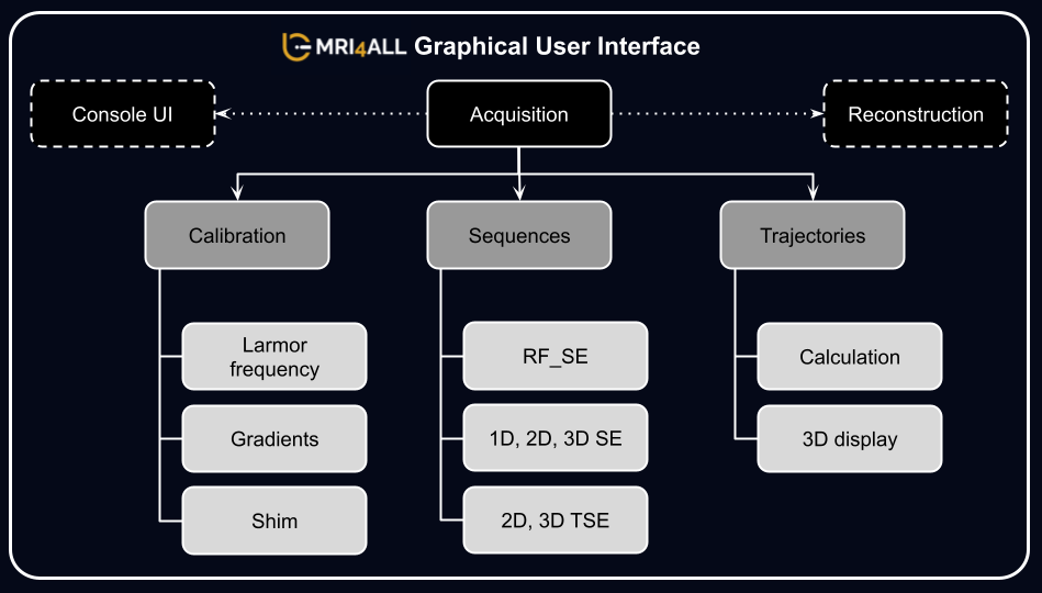
Figure 1 provides an overview of the acquisition modules of the MRI4ALL console5.Calibration sequences: The Larmor frequency can be calibrated using two methods: 1) finding the maximum echo peak over a range of RF excitations, or 2) finding the maximum signal-to-noise ratio (SNR). For both methods, the adjustment begins with a coarse search for the maximum, followed by a finer search, before refining the result using gradient-descent optimization. The gradients are adjusted by measuring a phantom of known dimensions for calibrating the strength of each gradient axis. The calibrated values are stored in the system configuration for subsequent use.
An interactive adjustment interface is available for linear B0 shimming. By manually selecting shim values, it is possible to see the effect on the signal linewidth in near real-time.
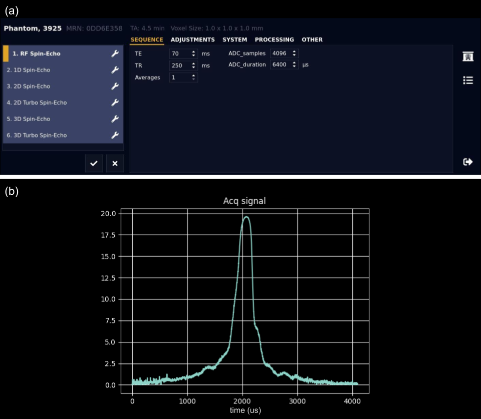
Acquisition Sequences: Multiple basic imaging sequences have been implemented to test the performance of the system. RF_SE is a Spin Echo (SE) sequence with only RF excitation and readout (ADC) events, designed to test the performance of RF coils. 1D_SE is a SE sequence with RF excitation pulse, readout, and frequency-encoding gradients, which can be used to test the performance of each gradient coil. 2D SE, 3D SE, 2D Turbo Spin Echo (TSE), and 3D TSE sequences have been implemented for imaging experiments. Sequence-specific parameters, such as FOV, resolution, orientation, TR, TE, and echo train length can be configured in the UI (Figure 3-a). In the current version, the default k-space sampling pattern is a Cartesian trajectory. Different ordering schemes are available for the phase-encoding steps, including linear-up and center-out. Furthermore, accelerated acquisition patterns such as partial Fourier and stack-of-stars have been implemented.
Trajectories: The UI provides an option to display the k-space trajectories for 2D and 3D sequences that are internally calculated using the PyPulseq library. This option is helpful both for debugging as well as educational purposes. Trajectories are estimated by integrating the gradient information over time after separating signal-encoding and spoiler gradients. The visualization functionality is available for TSE and SE sequences with 2D and 3D Cartesian trajectories.
Results
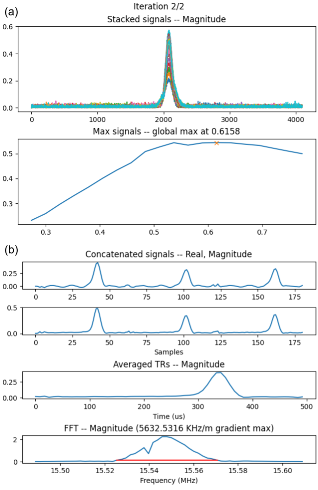
Several tests have been run to show the functionalities of the proposed tools on the MRI4ALL “Zeugmatron-Z1” scanner (42.87mT).Figure 2 shows results from the adjustment workflow for RF and gradient calibration, determining an optimal Larmor frequency of 1.8257MHz in this example (Figure 2a). Maximum gradient amplitudes and their evaluation graphs are provided in Figure 2b. The output of a 1D measurement and its Fourier transform are shown. The console automatically measures the perturbation of the signal profile produced by the presence of a calibration phantom to calculate the gradient strength.
Figure 3-a shows a screenshot of the user interface (which is not hardware-dependent). The available spin-echo-based pulse sequences are visible in the sequence queue on the left side. Sequence-specific parameters for RF_SE are shown in the protocol editor on the right side.
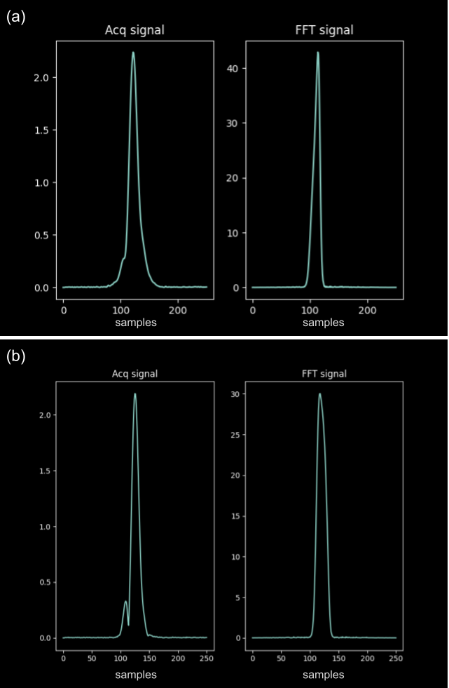
Figure 3b shows the echo acquired with the RF_SE sequence on a phantom. In the same way, and considering the current hardware limitations of the acquisition system, the 1D_SE sequence has been run along the x-axis, with and without turning the gradient amplifiers on. The corresponding acquired signals are presented in Figure 4ab.
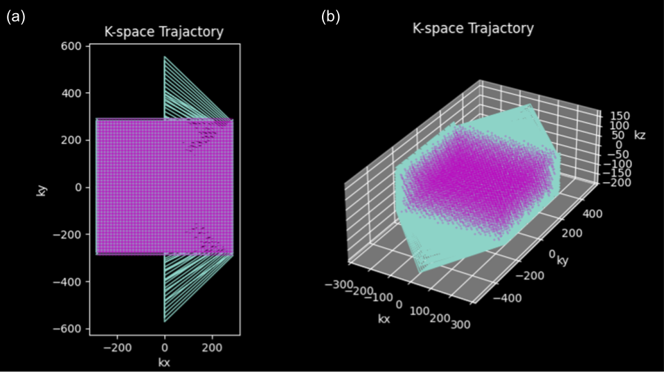
Figure 5 shows examples from the trajectory visualization for acquisitions with the 2D TSE and 3D SE sequences with linear ordering.
Discussion and Conclusion
The acquisition tools developed during the MRI4ALL Hackathon include a fully working calibration pipeline, a library of basic imaging sequences, and a tool to visualize calculated trajectories. These components were tested directly on the hardware system developed during the project and showed consistent results. However, they can also be used on other custom-built MRI systems that are using MaRCoS and PyPulseq. All source code is made publicly available in Git repositories.Future work includes testing of all components including the shim calibration, acquisition of 2D and 3D images using the developed sequences, and integration of additional acquisition schemes such as radial k-space sampling.
Acknowledgements
This work has been done in the frame of the MRI4ALL Hackathon hosted at New York University in October 2023. As such, it was supported by all the organizers of the event, as well as by NYU Langone Health.References
1. Ravi, K.S., Potdar, S., Poojar, P., Reddy, A.K., Kroboth, S., Nielsen, J.F., Zaitsev, M., Venkatesan, R. and Geethanath, S. Pulseq-Graphical Programming Interface: Open source visual environment for prototyping pulse sequences and integrated magnetic resonance imaging algorithm development. Magnetic resonance imaging 2018;52, pp.9-15.
2. Ravi, KS, Geethanath, S, Vaughan, JT: PyPulseq: A python package for mri pulse sequence design, Journal of Open Source Software, 2019; 4(42):1725.
3. Negnevitsky V, Vives-Gilabert Y, Algarín JM, Craven-Brightman L, Pellicer-Guridi R, O’Reilly T, Stockmann JP, Webb A, Alonso J, Menküc B. MaRCoS, an open-source electronic control system for low-field MRI. Journal of Magnetic Resonance 2023;350:107424.
4. https://redpitaya.com/
5. https://github.com/mri4all/console
Figures