4679
4Dflow-unwrap: A collection of python-based phase unwrapping algorithms1University and ETH Zurich, Zurich, Switzerland
Synopsis
Keywords: Software Tools, Velocity & Flow, Unwrapping
Motivation: Low-Venc PC-MRI acquisitions present the advantage of higher velocity-to-noise ratio (VNR). To the best of the author’s knowledge, no Python-based public repository exists with 4D flow MRI unwrapping algorithms and realistic examples.
Goal(s): To share a collection of simple unwrapping tools for 4D Flow MRI and test their performance.
Approach: To use three established unwrapping techniques to create a Python package to unwrap 4D flow MRI data.
Results: We demonstrate the performance of these algorithms on aortic data including multiple Venc values and undersampling factors.
Impact: The Python repository with a collection of simple unwrapping tools facilitates access to unwrapping techniques. These scripts can readily be used to unwrap any PC-MRI dataset.
Introduction
Low-Venc 4D flow acquisitions typically result in improved values of velocity-to-noise ratio (VNR) and in general, increased sensitivity to velocity. To leverage the advantages of low-Venc acquisitions, various multi-Venc approaches have been proposed to increase velocity quantification accuracy in blood vessels1-5. However, these multi-Venc approaches require either longer scans or higher undersampling factors. In order to circumvent this limitation, it is possible to algorithmically unwrap low-Venc data without the need of a high-Venc counterpart6,7. Although the unwrapping process might be trivial for well-behaved smooth flow (where the phase difference between adjacent spatial and temporal voxels lies between -π and π), phase maps from 4D flow MRI data suffer from limited spatial and temporal resolutions and are typically corrupted by noise and undersampling artifacts.In this work we collected and tested various unwrapping algorithms: Laplacian6, sorting by reliability following a noncontinuous path (NPRS)8 and graph-cut PU-max-flow (PUMA)9. All algorithms are freely available along with examples (https://gitlab.ethz.ch/pdirix/4dflow-unwrap).
Methods
Phase wrapping in 4D flow MRI can be presented as:$$ \psi = U(\phi) = \phi + 2n\pi$$
where $$$\phi$$$ is the wrapped phase, $$$n$$$ is an integer representing the number of wraps, $$$U$$$ is an unwrapping function and $$$\psi$$$ is the unwrapped phase. Unwrapping methods attempt to find the variable $$$n$$$. The Laplacian algorithm in 4D depends on parameter $$$ts$$$ representing temporal to spatial weighting. The NPRS version used in this work is sensitive to spatial resolution; we added an upsampling factor $$$uf$$$ that allows to control the image resolution prior to using the algorithm. The 3D Laplacian, and 3D and 4D PUMA algorithms do not depend on parameters. Because the PUMA algorithm is based on graph-cuts, it does not necessarily require a phase between -π and π, which allows to use it as a second step after unwrapping is performed by another algorithm (eg. NPRS + PUMA). The performance of NPRS and PUMA improved if the mask of the target vessel was provided.
In order to test these algorithms, a set of seven publicly available fully sampled 4-point 4D flow MRI scans (Venc=1.5ms-1) of healthy volunteers was used10. Wrapped velocity maps were generated for each encoding direction by retrospectively wrapping the original data with Venc=0.5 and 0.25ms-1. Additionally, all images were weighted by five virtual coils and undersampled using a pseudo-spiral Cartesian k-space trajectory11. Undersampling factors of R=2,4,8 and 16 were considered. Images were reconstructed using a locally low-rank approach12 (LLR) as implemented in the BART toolbox13. A grid search on reconstruction parameters was performed for each geometry to ensure optimal velocity reconstruction.
In order to further test these algorithms, we also generated a synthetic intracranial 4D flow scan. The right and left internal carotid arteries were manually segmented from the phase-contrast angiogram (PCA) of a healthy volunteer (0.8x0.8x0.8mm3) and flow and synthetic MRI were generated as described previously14.
Results
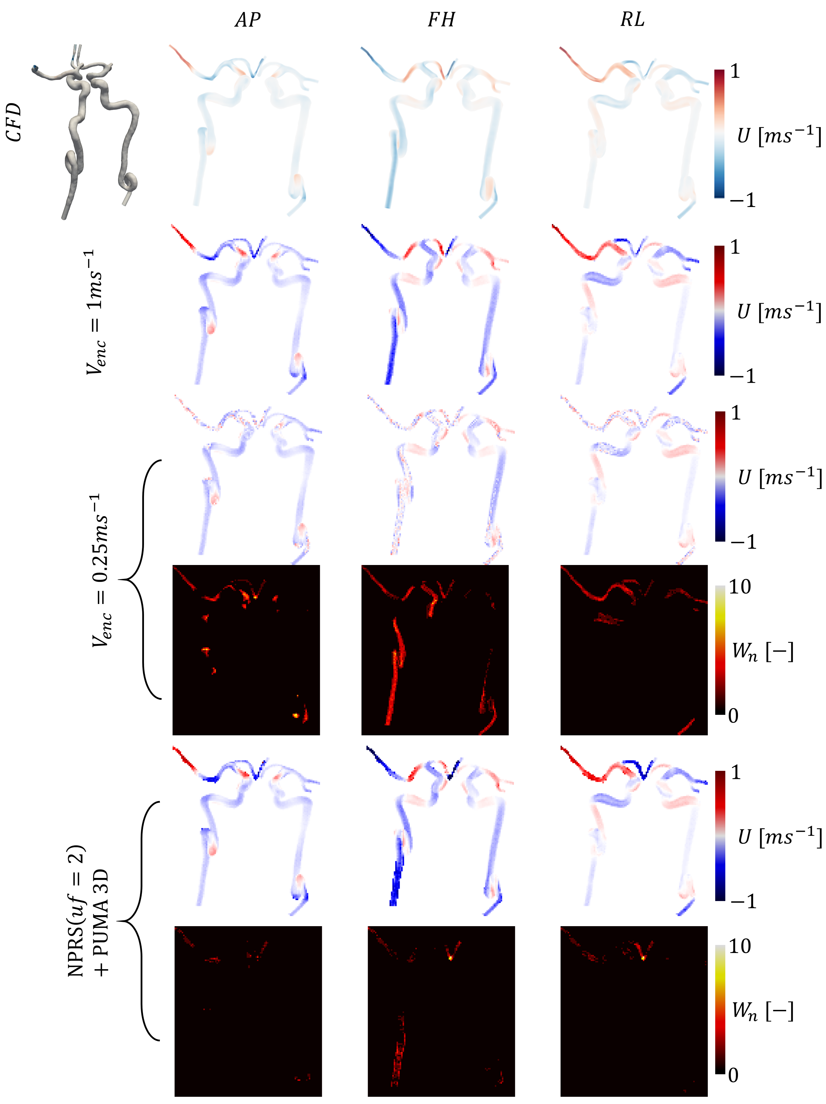
Unwrapped velocity fields and residual wrapped voxel maps are presented in Figure 1 for three volunteers and for all the methods described. Residual unwrapped voxels are visible for all images at the edges of the aorta. A summary of unwrapping quality based on the structural similarity index measure (SSIM) of velocity is available in Figure 2 for all simulations performed. Similarly, Figure 3 presents the percentage of residual wrapped voxels (WΦ). Unwrapping quality decreased with lower velocity encodings (Venc) and with higher undersampling factors, independently of the algorithm used. Laplacian approaches performed significantly worse than both NPRS and PUMA for all Venc and undersamplings. 4D Laplacian unwrapping was satisfactory in the ascending aorta but only for Venc=0.5ms-1. For Venc=0.25ms-1 the lowest amount of residual unwrapped voxels was around 14%, and decreased to 2% for Venc=0.5ms-1. Figure 4 presents an example of the NPRS + PUMA approach applied to flow in the right and left internal carotid arteries.Discussion
We tested three different commonly used unwrapping algorithms on 4D flow datasets to evaluate their performance. Although no algorithm was immune to case-specific unwrapping errors, we observed that combining NPRS and PUMA consistently resulted in high quality unwrapped velocity maps. 4D algorithms performed well for Venc=0.5ms-1 but their decrease in performance for cases with Venc=0.25ms-1 was significantly larger than when 3D algorithms were used. Problematic voxels were often located close to vessel boundaries, where partially wrapped data might have compromised the unwrapping process. While we focused on 4D and 3D data, it is possible to also unwrap 2D or 2D+t images. Ultimately our goal was to make the collection of unwrapping tools available in a repository that includes examples to facilitate their use in PC-MRI research.Acknowledgements
References
1. Ha, Hojin, et al. "Multi‐VENC acquisition of four‐dimensional phase‐contrast MRI to improve precision of velocity field measurement." Magnetic resonance in medicine 75.5 (2016): 1909-1919.
2. Callaghan, Fraser M., et al. "Use of multi‐velocity encoding 4D flow MRI to improve quantification of flow patterns in the aorta." Journal of Magnetic Resonance Imaging 43.2 (2016): 352-363.
3. Schnell, Susanne, et al. "Accelerated dual‐venc 4D flow MRI for neurovascular applications." Journal of Magnetic Resonance Imaging 46.1 (2017): 102-114.
4. Kroeger, Jan Robert, et al. "Velocity quantification in 44 healthy volunteers using accelerated multi-VENC 4D flow CMR." European Journal of Radiology 137 (2021): 109570.
5. Binter, Christian, et al. "Bayesian multipoint velocity encoding for concurrent flow and turbulence mapping." Magnetic resonance in medicine 69.5 (2013): 1337-1345
6. Loecher, Michael, et al. "Phase unwrapping in 4D MR flow with a 4D single‐step laplacian algorithm." Journal of Magnetic Resonance Imaging 43.4 (2016): 833-842.
7. Zhang, Jiacheng, et al. "Divergence-free constrained phase unwrapping and denoising for 4D flow MRI using weighted least-squares." IEEE transactions on medical imaging 40.12 (2021): 3389-3399.
8. Herráez, Miguel Arevallilo, et al. "Fast two-dimensional phase-unwrapping algorithm based on sorting by reliability following a noncontinuous path." Applied optics 41.35 (2002): 7437-7444.
9. Bioucas-Dias, Jos M., and Gonalo Valadao. "Phase unwrapping via graph cuts." IEEE Transactions on Image processing 16.3 (2007): 698-709.
10. Vishnevskiy, Valery, Jonas Walheim, and Sebastian Kozerke. "Deep variational network for rapid 4D flow MRI reconstruction." Nature Machine Intelligence 2.4 (2020): 228-235.
11. Peper, Eva S., et al. "Highly accelerated 4D flow cardiovascular magnetic resonance using a pseudo-spiral Cartesian acquisition and compressed sensing reconstruction for carotid flow and wall shear stress." Journal of Cardiovascular Magnetic Resonance 22.1 (2020): 1-15.
12. Zhang, Tao, John M. Pauly, and Ives R. Levesque. "Accelerating parameter mapping with a locally low rank constraint." Magnetic resonance in medicine 73.2 (2015): 655-661.
13. Uecker, Martin, et al. "Berkeley advanced reconstruction toolbox." Proc. Intl. Soc. Mag. Reson. Med. Vol. 23. No. 2486. 2015.
14. Dirix, Pietro, et al. "Synthesis of patient-specific multipoint 4D flow MRI data of turbulent aortic flow downstream of stenotic valves." Scientific Reports 12.1 (2022): 16004.
Figures
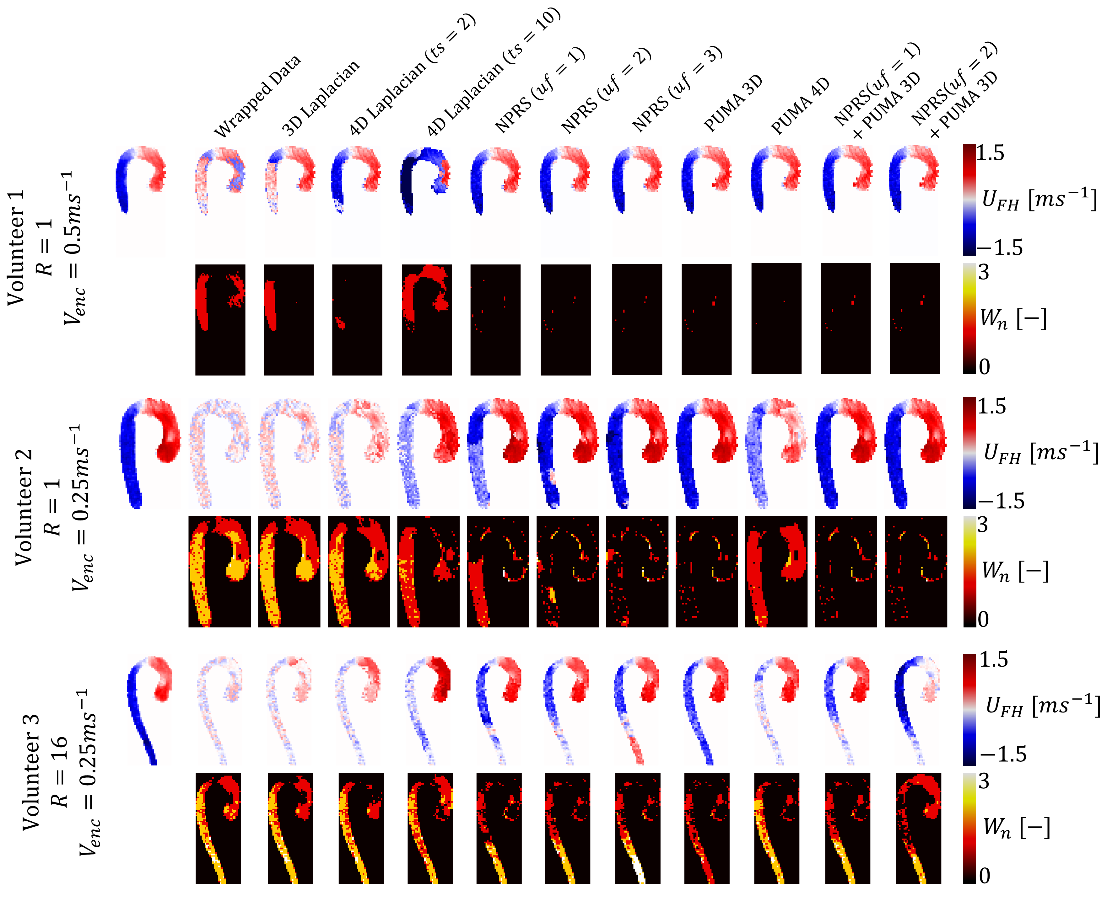
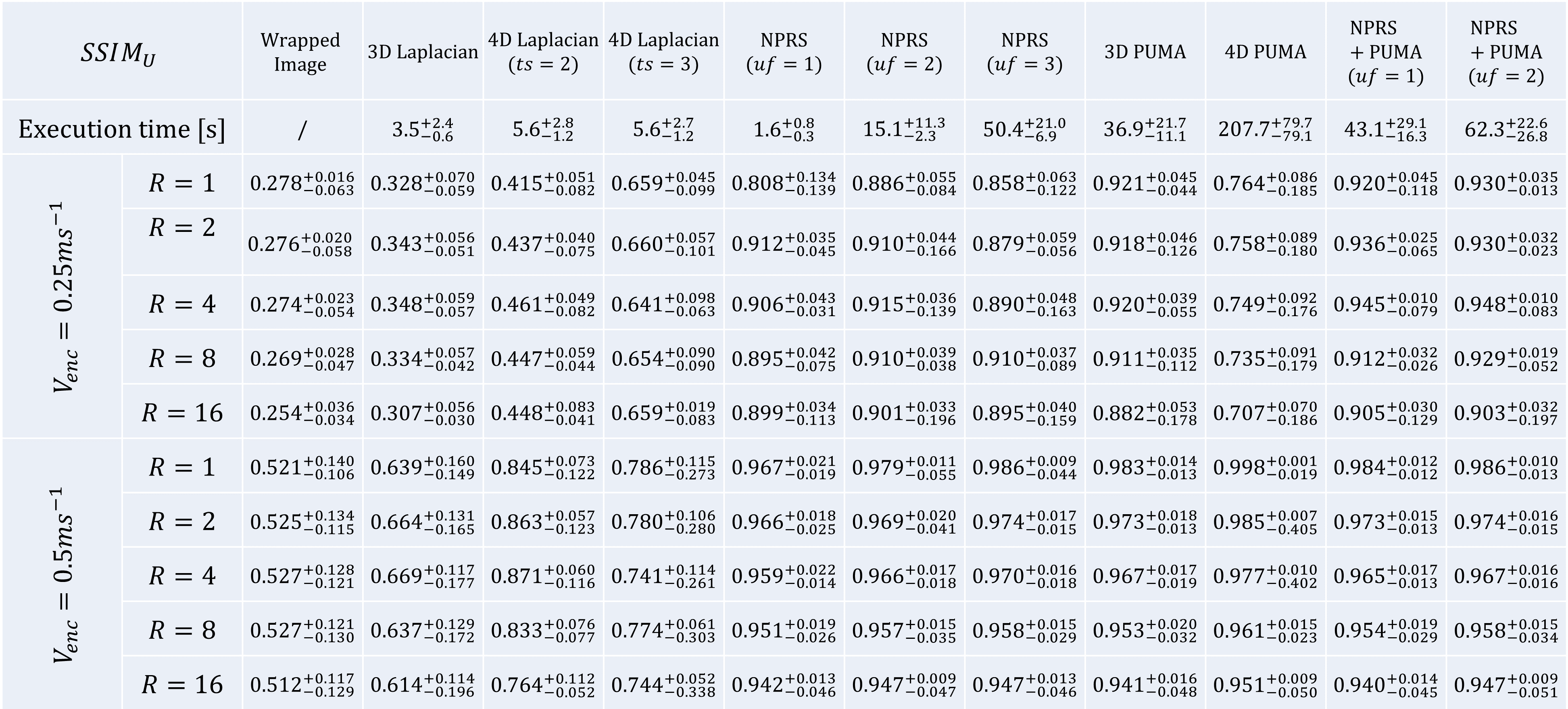
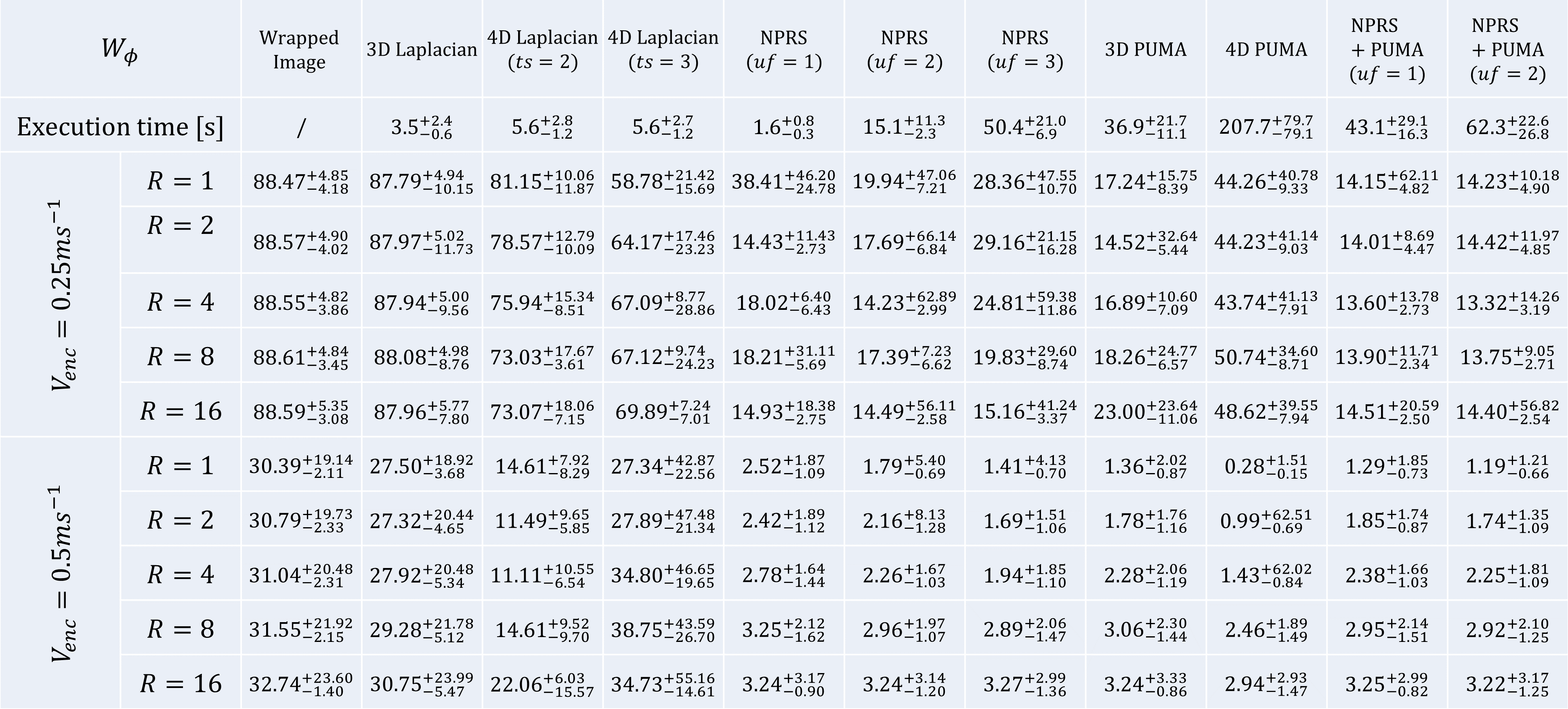
Figure 3: Quality of unwrapping based on percentage of residual wrapped voxels (WΦ). The table shows WΦ values (mean and range based on 7 healthy volunteers) for different algorithms and parameters as well as for Venc=0.25 and 0.5ms-1 and undersampling factor R=2,4,8 and 16. The second row shows the execution time required to unwrap a 4D frame.