4677
Tensor MP-PCA Denoising for Prostate MRI1University of Chicago, Chicago, IL, United States, 2Marmara University, Istanbul, Turkey
Synopsis
Keywords: Software Tools, Diffusion/other diffusion imaging techniques
Motivation: Prostate MRI primarily relies on diffusion-weighted imaging (DWI) but is notoriously challenged by low SNR, impacting the diagnostic process.
Goal(s): To implement the state-of-the-art tensor denoising method for prostate DWI
Approach: We applied the tMPPCA algorithm that makes use of the redundancy in multi-dimensional data to separate the most significant components (the diffusion-signal) and the remaining the thermal/scanner noise. We quantified the denoising efficacy with comprehensive qualitative and quantitative analysis.
Results: The tMP-PCA method, previously proved to be efficient on ex-vivo scans are extremely effective to enhance in-vivo prostate MRI images when a similar multi-dimensional protocol is followed.
Impact: The tMPPCA can effectively reduce noise without the trade-off of blurring—an achievement that has critical implications in cancer detection. This study is the first in-vivo implementation of tMPPCA for enhancing prostate DWI, employed under 10 minutes of scan time.
Introduction
Prostate magnetic resonance imaging (MRI) primarily relies on diffusion-weighted imaging (DWI) but is notoriously challenged by low signal-to-noise ratios (SNR), impacting the diagnostic process. Conventional denoising methods introduce unwanted blurring that compromises image resolution. On the other hand, the more recent generative deep learning approaches have their own pitfall of producing hallucinatory features, diminishing their clinical utility. Notably, the bulk of research has centered around brain imaging, leaving a knowledge gap in the context of prostate DWI denoising. Recently, Olesen et al. presented the tensor Marchenko-Pastur principal component analysis (tMPPCA) method, which exhibited outstanding denoising performance in ex-vivo mouse brain scans, surpassing existing denoising algorithms in preserving the integrity of multidimensional MRI data. Our study adapts this tMPPCA approach to in-vivo human prostate DWI acquired with varied echo time (TE) settings and evaluates its clinical value through a combination of objective metrics and subjective radiological evaluations. This study is the first in-vivo implementation of tMPPCA.Methods
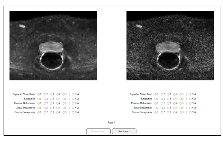
This study encompassed 20 prostate cancer patients, whose DWI scans were acquired with b-values of 0, 150, 1000, and 1500 sec/mm². Each diffusion-weighted image was acquired using four different echo time (TE) settings: 57ms, 70ms, 150ms, and 200ms. The resultant five-dimensional imaging data, with a matrix size of 128 × 128 × 24 × 4 × 4 (x × y × z × b × TE), presented a minimal dataset for the application of the tMPPCA algorithm, which requires high-order singular value decomposition for denoising. We applied the tMPPCA algorithm that makes use of the redundancy in multi-dimensional data to separate the most significant components (the diffusion signal) and the remaining the thermal/scanner noise. We quantified the denoising efficacy of the tMPPCA technique using the traditional SNR and Contrast-to-Noise Ratio (CNR), alongside advanced no-reference quality metrics Blind/Referenceless Image Spatial Quality Evaluator (BRISQUE) and Perceptual Image Quality Evaluator (PIQE). These metrics were calculated for images with and without tensor denoising. The statistical significance of our findings was established through paired sample t-tests. Additionally, for perceptual quality, we conducted an observer study involving three expert radiologists rated the images on a Likert scale based on “signal quality”, “resolution”, “prostate delineation”, “zonal delineation”, and “cancer conspicuity”. The order of the original and tMPPCA-processed images was randomized on each page to prevent any sequence bias.Results
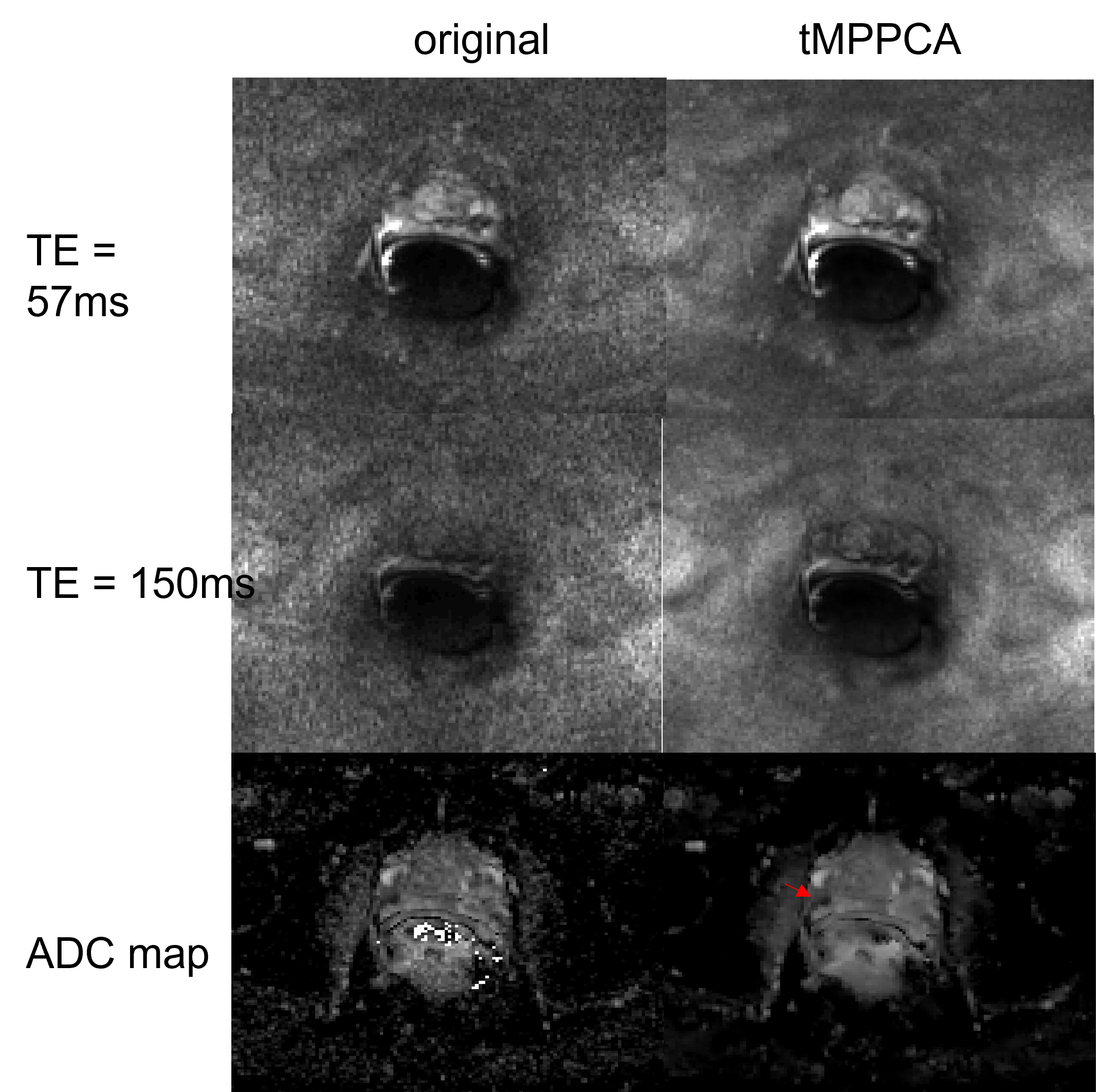
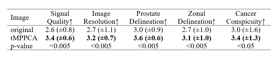
Application of the tMPPCA algorithm yielded significant improvements across all objective and subjective metrics (p<0.05). Furthermore, the objective quality metrics strongly correlated with the radiologists’ subjective evaluation criteria as evidenced by a strong positive Pearson Correlation Coefficient (PCC>0.7, p<0.005) between the two sets of evaluations. Enhanced images are exhibited in Figure 2, which visually underline the denoising efficiency, while Tables 1 and 2 detail the objective and subjective quality enhancements, respectively.Discussion
We applied the tMPPCA algorithm to multidimensional prostate DWI and showed that this method can effectively reduce noise without the trade-off of blurring—an achievement that has critical implications in the domain of cancer detection. Building upon Olesen et al.'s framework, this study is the first in-vivo implementation of tMPPCA for enhancing prostate DWI. Olesen et al. showed the efficiency of the method on ex-vivo mouse brain scans, taking 10-hours with multiple TEs, b-values and gradient encoding directions. We applied tMPPCA to our multi-dimensional DWI data that has 4 b-values × 4 TE values, which takes about 10 minutes per patient. This marked improvement in processing time, coupled with the noted increase in cancer conspicuity, primes this methodology for rapid clinical adoption, potentially enhancing diagnostic precision and patient management in prostate cancer care.Acknowledgements
Supported by the National Institutes of Health (R01 CA227036, 1R41CA244056-01A1, R01 CA17280, and 1S10OD018448-01), Sanford J. Grossman Charitable Trust and University of Chicago Medicine Comprehensive Cancer Center (P30 CA014599-37). The authors thank Dr. Sune Jespersen for sharing the source code of tMP-PCA with us.References
[1] J. L. Olesen, A. Ianus, L. Østergaard, N. Shemesh, and S. N. Jespersen, “Tensor denoising of multidimensional MRI data,” Magnetic Resonance in Med, vol. 89, no. 3, pp. 1160–1172, Mar. 2023, doi: 10.1002/mrm.29478.
[2] N. Wiest-Daesslé, S. Prima, P. Coupé, S. P. Morrissey, and C. Barillot, “Rician Noise Removal by Non-Local Means Filtering for Low Signal-to-Noise Ratio MRI: Applications to DT-MRI,” in Medical Image Computing and Computer-Assisted Intervention – MICCAI 2008, D. Metaxas, L. Axel, G. Fichtinger, and G. Székely, Eds., in Lecture Notes in Computer Science, vol. 5242. Berlin, Heidelberg: Springer Berlin Heidelberg, 2008, pp. 171–179. doi: 10.1007/978-3-540-85990-1_21.
[3] H. Cheng et al., “Denoising diffusion weighted imaging data using convolutional neural networks,” PLoS ONE, vol. 17, no. 9, p. e0274396, Sep. 2022, doi: 10.1371/journal.pone.0274396.
[4] J. Jurek et al., “Supervised denoising of diffusion-weighted magnetic resonance images using a convolutional neural network and transfer learning,” Biocybernetics and Biomedical Engineering, vol. 43, no. 1, pp. 206–232, Jan. 2023, doi: 10.1016/j.bbe.2022.12.006.
Figures