4676
MR software tools for real-time decision making and FOV prescription1Radiology, Martinos Center for Biomedical Imaging at MGH, Boston, MA, United States, 2Orchard Scientific, Yucca Valley, CA, United States, 3Radiology, Harvard Medical School, Boston, MA, United States, 4Fetal-Neonatal Neuroimaging & Developmental Science Center, Boston Children’s Hospital, Boston, MA, United States, 5AI in Medical Imaging, German Center for Neurodegenerative Diseases (DZNE), Bonn, Germany, 6McGovern Institute for Brain Research, Massachusetts Institute of Technology, Cambridge, MA, United States
Synopsis
Keywords: Software Tools, Software Tools, Data Acquisition; Neuroscience
Motivation: Many cutting-edge MR neuroimaging paradigms require real-time decision making and precise FOV positioning. We present two software tools to support such paradigms.
Goal(s): Develop two modules.
1) vSend: opens a socket and sends imaging data to another computer in a vendor-agnostic format, enabling real-time analysis.
2) AAhijack: reads a matrix from a socket and overwrites the Siemens AutoAlign matrix, enabling online slice prescription.
Approach: Modules are implemented as Siemens image reconstruction modules (ICE functors) in C++ and two slice prescription systems utilizing the modules are demonstrated.
Results: The slice prescription systems have comparable performance and various advantages and disadvantages.
Impact: The software tools presented have enabled a variety of cutting-edge MR neuroimaging paradigms including real-time fMRI, motion tracker calibration, real-time shimming, fetal head-pose detection and automated FOV prescription, reacquisition planning and single-slice BOLD imaging FOV prescription.
Introduction
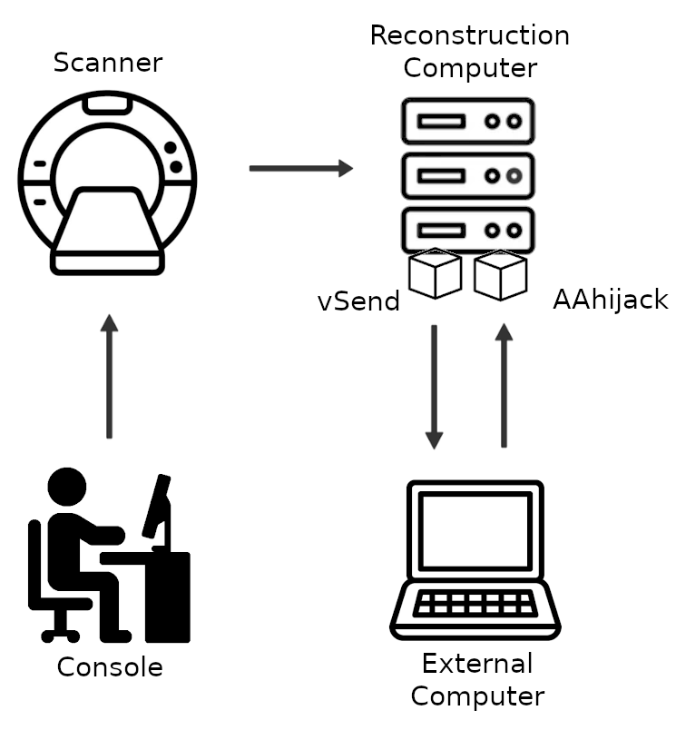
Many cutting-edge MRI neuroimaging paradigms require real-time decision making and precise FOV positioning. We present two software tools, implemented as Siemens image reconstruction modules, to support a variety of such paradigms. The first, called vSend, opens a socket and sends imaging data to another computer where it can be analysed in real-time. The second, called AAhijack, reads a matrix from a socket and overwrites the Siemens AutoAlign matrix, enabling online FOV prescription.The vSend module can support real-time fMRI (rtfMRI) neurofeedback experiments 1,2 and is compatible with the rtfMRI software murfi2 1,3. It can also be used with a python script 4 which can read the data sent by vSend and write a NIfTI 5 file, which allows it to be used in a variety of applications. To date it has been used to: calibrate motion trackers 6, update shims in real-time 7, detect fetal head-poses 8 and determine reacquisition priorities 9. vSend uses a vendor-agnostic data format 10.
AutoAlign is a Siemens’ tool to automate the alignment of slice positioning in head examinations 11. A scout image is acquired from which anatomical landmarks are extracted registered to a template. This creates a 4x4 rigid registration matrix which is saved on the scanner. Subsequent sequences for which AutoAlign is enabled will have their FOV multiplied by this matrix. The AAhijack module opens a socket and queries a remote server for a matrix which it then stores as an AutoAlign matrix. This tool has been used to prescribe FOVs across runs in single-slice BOLD imaging 12.
Methods
Both vSend and AAhijack have been implemented as Siemens ICE functors in IDEA using C++. vSend has been used on the following Siemens software baselines: VB17, VE11C, VE11E, VE12U, XA20A, XA50A. AAhijack has been used on VE11C and VE12U. All results in this abstract were obtained on a 7T Siemens Terra running VE12U.We validate the vSend module by comparing the NIfTI file derived conventionally from the DICOM images using mri_convert 13 with the NIfTI file generated via vSend.
We validate AAhijack by using it to store 5 arbitrary AutoAlign matrices and predicting the voxel to RAS (vox2ras) matrices of subsequent AutoAlign-enabled series.
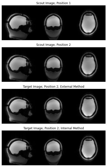
Finally, we demonstrate 2 prospectively updating slice prescriptions systems. The first, called the ‘external method’ (Figure 1), utilizes both vSend and AAhijack. A scout image of a head phantom 14 is acquired and vSend is used to send it to a remote computer. The phantom is then physically repositioned in the scanner and a second scout image is acquired and sent to the remote computer. The computer then registers the two volumes 15 and computes an AutoAlign matrix, which is stored on the scanner using AAhijack. A third AutoAlign-enabled target sequence is acquired and a voxel-wise registration between the first and third image is computed to determine the accuracy of the system. The second, called the ‘internal method’ is like the first, except the scout images are not sent to a remote computer but stored on the scanner and Siemens registration engine (PACE) is used to derive an AutoAlign matrix which is passed to AAhijack.
Results
vSend was validated with Gradient Echo and BOLD sequences. NIfTIs generated from DICOM using mri_convert 13 were compared to the NIfTIs from vSend using mri_diff 13. The files were identical except for minor differences in the header: TR, TE and flip angle were incorrect in the vSend volume and TE and flip angle were incorrect in the DICOM derived volume. These inaccuracies had no impact on the imaging paradigms that make use of vSend.We used 5 randomly selected AutoAlign matrices to validate AAhijack. In all cases, the predicted vox2ras matrices were accurate to at least 4 decimal places.
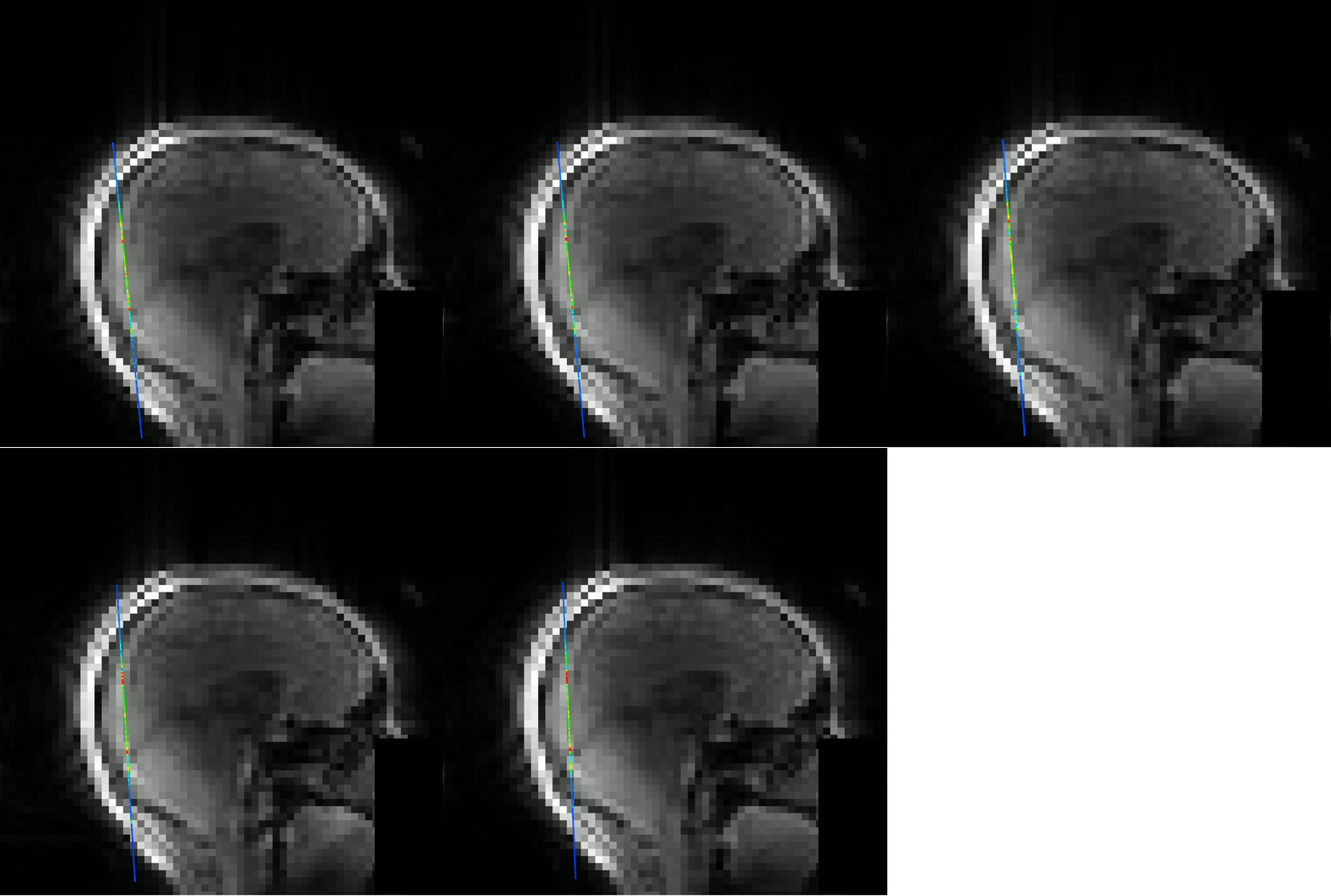
The slice prescription methods were validated visually (Figure 2) and by performing a voxel-wise residual registration 15 between the scout image of the phantom in the initial position and the target image after repositioning the phantom. The residual registration of the external method was 0.0069mm and 0.094 degrees. The residual registration of the internal method was 0.0082mm and 0.14 degrees. The displacement of the phantom was 1.79mm and 3.58 degrees. Figure 3 shows the external method prescribing a single BOLD slice in a human subject as they change position.
Conclusion and Discussion
The accuracies of the external and internal slice prescription methods are comparable. The internal method doesn't require external hardware and the external method allows for greater control of the registration process.The tools presented can be used to support a variety of cutting-edge MR imaging experiments.
Acknowledgements
This work was funded by the following NIH grants: U19NS123717, P41EB015896, R42CA183150, S10RR021110, R01HD093578, R01HD099846, R44MH124567, R21EB029641, R01HD110152, R00HD101553, S10OD023637References
[1]: Hinds, O., Ghosh, S., Thompson, T.W., Yoo, J.J., Whitfield-Gabrieli, S., Triantafyllou, C. and Gabrieli, J.D., 2011. Computing moment-to-moment BOLD activation for real-time neurofeedback. Neuroimage, 54(1), pp.361-368.
[2]: Stoeckel, L.E., Garrison, K.A., Ghosh, S.S., Wighton, P., Hanlon, C.A., Gilman, J.M., Greer, S., Turk-Browne, N.B., deBettencourt, M.T., Scheinost, D. and Craddock, C., 2014. Optimizing real time fMRI neurofeedback for therapeutic discovery and development. NeuroImage: Clinical, 5, pp.245-255.
[3]: murfi2: A realtime fMRI software platform. https://github.com/gablab/murfi2
[4]: A python script to receive images sent by vSend. https://github.com/gablab/murfi2/blob/master/util/python/receive_vsend/receive_nii.py
[5]: The nifti file format. https://nifti.nimh.nih.gov/nifti-1/documentation/
[6]: Frost, R., Wighton, P., Karahanoğlu, F.I., Robertson, R.L., Grant, P.E., Fischl, B., Tisdall, M.D. and van der Kouwe, A., 2019. Markerless high‐frequency prospective motion correction for neuroanatomical MRI. Magnetic resonance in medicine, 82(1), pp.126-144.
[7] Arango, N. S., Frost, R., Wighton, P., Stockmann, J., Ovidiu, C. A., and van der Kouwe, A. Motion-Compensated Slice-by-Slice ∆B0 Shimming with an AC/DC Shim Coil and Dual-Echo vNavs. Proceedings of ISMRM 2023.
[8]: Hoffmann, M., Abaci Turk, E., Gagoski, B., Morgan, L., Wighton, P., Tisdall, M.D., Reuter, M., Adalsteinsson, E., Grant, P.E., Wald, L.L. and van der Kouwe, A.J., 2021. Rapid head‐pose detection for automated slice prescription of fetal‐brain MRI. International journal of imaging systems and technology, 31(3), pp.1136-1154.
[9]: Gagoski, B., Xu, J., Wighton, P., Tisdall, M.D., Frost, R., Lo, W.C., Golland, P., van Der Kouwe, A., Adalsteinsson, E. and Grant, P.E., 2022. Automated detection and reacquisition of motion‐degraded images in fetal HASTE imaging at 3 T. Magnetic resonance in medicine, 87(4), pp.1914-1922.
[10]: vSend’s OpenHeader format specification https://github.com/gablab/murfi2/blob/master/src/io/RtExternalSenderImageInfo.h
[11]: van der Kouwe, A.J., Benner, T., Fischl, B., Schmitt, F., Salat, D.H., Harder, M., Sorensen, A.G. and Dale, A.M., 2005. On-line automatic slice positioning for brain MR imaging. Neuroimage, 27(1), pp.222-230.
[12]: Varadarajan, D., Wighton, P., Chen, J., Proulx, S., Frost, R., van der Kouwe, A., Berman, A. and Polimeni, J. 2023 Measuring individual vein and artery BOLD responses to visual stimuli in humans with multi-echo single-vessel functional MRI at 7T. Proceedings of ISMRM 2023.
[13]: FreeSurfer software suite v7.3.2. https://surfer.nmr.mgh.harvard.edu/fswiki/FreeSurferMethodsCitation
[14]: A.A. Martinos Center / Wald group anthropomorphic phantom builder's Wiki https://phantoms.martinos.org/
[15]: Reuter, M., Rosas, H.D. and Fischl, B., 2010. Highly accurate inverse consistent registration: a robust approach. Neuroimage, 53(4), pp.1181-1196.
Figures