4673
MaRGA: a Graphical and Application Interface for the MaRCoS open-source console1i3M, CSIC, Valencia, Spain, 2Universitat Politècnica de València, Valencia, Spain, 3Tesoro Imaging SL, Valencia, Spain
Synopsis
Keywords: Software Tools, Software Tools, Open-source, MaRCoS
Motivation: MaRCoS integrates hardware, firmware, and software for MRI scanner control, focusing on system reliability but overlooking user experience.
Goal(s): Develop MaRGA (MaRCoS Graphical Application), a user-friendly Graphical User Interface (GUI) for low-field MRI community, ensuring intuitive MaRCoS control and clinical environment compatibility.
Approach: Designed MaRGA with simplified panels and updated API, enabling features like DICOM image export, clinical protocol management, and streamlined image reconstructions.
Results: Tested MaRGA on 0.2 T and 72 mT scanners in lab and hospital settings, enhancing workflow efficiency and user satisfaction through customizable controls and improved overall experience.
Impact: The introduction of MaRGA, a user-friendly Graphical User Interface (GUI) for low-field MRI scanner control, enhances user experience and workflow efficiency with MaRCoS. MaRGA significantly improves the operation of low-field MRI scanners in both laboratory and clinical settings.
Introduction
The electronic control system or 'console' is pivotal to any MRI scanner. A prominent open-source initiative is Magnetic Resonance Control System (MaRCoS) [1, 2], a sophisticated and versatile open-source platform for MRI control with high-performance attributes and low-cost. A relevant milestone regarding MaRCoS is to develop a Graphical User Interface (GUI) that facilitates scanner operation in laboratories and hospital environments. In this work, we introduce MaRGA (MaRCoS Graphical Application) a new graphical user interface (GUI) and application programming interface (API) [3] designed to ease the interaction between users and MaRCoS.Methods
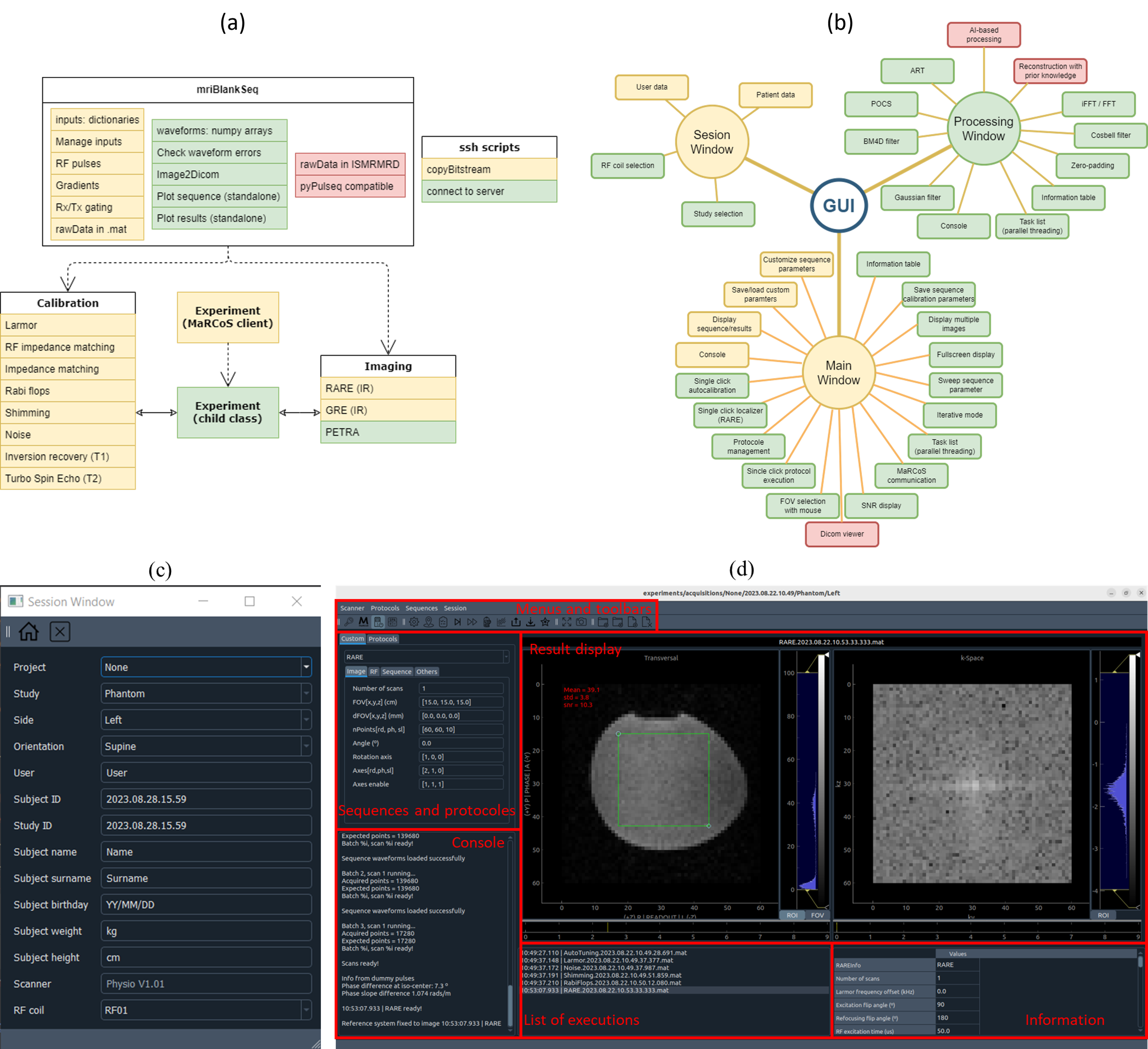
The MaRGA API and the GUI are both written in Python. The former (Figure 1.a) includes methods to handle communication with MaRCoS, a parent class that provides a structured framework for creating, executing, and managing complex MRI sequences, and different child classes corresponding to specific sequences. The GUI (Figure 1.b) has been developed using the PyQt5 library and integrates with the underlaying API, allowing users to communicate with the MaRCoS server and to initiate sequences, adjust parameters, and oversee data acquisition.The GUI is based on three different windows. The Session Window (Figure 1.c) serves as the initial point of interaction, allowing users to input essential information related to the imaging session. This window takes in details such as user credentials, patient information, project identifiers, and other relevant metadata. In the Main Window (Figure 1.d), users interact with the sequences and results. It includes a menu bar and toolbars for interacting with the MaRCoS server, running calibrations, visualizing or executing pulse sequences, saving or loading input parameters, and creating or deleting protocols. Additionally, there is a space to show previously executed or pending sequences and the parameters used in those sequences. Finally, the Post-Processing Window can be used to analyze and refine the acquired data, both in k-space and image space.
To test the usability of MaRGA, we conducted experiments in two distinct setups: a laboratory-based MRI scanner used for preclinical dental imaging [4] and a hospital-based MRI scanner specializing in in-vivo extremity imaging [5]. In the laboratory setup we run Pointwise Encoding Time Reduction with Radial Acquisition (PETRA) [6], typically used for hard tissue imaging, in a home-made phantom. In the system installed in the hospital we run a protocol including an autocalibration and four RARE sequences with different contrasts in a volunteer.
Results
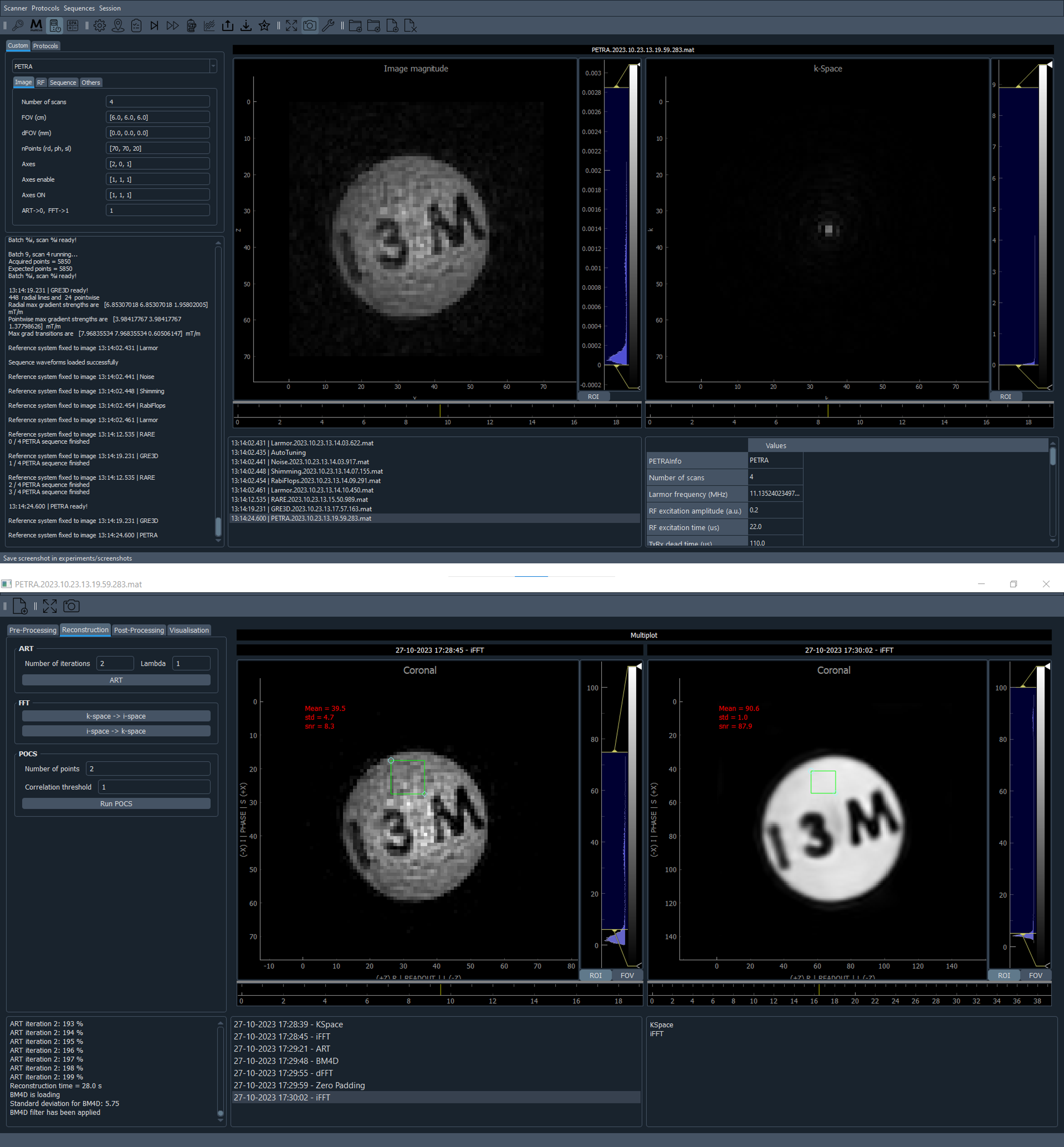
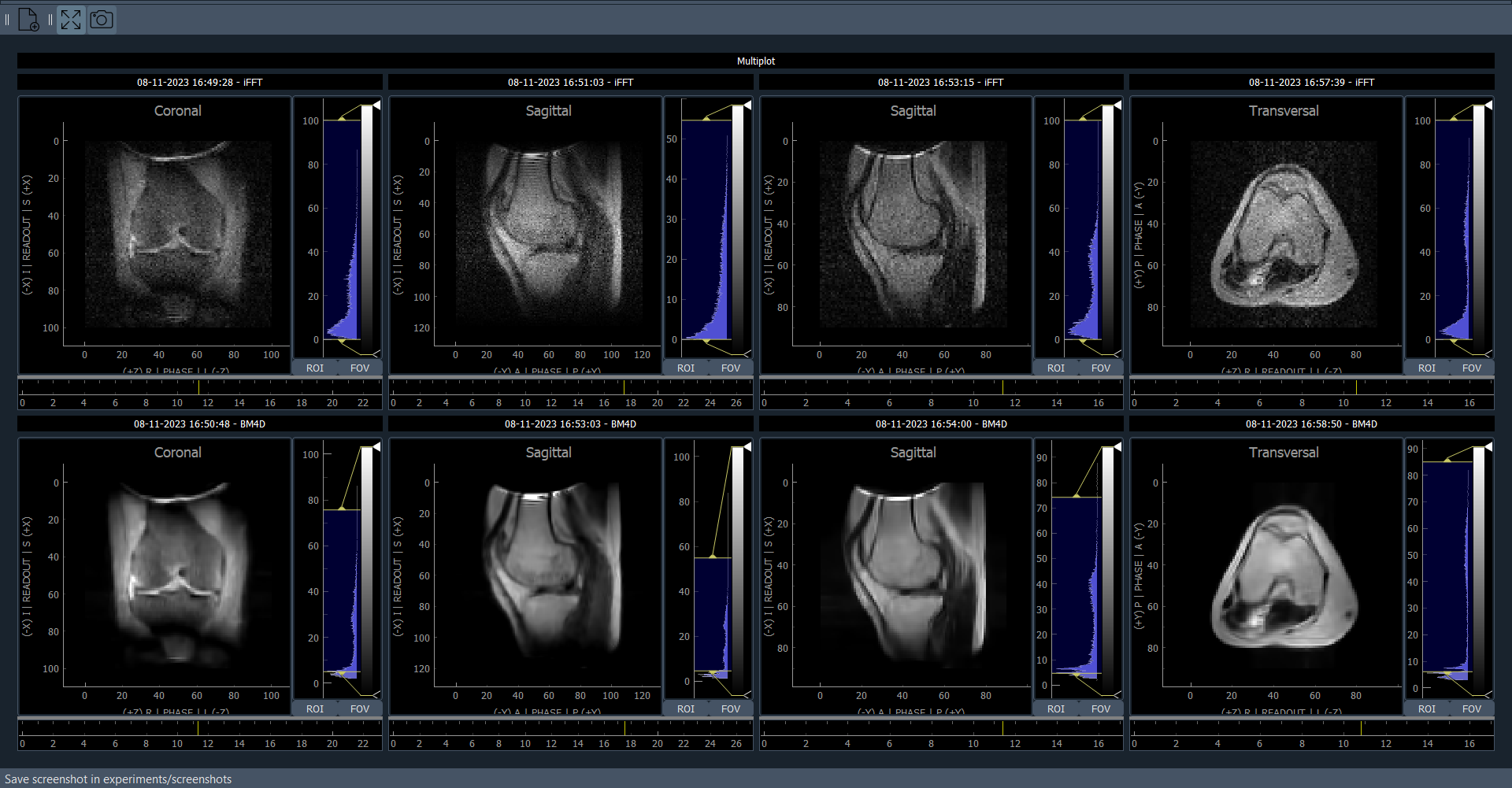
Figure 2 shows a screenshot of the Main Window (top) and Post-Processing Window (bottom) with the results obtained using PETRA in the laboratory-based scanner. By default, the PETRA sequence is configured in the API to reconstruct the images with FFT after regridding. Then, we use the postprocessing window to apply Algebraic Reconstruction Technique (ART) [7], apply a BM4D [8] filter and upscale the matrix size of the image.In Figure 3, a screenshot of the Post-Processing Window illustrates the four acquired images in the hospital-based scanner within the protocol, both before (top) and after (bottom) post-processing. Here we apply a BM4D filter to all images. For Inversion Recovery RARE and T1 RARE sequences, we apply a cos-bell filter in k-space along the readout direction before Fourier reconstruction, followed by BM4D.
Discussion
MaRGA contains sequences for imaging (e.g. RARE, GRE or PETRA) as well as sequences for calibration (Rabi flopping, shimming, Larmor frequency and others). From MaRGA we can also run autocalibration sequences. To this end, we set the field of view directly from a scout image, we select the region of interest, we show the mean value and standard deviations to get a quick estimation of the signal-to-noise ratio, and we develop protocols to generate standardized sets of images from different sequences to make the GUI accessible to non-expert users.As we look forward, we plan to make MaRGA fully compatible with Pulseq based on prior developments for MaRCoS [9]. Another area for future development is to save raw data using the ISMRMRD standard [10] to ensure uniformity in data representation and further enhances interoperability across various MRI systems. An important aspect in the evolution of MaRGA lies in certifying the software for clinical use to validate the reliability and accuracy of the system and to provide confidence to users.
Conclusion
In this work we present MaRGA, a graphical and application interface to easily interact with MaRCoS, execute pulse sequences and visualize the results.Acknowledgements
Project funded by: the EU (EIC Transition, 101136407), EURAMET (22HLT02), Spanish MICINN (PID2022-142719OB-C22), and the Valencian Innovation Agency (INNVA1/2022/4, INNVA1/2023/30).
References
[1] T. Guallart‐Naval et al., “Benchmarking the performance of a low‐cost magnetic resonance control system at multiple sites in the open MaRCoS community,” NMR Biomed, no. March, pp. 1–13, 2022, doi: 10.1002/nbm.4825.
[2] V. Negnevitsky et al., “MaRCoS, an open-source electronic control system for low-field MRI,” Journal of Magnetic Resonance, p. 107424, 2023, doi: 10.1016/j.jmr.2023.107424.
[3] https://github.com/yvives/PhysioMRI_GUI
[4] Algarín, J. M., Díaz-Caballero, E., Borreguero, J., Galve, F., Grau-Ruiz, D., Rigla, J. P., Bosch, R., González, J. M., Pallás, E., Corberán, M., Gramage, C., Aja-Fernández, S., Ríos, A., Benlloch, J. M., & Alonso, J. (2020). Simultaneous imaging of hard and soft biological tissues in a low-field dental MRI scanner. Scientific Reports, 10, 21470. https://doi.org/https://doi.org/10.1038/s41598-020-78456-2
[5] Guallart-Naval, T., Algarín, J. M., Pellicer-Guridi, R., Galve, F., Vives-Gilabert, Y., Bosch, R., Pallás, E., González, J. M., Rigla, J. P., Martínez, P., Lloris, F. J., Borreguero, J., Marcos-Perucho, A., Negnevitsky, V., Martí-Bonmatí, L., Ríos, A., Benlloch, J. M., & Alonso, J. (2022). Portable magnetic resonance imaging of patients indoors, outdoors and at home. Scientific Reports, 12. https://doi.org/10.1038/s41598-022-17472-w
[6] Grodzki, D. M., Jakob, P. M., & Heismann, B. (2012). Ultrashort echo time imaging using pointwise encoding time reduction with radial acquisition (PETRA). Magnetic Resonance in Medicine, 67(2), 510–518. https://doi.org/10.1002/mrm.23017
[7] Gordon, R., Bender, R., & Herman, G. T. (1970). Algebraic Reconstruction Techniques (ART) for three-dimensional electron microscopy and X-ray photography. Journal of Theoretical Biology, 29(3), 471–481. https://doi.org/10.1016/0022-5193(70)90109-8
[8] Maggioni, M., Katkovnik, V., Egiazarian, K. & Foi, A. Nonlocal transform-domain filter for volumetric data denoising and reconstruction. IEEE Trans. Image Process. 22, 119–133. https ://doi.org/10.5041/rmmj.10355 5 (2013).
[9] Layton, K. J., Kroboth, S., Jia, F., Littin, S., Yu, H., Leupold, J., Nielsen, J. F., Stöcker, T., & Zaitsev, M. (2017). Pulseq: A rapid and hardware-independent pulse sequence prototyping framework. Magnetic Resonance in Medicine, 77(4), 1544–1552. https://doi.org/10.1002/mrm.26235
[10] Inati, S. J., Naegele, J. D., Zwart, N. R., Roopchansingh, V., Lizak, M. J., Hansen, D. C., Liu, C. Y., Atkinson, D., Kellman, P., Kozerke, S., Xue, H., Campbell-Washburn, A. E., Sørensen, T. S., & Hansen, M. S. (2017). ISMRM Raw data format: A proposed standard for MRI raw datasets. Magnetic Resonance in Medicine, 77(1), 411–421. https://doi.org/10.1002/mrm.26089
Figures