4672
End-to-end polar analysis software package for radially acquired fMRI1Computer Science and Artificial Intelligence Laboratory, Massachusetts Institute of Technology, Cambridge, MA, United States, 2Department of Biomedical Engineering, Amirkabir University of Technology (Tehran Polytechnic), Tehran, Iran (Islamic Republic of), 3Aerospace & Mechanical Engineering, University of Southern California, Los Angeles, CA, United States
Synopsis
Keywords: Software Tools, Brain
Motivation: Achieving full advantage of radial acquisition and reconstruction by conducting the complete analysis in the polar coordinate system.
Goal(s): Implement a comprehensive end-to-end fMRI analysis software package with a graphical user interface.
Approach: Radially acquired fMRI was reconstructed natively in the polar coordinate system using the Polar Fourier Transform, followed by utilizing preprocessing steps (motion correction and brain extraction) implemented in the polar coordinate system to perform statistical analysis.
Results: The software package performs reconstruction, preprocessing, and statistical analyses of radially acquired fMRI data in the polar coordinate system and is validated using fMRI of 31 healthy subjects.
Impact: This software package facilitates the native polar analysis of radially acquired fMRI that increases the specificity and/or improves the spatial resolution, particularly in task-based brain mapping studies with the region of interest considerably smaller than the field of view
Introduction
Functional magnetic resonance imaging (fMRI) is a ubiquitous high-resolution brain mapping tool that has been used extensively in neuroscience studies and interventional neuroradiology1. Polar fMRI with Radial acquisition/reconstruction has been recently introduced and showed values in specific applications that require higher specificity, better resolution in brain mapping, or when the region of interest is considerably smaller than the field of view2. It was shown that the efficiency of this approach requires including the Polar Fourier Transform (PFT)3 algorithm as the reconstruction technique within the entire polar imaging process. Although they hypothesized some advantages for fully polar fMRI, statistical analysis required that the reconstructed images be transformed to the Cartesian coordinates. Here, we propose a comprehensive end-to-end fMRI analysis software package with a graphical user interface that performs reconstruction, preprocessing, and statistical analyses of radially acquired fMRI data in the polar coordinate system, so the aforementioned hypothesis can be verified. The final output can be taken to Cartesian for visualization purposes.Method
Polar reconstructionPFT is a new technique to reconstruct radially acquired data directly in the polar coordinate system without Cartesian regridding using Hankel transform3, where F(ρ,φ) is the radially acquired k-space and Hn is the n-order Hankel transform. To reduce the computation time, the Bessel function of the first kind required for the Hankel transform was calculated and cached once to avoid recalculation for subsequent iterations. Furthermore, the option to visualize the intermediate reconstruction steps is implemented to facilitate detailed assessments.
Motion Correction
Voluntary and involuntary patient movements cause motion artifacts that jeopardize the accuracy of the final fMRI statistical analysis. Therefore, rigid body motion correction and registration is an essential preprocessing step toward statistical analysis. Here, we adopted the polar local scale-invariant feature (SIFT) method4 to estimate the movement parameters, namely 2D translations, and rotations in the polar coordinate system. In short, the local extremums of high-pass-filtered versions of fixed and moved images and their gradients are detected. The matched pairs of feature points were utilized to solve the linear equations of rigid body transformation. The registration was applied natively in the polar coordinate system to avoid unnecessary additive interpolation errors.
Brain Extraction and statistical analysis
Inspired by the work of Jenkinson et al.5, we devised an algorithm to tune a threshold between the 2 and 98 percentiles of pixel intensity values, followed by morphological operators imposing topological priors to segment the brain in polar fMRI images. Furthermore, after all preprocessing steps, the statistical analysis using a general linear model (GLM) was implemented to generate brain activation maps.
Validation Data
The dataset acquired by Malekian et al.2 was adopted to validate the software package. In brief, 31 healthy subjects (31 ± 12 years old, Gender: 8 female & 23 male) were images using a 3-Tesla Siemens TIM Trio MRI scanner (Erlangen, Germany) to acquire radial trajectories of balanced steady-state free precession (bSSFP) fMRI with TR/TE = 6.12/3.06 ms, squared FOV = 224 mm, number of Phase-Encoding/Spokes = 112, Flip Angle = 30°, Voxel size = 2×2×3 mm, and 72 temporal measurements.
Results
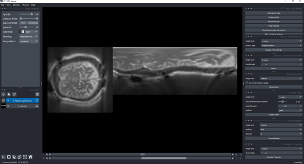
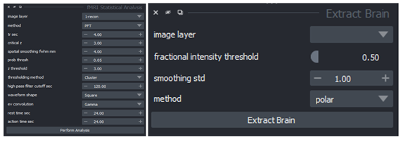
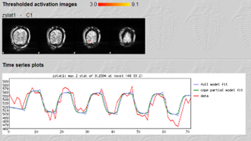
The full pipeline to perform end-to-end native polar analysis of radially acquired fMRI can be done through the graphical user interface of the proposed software package implemented in Python. Figure 1 demonstrates the data management tools in the left pane, multidimensional view options in the bottom pane, and analysis sub-panes in the right pane. Analysis sub-panes include six groups of tasks, i.e., sample data, data preparation, reconstruction, preprocessing, coordinate system conversion, and fMRI statistical analysis (Figure 3). The thorough validation of all modules and the whole process was successfully performed using the validation data.Discussion and Conclusion
A comprehensive end-to-end analysis software package to load, reconstruct, pre-process, analyze, and visualize fMRI in polar coordinates with the graphical user interface is developed and validated in the current work. This tool facilitates the native polar analysis of radially acquired fMRI that increases the specificity and/or improves the spatial resolution, particularly in task-based brain mapping studies with the region of interest considerably smaller than the field of view. The final results are visualized in polar and Cartesian coordinate systems. Future works might continue this work to study the noise considerations of polar fMRI and the advantages of non-uniform characteristics of PFT reconstructions.Acknowledgements
No acknowledgement found.References
- Kriegeskorte N, Bandettini P. Analyzing for information, not activation, to exploit high-resolution fMRI. Neuroimage 2007;38(4):649–62.
- Malekian, Vahid, et al. SSFP fMRI at 3 tesla: Efficiency of polar acquisition-reconstruction technique. Magnetic resonance imaging 2020;74:171-180.
- Golshani S, Nasiraei-Moghaddam A. Efficient radial tagging CMR exam: a coherent k-space reading and image reconstruction approach. Magn Reson Med 2017;77(4):1459–72.
- Lowe, David G. "Distinctive image features from scale-invariant keypoints." International journal of computer vision 60 (2004): 91-110.
- M. Jenkinson, M. Pechaud, and S. Smith. BET2: MR-based estimation of brain, skull and scalp surfaces. In Eleventh Annual Meeting of the Organization for Human Brain Mapping, 2005.
Figures