4665
Improved Fat-Water separation with Deep Learning-based ad-hoc MRI reconstruction incorporating spatial smoothing.1Centre for Biomedical Engineering, Indian Institute of Technology - Delhi, New Delhi, India, 2Department of Radio Diagnosis, All India Institute of Medical Sciences, New Delhi, India, 3Department of Biomedical Engineering, All India Institute of Medical Sciences, New Delhi, India
Synopsis
Keywords: AI/ML Image Reconstruction, Quantitative Imaging, Fat-water seperation, PDFF, Deep Learning, Fat Quantification, Physics Informed Deep Learning
Motivation: The novel Deep Learning (DL)-based Ad-Hoc Reconstruction (AHR) method for fat-water separation in Multi Echo-Magnetic Resonance Imaging (ME-MRI) has absolute generalizability. It can perform fat-water separation with the ME-MRIs from any anatomical region and views with varied numbers of echoes.
Goal(s): This research investigates the fat-water separation performance of spatial smoothing incorporated DL-based AHR method in ME-MRIs with and without noise.
Approach: The fat-water separation biophysical model based loss in AHR is added with spatial smoothing constraints.
Results: Results demonstrate that incorporating spatial smoothing in AHR improves the fat-water separation performance in ME-MRIs without noise, however, no performance improvements in ME-MRIs containing noise.
Impact: The PDFF maps obtained from fat-water separation in Multi Echo-MRI (ME-MRI) are of diagnostic and prognostic value in many diseases. This study investigates the performance of a novel Deep Learning-based Ad-Hoc Reconstruction method with spatial smoothing for fat-water separation.
Introduction
Multi Echo-Magnetic Resonance Imaging (ME-MRI) signal is used for fat-water separation to produce individual fat and water maps of any region of interest in the body. The Proton Density Fat Fraction (PDFF) maps calculated using these fat-water maps have both diagnostic and prognostic value in many diseases like Non-alcoholic fatty liver, Cancers, etc.1,2. Various studies have shown that the trained Deep Learning (DL) models can perform as accurately as the traditional optimization methods in fat-water separation3-5. However, these trained DL models have problems such as generalizability issues6,7. Jafari et al.8 proposed a DL-based Ad-Hoc Reconstruction (AHR) method for fat-water separation that overcomes these generalizability issues in conventionally trained DL models. DL-based AHR does not involve training with a large dataset and can fat-water separate ME-MRI from any anatomical region and their views with varied numbers of echoes8. Spatial smoothing constraints have been used in the reliable estimation of various parameter maps from different imaging modalities9. This research investigates the fat-water separation performance of spatial smoothing incorporated DL-based AHR in ME-MRIs with and without noise.Methods
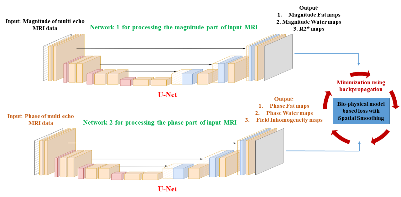
DL-based AHR method for fat-water separation incorporating spatial smoothing:Figure 1 describes the DL-based AHR method for fat-water separation in ME-MRI. Two U-Net models were used to process ME-MRI's magnitude and phase components separately. The three outputs from the first U-Net were fat $$$|F|$$$ and water $$$|W|$$$ magnitude maps, along with $$$R_2^*$$$ maps. The three outputs from the second U-Net were fat $$$\varphi(F)$$$ and water $$$\varphi(W)$$$ phase maps and field inhomogeneity $$$\psi$$$ maps . The outputs from both the networks were given as input to the fat-water separation biophysical model-based loss function along with the Total Variation (TV) penalty10 given in equation (1):
$$\text{Loss}=\left\|I_n-\left(W+F \cdot e^{j 2 \pi f t_n}\right)\cdot e^{j 2 \pi \psi t_n}e^{-R_2^* t_n}\right\|_2^2+\alpha(T V(W)+T V(F))\tag{1}$$
$$$I_n$$$ is the signal from $$$n$$$th echo image. $$$W$$$ is the signal from water protons, $$$F$$$ is the signal from fat protons, $$$f$$$ is the chemical‐shift frequency between fat and water protons, $$$t_n$$$ is the TE of $$$n$$$th echo, $$$\psi$$$ is the field inhomogeneity and $$$R_2^*$$$ is the inverse of $$$T_2^*$$$. $$$TV$$$ is the Total Variation spatial smoothing constraint and $$$\alpha$$$ is the smoothing weight10. The loss described in equation (1) is continuously minimized in both the U-Nets by backpropagating for 10,000 epochs with a learning rate 6x10⁻⁴ on a batch of ME-MRI estimating the required Fat $$$F$$$ and $$$W$$$ Water maps. $$$\alpha$$$ is set to 1×10⁻⁶.
Performance Analysis:
Seven batches of 12-echo MRIs hosted in the ISMRM 2012 fat-water separation workshop11 were used in this study. ME-MRIs were fat-water separated using the AHR with and without spatial smoothing to produce fat, water and PDFF maps, and they were compared against the ground maps by computing Structural Similarity Index Measure (SSIM)12 and Normalized Root Mean Square Error (NRMSE). Ground truth maps were generated using the widely used hierarchical IDEAL method13. Additional experiments were conducted by introducing noises in ME-MRIs: a Gaussian noise of mean 120 and standard deviation 20 is added in the magnitude component, the signal is converted to cartesian form, and the same Gaussian noise is added in the real and imaginary components of the ME-MRI signal and the phase retrieved from this noisy signal is utilized instead of the original phase.
Results
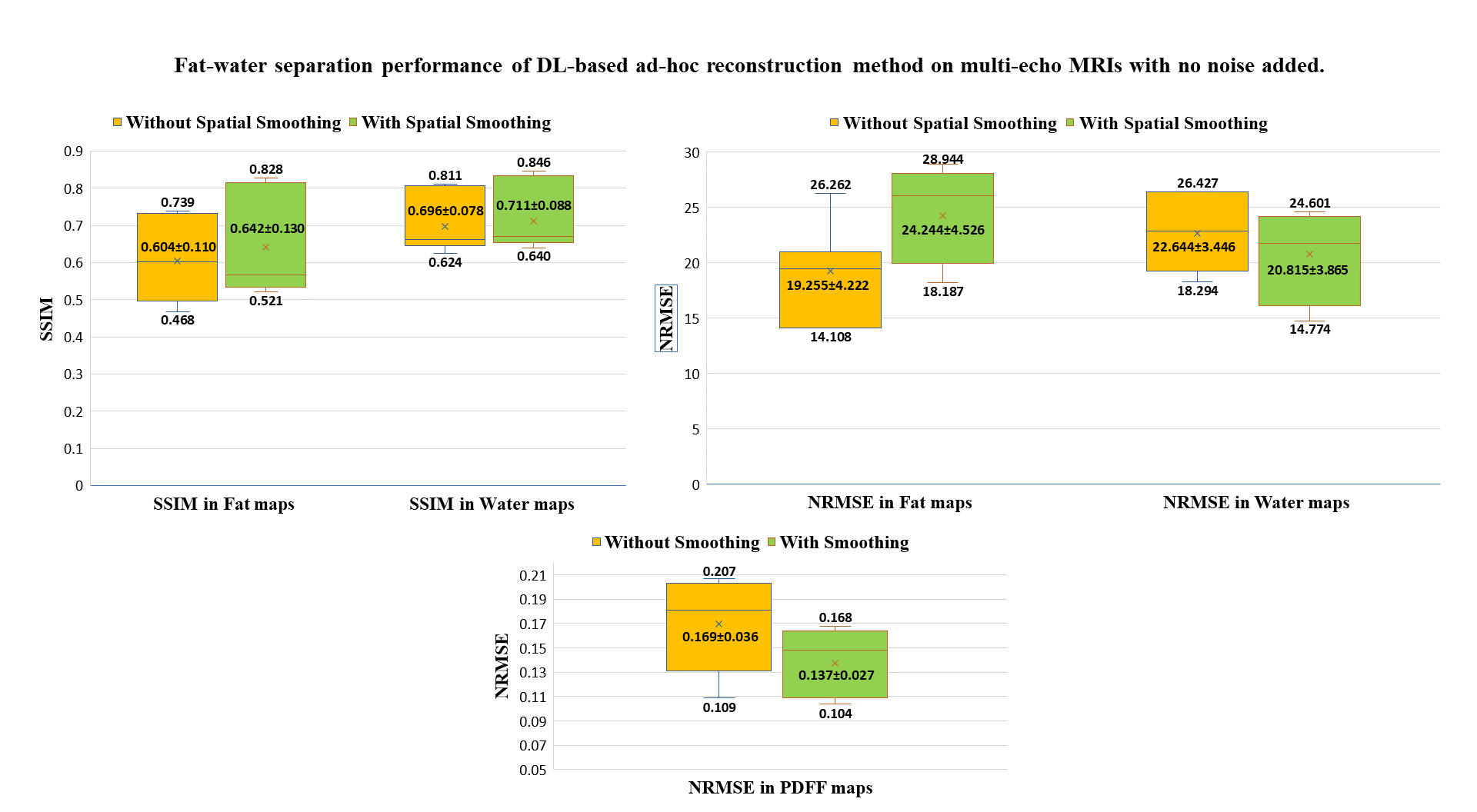
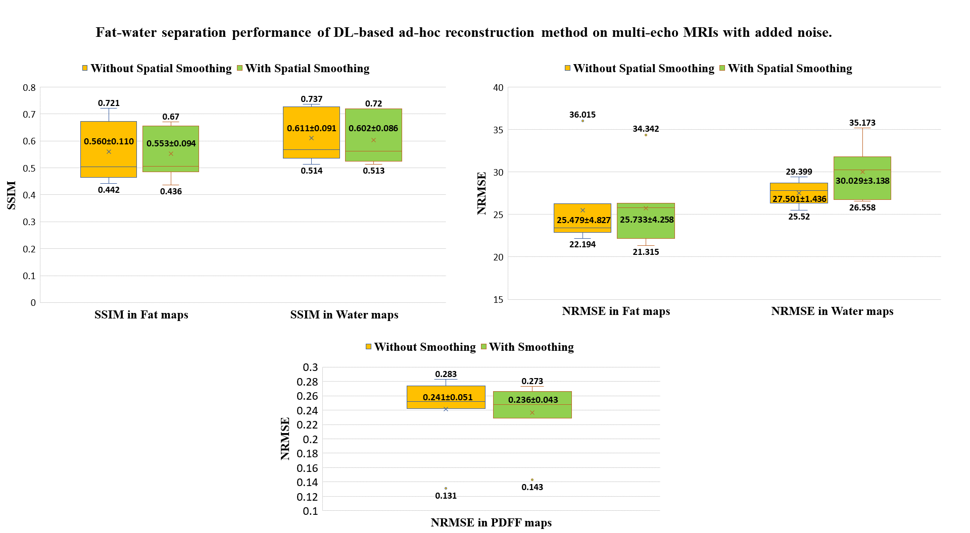
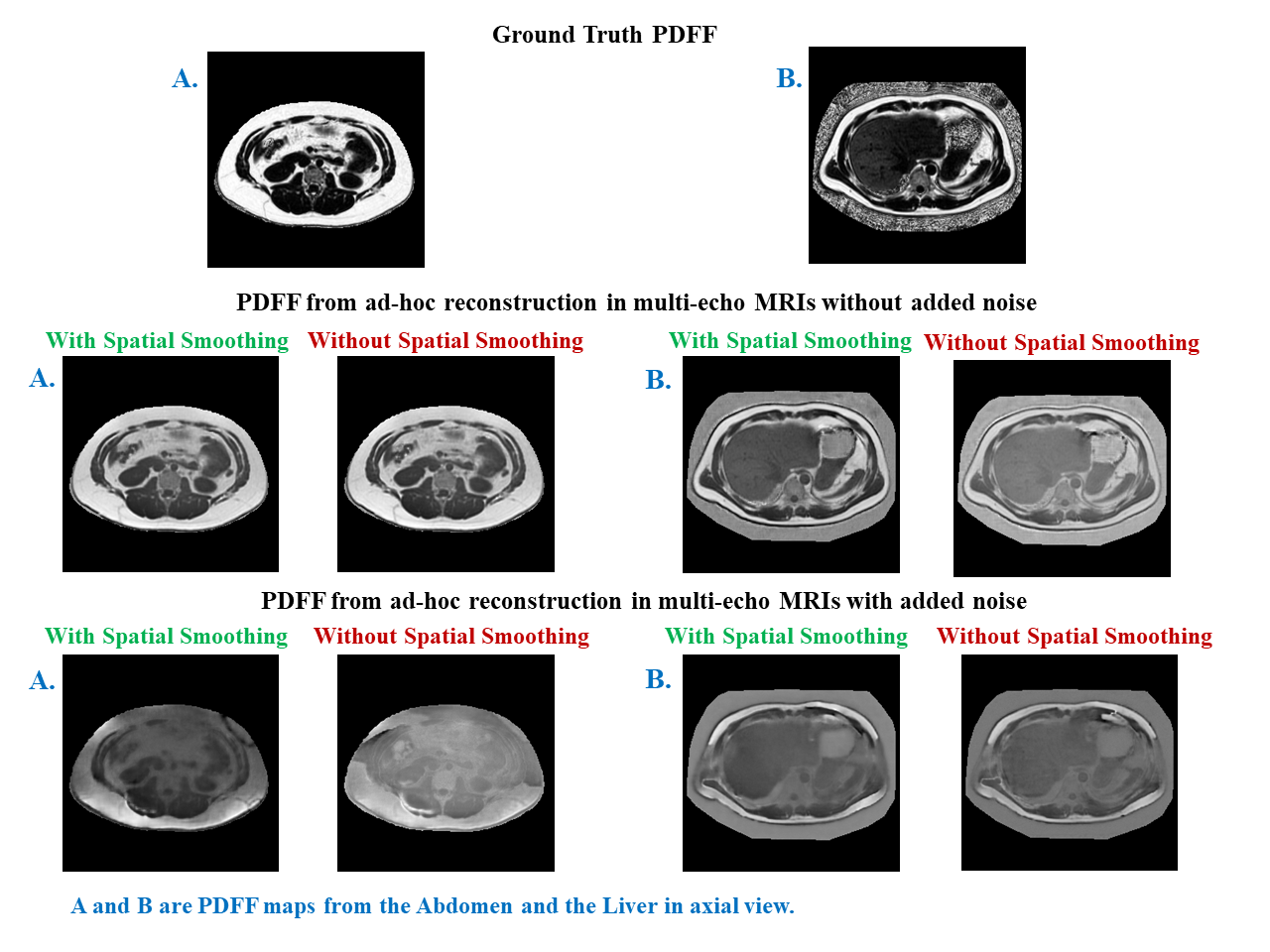
Plots in Figure 2 and Figure 3 present the fat-water separation performance of the AHR without and with spatial smoothing in ME-MRIs without and with noise added, respectively. In ME-MRIs without added noise, adding spatial homogeneity constraints to AHR improved the SSIMs from 0.604±0.110 to 0.642±0.130 and from 0.696±0.078 to 0.711±0.088 in fat and water images, respectively, the NRMSE in the water images improved from 22.644±3.446 to 20.815±3.865 and the NRMSE in clinically significant PDFF maps improved from 0.169±0.036 to 0.137±0.027. However, adding spatial smoothing in AHR gave no significant improvements in the fat-water separation performance in ME-MRIs with added noise. Figure 4 visually compares the PDFFs obtained from the AHR method with and without spatial smoothing in ME-MRIs of two representative subjects with and without added noise against the ground truth.Discussion and Conclusion
Incorporating spatial smoothing in DL-based AHR improves fat-water separation performance in ME-MRIs without noise, however, no improvements were observed in ME-MRIs containing noise. Studies report that spatial smoothing improves parameter estimations in noisy MRIs9,14. The reason for contradictory results could be the large magnitude of noise improvised in this study. Experiments with a spectrum of signal noises are required for further studies. There is dearth of literature on quality and appropriate level of simulated noise in phase images, which also needs further study.Acknowledgements
The human subject multi-echo MRI data utilized in this study was hosted in the 2012 ISMRM Fat-Water separation workshop11.References
1. Thomas, E.L., Fitzpatrick, J.A., Malik, S.J., Taylor-Robinson, S.D. and Bell, J.D., 2013. Whole body fat: content and distribution. Progress in nuclear magnetic resonance spectroscopy, 73, pp.56-80.
2. Yip, C., Dinkel, C., Mahajan, A., Siddique, M., Cook, G.J. and Goh, V., 2015. Imaging body composition in cancer patients: visceral obesity, sarcopenia and sarcopenic obesity may impact on clinical outcome. Insights into imaging, 6, pp.489-497.
3. J. W. Goldfarb, J. Craft, and J. J. Cao, “Water-fat separation and parameter mapping in cardiac MRI via deep learning with a convolutional neural network: MR Water-Fat Separation,” J. Magn. eason. Imaging, vol. 50, no. 2, pp. 655–665, Aug. 2019, doi: 10.1002/jmri.26658.
4. J. Cho and H. Park, “Robust water–fat separation for multi‐echo gradient‐recalled echo sequence using convolutional neural network,” Magn. eason. Med., vol. 82, no. 1, pp. 476–484, Jul. 2019, doi: 10.1002/mrm.27697.
5. J. Andersson, H. Ahlström, and J. Kullberg, “Separation of water and fat signal in whole‐body gradient echo scans using convolutional neural networks,” Magn. eason. Med., vol. 82, no. 3, pp. 1177–1186, Sep. 2019, doi: 10.1002/mrm.27786.
6. X. Liang, D. Nguyen, and S. B. Jiang, “Generalizability issues with deep learning models in medicine and their potential solutions: illustrated with cone-beam computed tomography (CBCT) to computed tomography (CT) image conversion,” Mach. Learn. Sci. Technol., vol. 2, no. 1, p. 015007, Mar. 2021, doi: 10.1088/2632-2153/abb214.
7. J. Krois et al., “Generalizability of deep learning models for dental image analysis,” Sci. Rep., vol. 11, no. 1, p. 6102, Mar. 2021, doi: 10.1038/s41598-021-85454-5.
8. R. Jafari et al., “Deep neural network for water/fat separation: Supervised training, unsupervised training, and no training,” Magn. eason. Med., vol. 85, no. 4, pp. 2263–2277, Apr. 2021, doi: 10.1002/mrm.28546.
9. Kayal EB, Kandasamy D, Khare K, Alampally JT, Bakhshi S, Sharma R, Mehndiratta A. Quantitative Analysis of Intravoxel Incoherent Motion (IVIM) Diffusion MRI using Total Variation and Huber Penalty Function. Med Phys. 2017 Nov;44(11):5849-5858. Doi: 10.1002/mp.12520. Epub 2017 Oct 11. PMID: 28817196.
10. Rodríguez, P., 2013. Total variation regularization algorithms for images corrupted with different noise models: a review. Journal of Electrical and Computer Engineering, 2013, pp.10-10.
11. Hu, H.H., Börnert, P., Hernando, D., Kellman, P., Ma, J., Reeder, S. and Sirlin, C., 2012. ISMRM workshop on fat–water separation: insights, applications and progress in MRI. Magnetic resonance in medicine, 68(2), pp.378-388.
12. Zhou Wang, A. C. Bovik, H. R. Sheikh and E. P. Simoncelli, “Image quality assessment: from error visibility to structural similarity,” in IEEE Transactions on Image Processing, vol. 13, no. 4, pp. 600-612, April 2004, doi: 10.1109/TIP.2003.819861.
13. Jiang, Y. and Tsao, J., 2012. Fast and robust separation of multiple chemical species from arbitrary echo times with complete immunity to phase wrapping. In Proceedings of the 20th Annual Meeting of ISMRM (Vol. 388).
14. Vij, M., Malagi, A.V., Baidya Kayal, E., Saini, J. and Mehndiratta, A., 2020. IVIM analysis using Total Variation Penalty Regularization Based Model for Brain Tumor Analysis. In Proc Intl Soc Mag Reson Med (Vol. 28, pp. 1-4).
Figures