4659
AI-Integrated MRS Scan Identifies and Updates Scan Parameters in the Presence of OOV Artifacts1Russell H. Morgan Department of Radiology and Radiological Sciences, Johns Hopkins School of Medicine, Baltimore, MD, United States, 2F. M. Kirby Research Center for Functional Brain Imaging, Kennedy Krieger Institute, Baltimore, MD, United States, 3Philips Healthcare, Rochester, MN, United States
Synopsis
Keywords: Acquisition Methods, Machine Learning/Artificial Intelligence, Spectroscopy, Brain, Artifacts, Convolutional Neural Network
Motivation: Deep learning is a promising new tool for post-processing MRS data. Neural network-based MRS acquisition methods do not yet exist, but should lead to higher-quality data.
Goal(s): The goal of this work was to create an “intelligent MRS scan” by integrating a Convolutional Neural Network (CNN) directly into a MRS acquisition protocol.
Approach: Here, a CNN-powered pre-scan collects a single-transient from 48 different gradient geometries, and updates future scans, without human intervention, to avoid out-of-voxel (OOV) artifacts.
Results: The AI-informed scan produced high-quality data for all participants while the control parameters failed half of the time in the artifact-rich mPFC region.
Impact: The work demonstrates the first AI-integrated MRS scan protocol in which an intelligent pre-scan modifies scan parameters to improve data quality, here reducing out-of-voxel artifacts.
Introduction
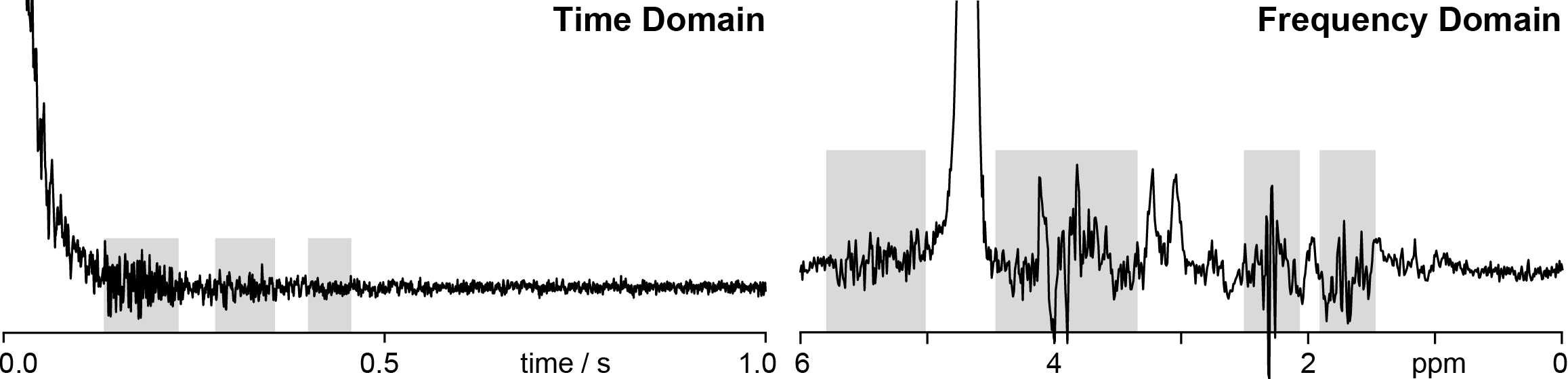
The MRS pre-scan phase has not changed substantially in decades, consisting of steps to optimize shimming, transmit/receive gain and center frequency F0. Each step is optimized to reduce non-data-acquisition time. Studies often acquire data of less-than-perfect quality in aspects that are ignored by this pre-scan, such as lipid signal excitation or out-of-voxel (OOV) echoes. Such artifacts are only revealed at the point of analysis – intelligent MRS scans should be able to recognize a range of artifacts and adjust parameters to mitigate them.OOV artifacts, commonly found within in vivo spectra1, appear as broad signals with strong first-order phase (as in Figure 1) that obstruct processing and quantification, often leading to unusable data. They arise from gradient echoes – water signals from outside the voxel are refocused by evolution in local field gradients2-4. In the k-space representation, OOV echoes are dephased at the start of the acquisition, starting away from the k-origin. Evolution in a local gradient then moves them across k-space, and if that trajectory passes through the origin, they are refocused as an echo. The presence of OOV artifacts can thus be altered by changing the starting-point of signals in k-space.
Although OOV artifacts are usually discovered during analysis (when it is too late), a better strategy is to acquire data that avoids them. Furthermore, selecting from several possible options under the time constraints of a pre-scan is not possible for a human operator. Here, a Convolutional Neural Network (CNN), developed to identify OOV echoes with high fidelity in single transients5, was integrated into a pre-scan to select the best crusher gradient scheme and adjust the parameters for all future scans, providing the first artificially intelligent (AI) MRS scan protocol.
Methods
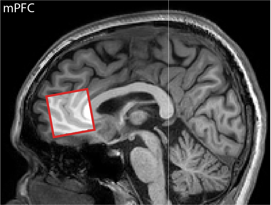
Four participants (aged 45.3±10.7 years) were scanned with a 3.0 T Philips scanner. A 27-mL cubic voxel was placed on the midline in medial prefrontal cortex (mPFC) as shown in Figure 2.The starting-point of OOV signals in k-space can be changed by changing their crusher-gradient history both by reassigning the three crusher axes of the sequence to the spatial axes of the voxel, and changing the polarity of crusher-gradients on each axis. This results in a total of 48 distinct permutations of 6 slice orders (e.g., [x,y,z], [y,x,z], etc.) and 8 crusher polarity combinations (e.g., [+,+,+], [+,+,-], etc.). A single transient of each of these is collected as a pre-scan.
Data from the pre-scan were then passed through a CNN, previously trained to recognize OOV artifacts in single-transient data5, returning an ‘OOV prevalence’ score for each transient. A GABA-edited MEGA-PRESS (TR/TE = 2000/80 ms; 2 kHz spectral width; 2048 samples) scan was then acquired with the crusher geometry that gave the lowest OOV score (denoted AI-informed) and as a fair comparator, a second geometry randomly selected from among the 48 (denoted Control).
Data were analyzed using Osprey6 version 2.5.0. The linear combination model fit residual for the GABA difference spectrum (relResdiff1) was used to determine whether the AI-informed scan provided usable and/or higher quality data. This quality metric compares the relative amplitude of the fit residual against the standard deviation of the noise.
Results
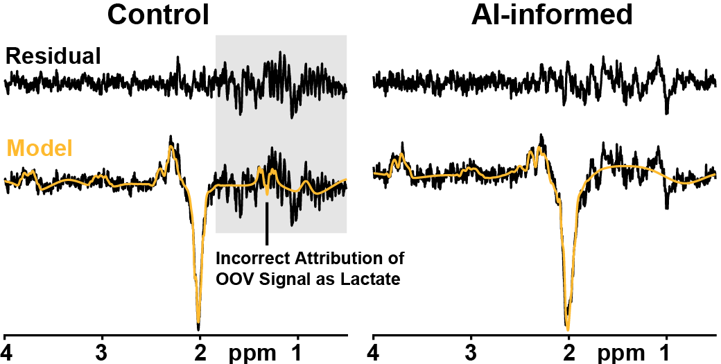
Data were successfully acquired in 4 subjects, including CNN processing of pre-scan data on the scanner. Given the challenging prefrontal voxel location, OOV artifacts were observed throughout the acquired spectra. In 2 of the 4 participants, the randomly selected geometry resulted in data of such poor quality that it resulted in failure of the processing and model fitting steps. All the AI-informed datasets were usable. This was seen in the quality metric with a lower normalized residual for AI-informed scans (mean±SD: 9.80±3.28) compared with the randomly selected control geometry (mean±SD: 128.96±129.67) shown in Figure 3.Discussion
This proof-of-concept work illustrates the potential of on-scanner CNN deployment for MRS sequence optimization, either as a pre-scan and eventually as a real-time sequence adaptation. In the future, it will be possible to train CNNs to recognize all the common failure-modes of MRS, and adapt parameters during the acquisition itself to dramatically improve data quality and reduce failures.Importantly, many brain regions of strong clinical and cognitive interest (e.g., mPFC, hippocampus) lie in areas of relatively poor field homogeneity, susceptible to OOV artifacts3,4. This is especially problematic for edited-MRS sequences because they contain additional pulses that are not spatially selective and target lower-concentration metabolites with weaker signals and lower artifact tolerance. Further work is required to understand which elements of sequence design most effectively reduce their appearance.
Conclusion
This CNN-powered pre-scan demonstrates a new era of intelligent MRS acquisition.Acknowledgements
This work has been supported by The National Institute of Health, grants T32 AG00096, R00 AG062230, R21 EB033516, R01 EB016089, R01 EB023963, K00 AG068440, R21 AG053040, R01 AG076942, and P41 EB031771.References
- Kreis, R. (2004). Issues of spectral quality in clinical 1H‐magnetic resonance spectroscopy and a gallery of artifacts. NMR in Biomedicine, 17(6), 361-381.
- Starck, G., Carlsson, Å., Ljungberg, M., & Forssell‐Aronsson, E. (2009). k‐space analysis of point‐resolved spectroscopy (PRESS) with regard to spurious echoes in in vivo 1H MRS. NMR in Biomedicine: An International Journal Devoted to the Development and Application of Magnetic Resonance In vivo, 22(2), 137-147.
- Ernst, T., & Chang, L. (1996). Elimination of artifacts in short echo time 1H MR spectroscopy of the frontal lobe. Magnetic resonance in medicine, 36(3), 462-468.
- Carlsson, Å., Ljungberg, M., Starck, G., & Forssell-Aronsson, E. (2011). Degraded water suppression in small volume 1 H MRS due to localised shimming. Magnetic Resonance Materials in Physics, Biology and Medicine, 24, 97-107.
- Gudmundson, A. T., Davies-Jenkins, C. W., Özdemir, İ., Murali-Manohar, S., Zöllner, H. J., Song, Y., Hupfeld, K.E., Schnitzler, A., Oeltzschner, G., Stark, C.E. and Edden, R. A. (2023). Application of a 1H Brain MRS Benchmark Dataset to Deep Learning for Out-of-Voxel Artifacts. Imaging Neuroscience.
- Oeltzschner, G., Zöllner, H. J., Hui, S. C., Mikkelsen, M., Saleh, M. G., Tapper, S., & Edden, R. A. (2020). Osprey: Open-source processing, reconstruction & estimation of magnetic resonance spectroscopy data. Journal of neuroscience methods, 343, 108827.
Figures