4654
Motion-insensitive multi-contrast intracranial vascular imaging:Feasibility of self-navigated motion-detection from iSNAP sequence1Institute of Science and Technology for Brain-inspired Intelligence, Fudan University, Shanghai, China, 2Philips Healthcare, Shanghai, China, 3Key Laboratory of Computational Neuroscience and Brain-Inspired Intelligence (Fudan University), Ministry of Education, Shanghai, China
Synopsis
Keywords: Motion Correction, Vessels
Motivation: The multi-contrast intracranial vascular imaging sequence iSNAP is less sensitive to motion due to the use of 3D radial readout, but image blurring and thus decreased visibility of distal small vessels may still occur in the case of large head motion.
Goal(s): To evaluate the feasibility of estimating the continuous head motion from iSNAP k-space data under different temporal resolutions.
Approach: Subsets of iSNAP's 3D radial k-space data were used to reconstruct a series of low-resolution images.Then the SPM toolbox was used to perform rigid registration,between the image volumes.
Results: The temporal profiles of the motion parameters are well estimated.
Impact: The results indicate the head motion can be well estimated from iSNAP. The estimated motion parameters can be used to correct the k-space data in the future, thus allowing reconstruction of motion-free iSNAP images.
Introduction
Tissue motion will cause artifacts or image blurring in MR imaging, and is difficult to avoid in many clinical settings, such as scanning unconscious stroke patients. The iSNAP is an efficient intracranial MR vascular imaging sequence, which is based on a combination of arterial spin labeling (ASL) preparation and 3D golden angle radial gradient echo readout, for simultaneous obtainment of brain dynamic MRA (dMRA), static MRA (sMRA), intracranial vascular wall images and t1-weighted (T1W) brain structure images, allowing a comprehensive and efficient assessment of intracranial vascular health[1]. Although iSNAP is less sensitive to motion than the conventional MR vascular imaging sequences due to the use of 3D radial acquisition, image blurring and thus decreased visibility of distal small vessels may still occur in the case of large head motion. Therefore, motion correction is important to improve usability of iSNAP sequence. We thus propose to retrospectively estimate the continuous head motion from the iSNAP k-space data based on a highly-undersampled image reconstruction and image registration, and then correct the k-space data with the estimated motion parameters followed by a motion-insensitive image reconstruction. In this preliminary study, we evaluated the feasibility of estimating the continuous head motion from iSNAP k-space data under different temporal resolutions.Methods
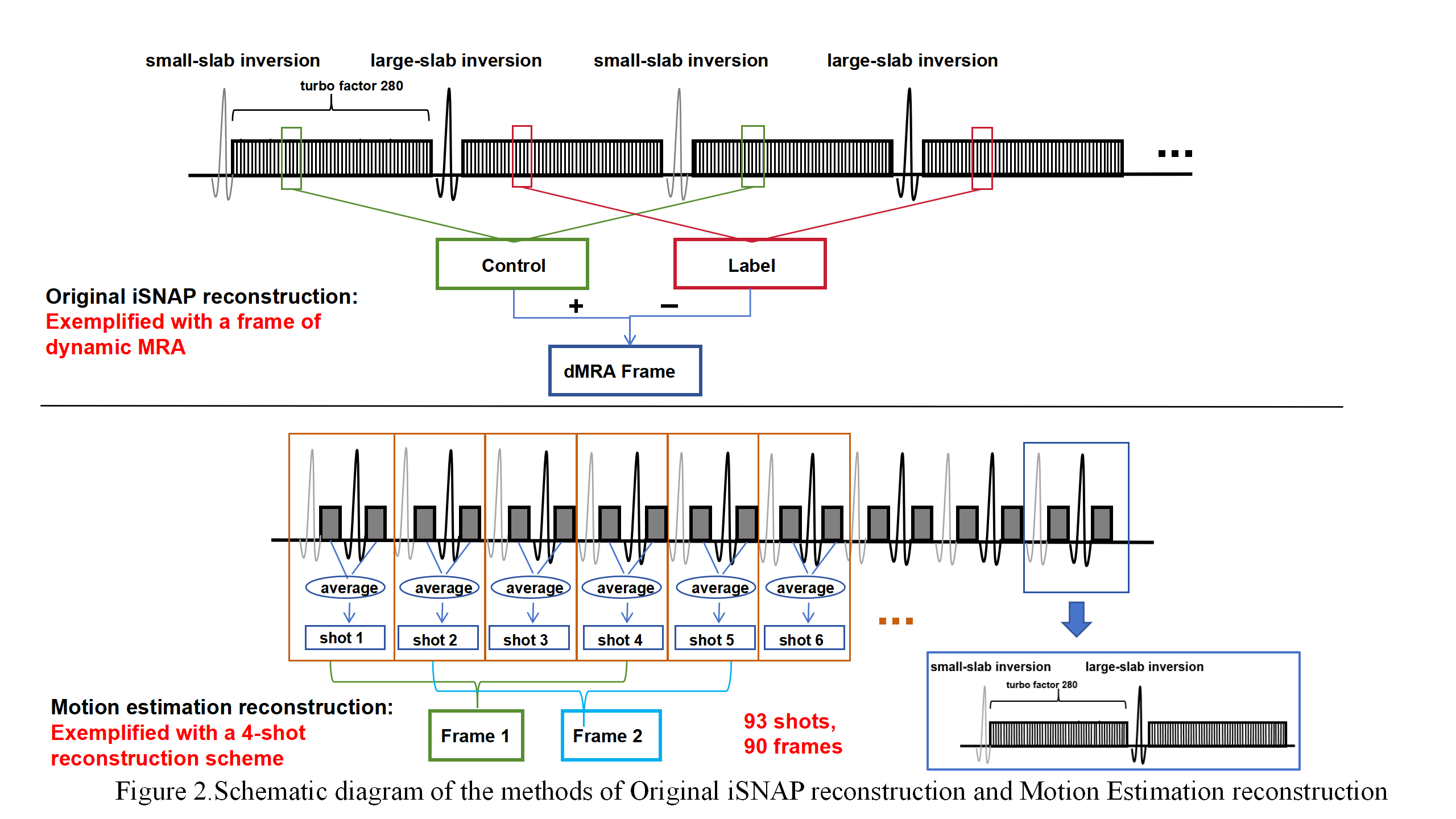
As shown in Figure 1, the iSNAP sequence employs multi-shot readout with interleaved ASL Control and Label acquisitions, which have the same trajectory, and the radial spokes were designed to have a golden angle increment along the shot dimension[1]. The main imaging parameters for iSNAP are as follows: turbo factor of 280, a total of 186 shots (93 for Control and 93 for Label), a shot interval of 2139 ms, an FA of 6 degrees, a TR of 7.6 ms, a TE of 2.7 ms, voxel size 0.8 mm isotropic, a FOV of 204.8 × 179.2 × 144 mm³ was reconstructed, total scan time 6 min 35 sec. Two healthy subjects (female, age 24 and 27 years) were scanned using a 3T Philips Ingenia CX scanner equipped with a 32-channel head coil. The iSNAP sequence was scanned 3 times, with the subjects asked to keep still, nod, and rotate the head back and forth in the right-left direction, respectively. For the latter two scenarios, the extent of head motion increased from small to large.As shown in Figure 1, to estimate the continuous head motion, we first averaged the k-space data of the paired Control and Label shots, and then reconstructed a series of 3D image volumes, each from k-space data of N consecutive shots, using nonuniform FFT algorithm. Then the SPM toolbox was used to perform rigid registration, represented by 3 translation and 3 angulation parameters, between the image volumes.
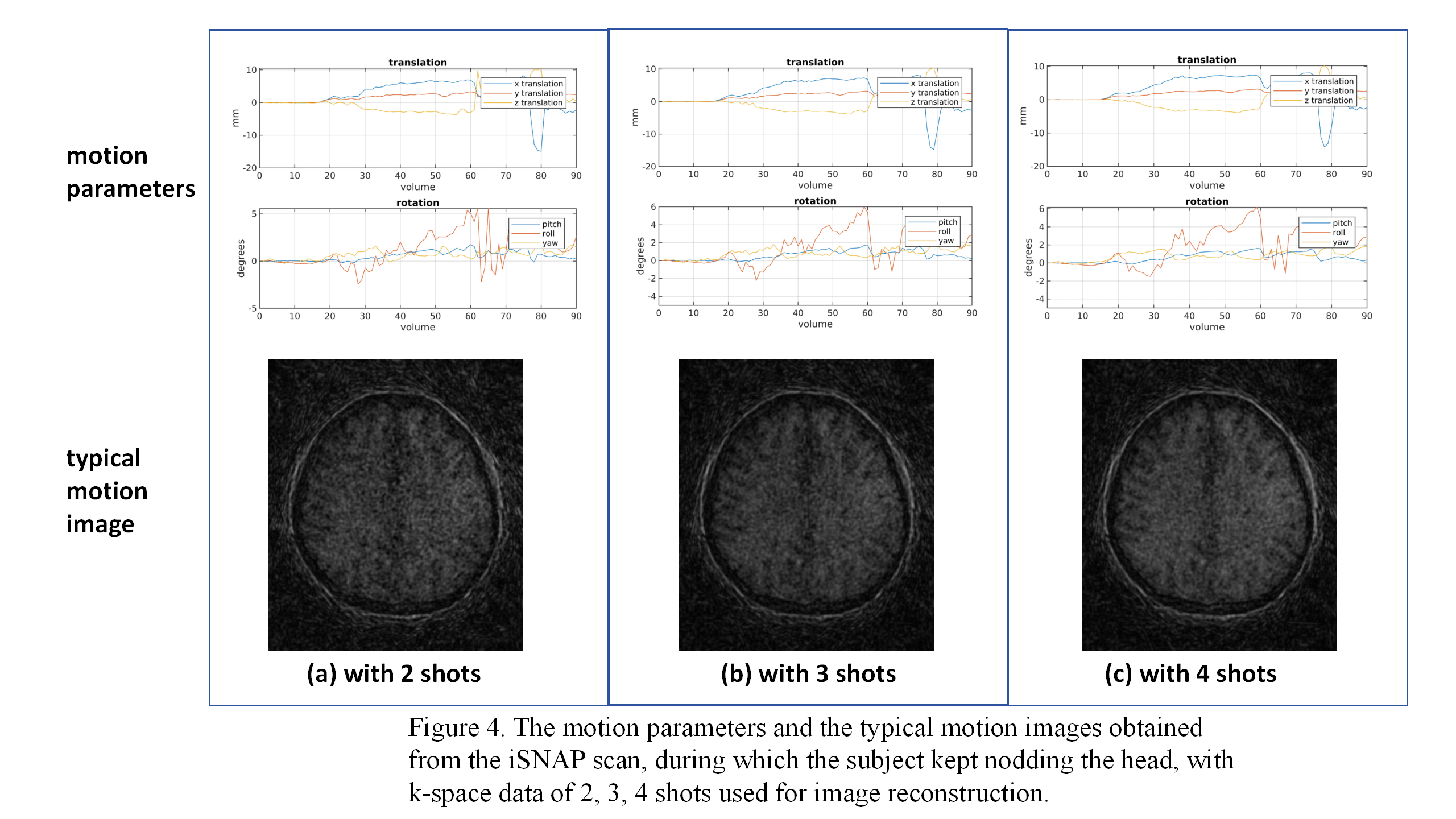
For comparison purposes, the reconstruction and registration were performed for an N of 2, 3, and 4.
Results
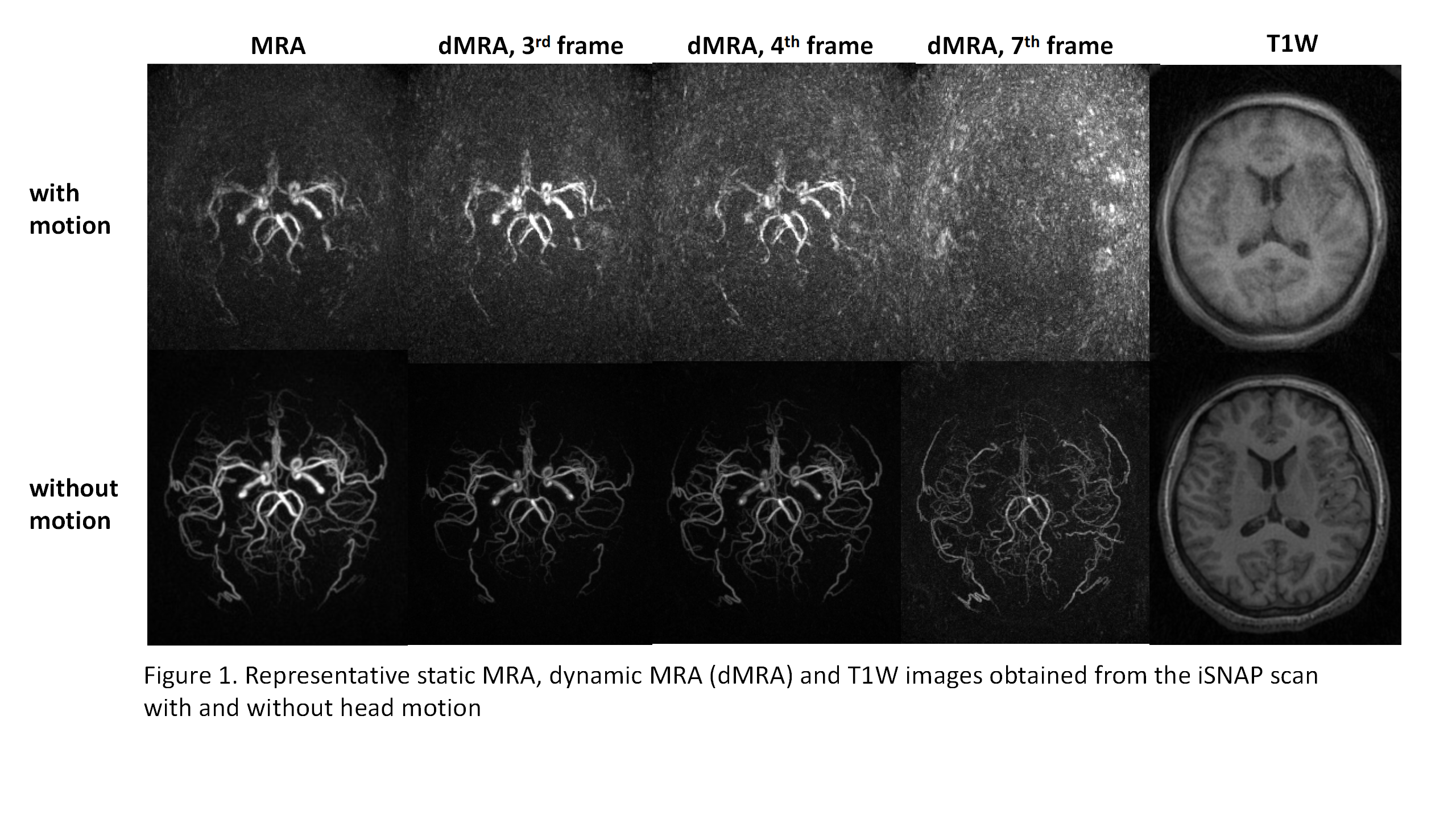
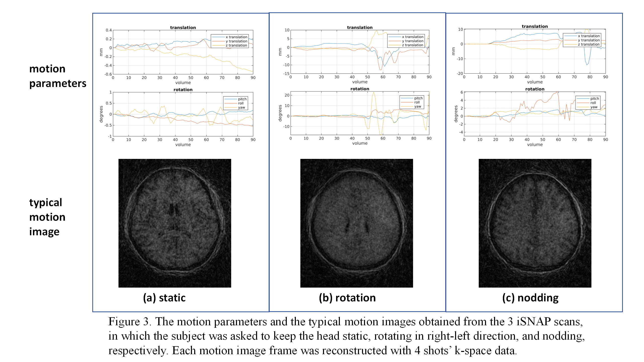
Figure 2 shows the original iSNAP images (static MRA, dMRA, T1W) with and without motion, indicating that motion can severely reduce the quality of iSNAP images. Figure 3 shows the motion parameters and typical motion images reconstructed with 4 shots’ data for the 3 iSNAP scans The results indicate that the head motion generally can be estimated from the motion images, although the images are noisy and blurred.Figure 4 shows the motion parameters and typical motion images reconstructed with 2, 3, and 4 shots’ data. Overall, the temporal profiles of the motion parameters are similar between the 3 reconstructions.Discussion and Conclusion
The golden angle increment of the radial spokes applies to the original iSNAP image reconstruction, but not the motion reconstruction. Therefore, the motion reconstruction is expected to have a degraded image quality and maybe only the large brain structures are detectable, since the k-space data were not evenly distributed, especially when few shots are used. However, our results indicate the head motion still can be well estimated. This probably is because the rigid registration of head images relies largely on the overall brain structure and less on the fine details. One limitation of the study is that there is no gold standard for the motion measurement, and thus it is difficult to quantitatively evaluate the accuracy of the estimated motion parameters. Our future work is to use the estimated motion parameters to correct the k-space data and then use the new data to reconstruct motion-free iSNAP images.Acknowledgements
This work was supported by Natural Science Foundation of Shanghai (22ZR1403900).References
[1] Chen Z, Zhou Z, Qi H, et al. A novel sequence for simultaneous measurement of whole‐brain static and dynamic MRA, intracranial vessel wall image, and T1‐weighted structural brain MRI[J]. Magnetic resonance in medicine, 2021, 85(1): 316-325.
Figures