4648
A Dictionary Matching-Based Motion Correction Method for Cardiac Multi-Parametric Mapping1Shanghai Jiao Tong University, Shanghai, China
Synopsis
Keywords: Motion Correction, Motion Correction, multi-parametric mapping
Motivation: Motion correction (MoCo) for cardiac parametric mapping can be challenging due to the dynamic signal variations. Traditional model-based methods need an analytical model, which is often unavailable for multi-parametric mapping applications.
Goal(s): To propose a model-free dictionary matching-based MoCo method for cardiac multi-parametric mapping.
Approach: The method alternates between dictionary matching and image registration. In vivo validation was performed in 10 healthy subjects for cardiac joint T1 and T2 mapping with controlled breathing.
Results: Compared with non-MoCo, the proposed method significantly reduced inter-image misalignment and improved the quality of the T1 and T2 maps.
Impact: The proposed MoCo method can be applied to any quantitative MRI application with a signal dictionary, which includes both single-parametric and multi-parametric mapping.
Introduction
Retrospective motion correction (MoCo) is imperative for accurate cardiac parametric mapping1,2. However, the image contrast variations pose a significant challenge to image-based MoCo methods1,3–9, which solely rely on image registration algorithms to estimate the motion. Model-based methods10–13 use physical models to synthesize images of similar contrasts as the acquired images, transforming the original multi-contrast registration problem into a single-contrast registration problem, which is easier to solve. However, current model-based methods need an analytical signal model for image synthesis, which is often absent for multi-parametric mapping14–16. Here we propose a model-free dictionary matching-based MoCo method for cardiac multi-parametric mapping. We validated the method for cardiac Multimapping16 in 10 healthy subjects with controlled breathing.Methods
TheoryLet $$$\mathbf{D}$$$ be the dictionary, with each unit-length entry (column) $$$\mathbf{d}_{\mathrm{n}}$$$ corresponding to a simulated signal determined by tissue and imaging parameters. We formulate the proposed method as an optimization problem
$$\min _{\boldsymbol{\alpha}, \boldsymbol{\theta}} \sum_{\mathbf{r}}\|\mathbf{M}(\mathbf{y}, \mathbf{r}, \boldsymbol{\theta})-\mathbf{D} \boldsymbol{\alpha}(\mathbf{r})\|_{2}^{2}+\lambda \mathrm{R}(\boldsymbol{\theta}) \text { s.t. }\|\boldsymbol{\alpha}(\mathbf{r})\|_{0} \leq 1 \text {, }\tag{1}$$
where $$$\mathbf{r}$$$ is the spatial coordinate, $$$\mathbf{y}$$$ the acquired image signal, $$$\boldsymbol{\alpha}$$$ the sparse code, $$$\mathbf{M}$$$ the deformation operator, $$$\boldsymbol{\theta}$$$ the deformation parameters, $$$\mathrm{R}$$$ the spatial regularization term, and $$$\lambda$$$ the regularization coefficient. We solve the problem by alternating minimization with two steps.
Step 1: By fixing $$$\boldsymbol{\theta}$$$, we obtain a dictionary matching subproblem. The optimal sparse code $$$\boldsymbol{\alpha}^{*}(\mathbf{r})$$$ is computed by finding the entry that best matches the deformed image signal $$$\mathbf{M}(\mathbf{y}, \mathbf{r}, \boldsymbol{\theta})$$$ based on the inner product:
$$\left[\boldsymbol{\alpha}^{*}(\mathbf{r})\right]_{\mathrm{n}}=\left\{\begin{array}{l}\left\langle\mathbf{M}(\mathbf{y}, \mathbf{r}, \boldsymbol{\theta}), \mathbf{d}_{\mathrm{n}}\right\rangle, \text { for } \mathrm{n}=\underset{\mathrm{m}}{\arg \max }\left\langle\mathbf{M}(\mathbf{y}, \mathbf{r}, \boldsymbol{\theta}), \mathbf{d}_{\mathrm{m}}\right\rangle, \\0, \text { otherwise, }\end{array}\right.\tag{2}$$
where $$$\left[\boldsymbol{\alpha}^{*}(\mathbf{r})\right]_{\mathrm{n}}$$$ is the $$$\mathrm{n}$$$-th component of $$$\boldsymbol{\alpha}^{*}(\mathbf{r})$$$. The constructed synthetic images $$$\mathbf{D} \boldsymbol{\alpha}^{*}(\mathbf{r})$$$ are motion-free and have consistent contrasts with the acquired images.
Step 2: By fixing $$$\boldsymbol{\alpha}$$$, we obtain an image registration subproblem, i.e., registering the acquired images $$$\mathbf{y}(\mathbf{r})$$$ to the synthetic images $$$\mathbf{D} \boldsymbol{\alpha}(\mathbf{r})$$$. We used the free-form deformations and bending energy as the deformation model and spatial regularization term, respectively17. $$$\boldsymbol{\theta}$$$ was the collection of control point meshes for all images. We implemented a gradient descent algorithm to solve the subproblem.
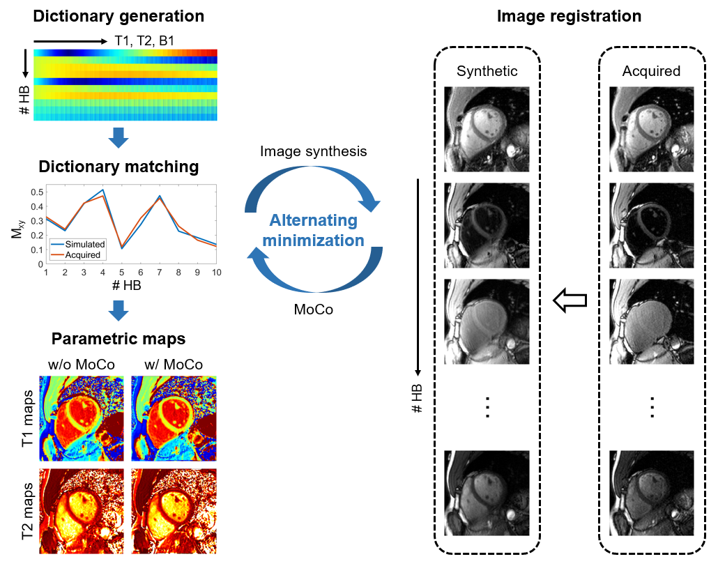
As illustrated in Figure 1, the proposed methods alternately construct motion-free synthetic images by dictionary matching and register the acquired images to the synthetic images until convergence. The motion-corrected parametric maps are generated by the last dictionary matching.
Experiments
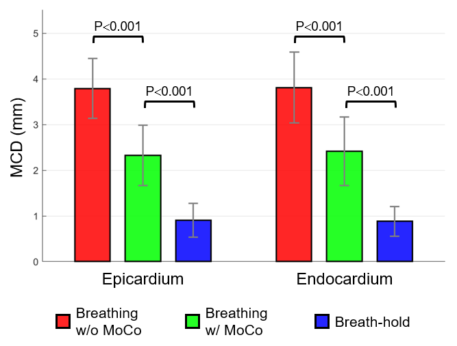
The institutional review board approved the study. We performed cardiac Multimapping16 over 10 healthy subjects in a 3T scanner (uMR 790, United Imaging Healthcare, Shanghai, China) after obtaining written informed consent from each subject. For each subject, we acquired 3 short-axis slices in both controlled-breathing and breath-hold states. We constructed the dictionary for each scan using the extended phase graph (EPG)18 for ranges of T1, T2, and B116. To evaluate the MoCo performance, we measured the mean contour distances (MCD) of the epicardium and endocardium between each pair of images from the image sequence. Two readers qualitatively evaluated the quality of the reconstructed T1 and T2 maps using a 5-point scale.
Results
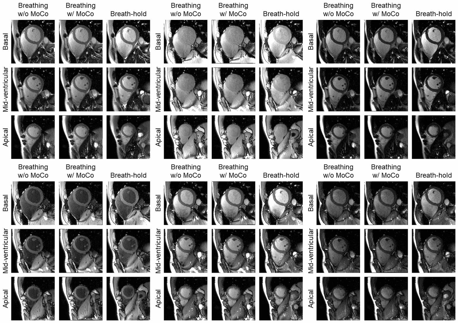
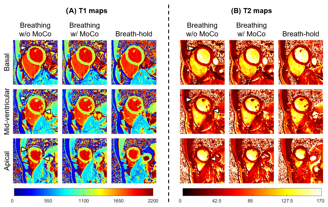
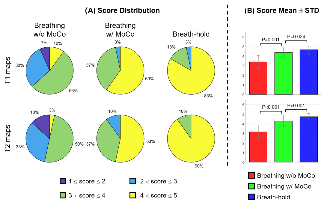
Figure 2 shows the multi-contrast images of one representative subject without and with our MoCo method. The method considerably reduced the motion, especially in the left ventricular region. Figure 3 shows the parametric maps of the same subject in Figure 2 before and after MoCo. Without MoCo, the reconstructed T1 and T2 maps exhibited relatively severe motion artifacts (white arrows). Our MoCo method considerably improved the mapping quality, which was similar to that of the breath-hold scan. Figure 4 shows comparisons of average epicardial and endocardial MCDs between reconstructions of the non-MoCo, MoCo, and breath-hold datasets. For both epicardial and endocardial contours, the motion-corrected images exhibited significantly lower MCDs than the uncorrected images. The breath-hold images achieved the lowest MCDs. Figure 5 shows the result of qualitative comparisons. After MoCo, the proportion of maps with poor or fair qualities substantially declined (T1: 3% vs 37%; T2: 10% vs 46%), while that of excellent qualities substantially increased (T1: 60% vs 10%; T2: 53% vs 3%). Based on the Wilcoxon signed-rank test, the proposed method significantly improved the rating scores for both T1 and T2 maps.Discussion and Conclusions
We developed a model-free dictionary matching-based MoCo method for cardiac multi-parametric mapping. The preliminary results showed that the method reduced the respiratory motion and significantly improved the mapping quality relative to non-MoCo in the presence of controlled breathing. In principle, the method can be applied to any MRI application where a dictionary of the signal is available. Future investigations will focus on the use of deep learning-based registration methods to further improve the MoCo quality.Acknowledgements
No acknowledgements found.
References
1. Roujol S, Basha TA, Weingärtner S, et al. Impact of motion correction on reproducibility and spatial variability of quantitative myocardial T2 mapping. J Cardiovasc Magn Reson. 2015;17(1):46. doi:10.1186/s12968-015-0141-1
2. Sheagren CD, Cao T, Patel JH, et al. Motion-compensated T1 mapping in cardiovascular magnetic resonance imaging: a technical review. Front Cardiovasc Med. 2023;10:1160183. doi:10.3389/fcvm.2023.1160183
3. Giri S, Shah S, Xue H, et al. Myocardial T2 mapping with respiratory navigator and automatic nonrigid motion correction. Magn Reson Med. 2012;68(5):1570-1578. doi:10.1002/mrm.24139
4. Odille F, Escanyé JM, Atkinson D, Bonnemains L, Felblinger J. Nonrigid registration improves MRI T2 quantification in heart transplant patient follow-up. J Magn Reson Imaging. 2015;42(1):168-174. doi:10.1002/jmri.24741
5. Roujol S, Foppa M, Weingärtner S, Manning WJ, Nezafat R. Adaptive registration of varying contrast-weighted images for improved tissue characterization (ARCTIC): Application to T1 mapping. Magn Reson Med. 2015;73(4):1469-1482. doi:10.1002/mrm.25270
6. Huizinga W, Poot DH, Guyader JM, et al. PCA-based groupwise image registration for quantitative MRI. Med Image Anal. 2016;29:65-78.
7. Guyader JM, Huizinga W, Fortunati V, et al. Total Correlation-Based Groupwise Image Registration for Quantitative MRI. In: 2016 IEEE Conference on Computer Vision and Pattern Recognition Workshops (CVPRW). ; 2016:626-633. doi:10.1109/CVPRW.2016.84
8. Tao Q, van der Tol P, Berendsen FF, Paiman EHM, Lamb HJ, van der Geest RJ. Robust motion correction for myocardial T1 and extracellular volume mapping by principle component analysis-based groupwise image registration. J Magn Reson Imaging. 2018;47(5):1397-1405. doi:10.1002/jmri.25863
9. Zhang S, Le TT, Kabus S, et al. Cardiac magnetic resonance T1 and extracellular volume mapping with motion correction and co-registration based on fast elastic image registration. Magn Reson Mater Phys Biol Med. 2018;31(1):115-129. doi:10.1007/s10334-017-0668-2
10. Xue H, Shah S, Greiser A, et al. Motion correction for myocardial T1 mapping using image registration with synthetic image estimation. Magn Reson Med. 2012;67(6):1644-1655.
11. Kellman P, Wilson JR, Xue H, Ugander M, Arai AE. Extracellular volume fraction mapping in the myocardium, part 1: evaluation of an automated method. J Cardiovasc Magn Reson. 2012;14(1):63. doi:10.1186/1532-429X-14-63
12. van de Giessen M, Tao Q, van der Geest RJ, Lelieveldt BP. Model-based alignment of look-locker MRI sequences for calibrated myocardical scar tissue quantification. In: 2013 IEEE 10th International Symposium on Biomedical Imaging. IEEE; 2013:1038-1041.
13. Tilborghs S, Dresselaers T, Claus P, et al. Robust motion correction for cardiac T1 and ECV mapping using a T1 relaxation model approach. Med Image Anal. 2019;52:212-227. doi:10.1016/j.media.2018.12.004
14. Cruz G, Jaubert O, Botnar RM, Prieto C. Cardiac Magnetic Resonance Fingerprinting: Technical Developments and Initial Clinical Validation. Curr Cardiol Rep. 2019;21(9):91. doi:10.1007/s11886-019-1181-1
15. Milotta G, Bustin A, Jaubert O, Neji R, Prieto C, Botnar RM. 3D whole-heart isotropic-resolution motion-compensated joint T1/T2 mapping and water/fat imaging. Magn Reson Med. 2020;84(6):3009-3026. doi:10.1002/mrm.28330
16. Henningsson M. Cartesian dictionary-based native T1 and T2 mapping of the myocardium. Magn Reson Med. 2022;87(5):2347-2362. doi:10.1002/mrm.29143
17. Rueckert D, Sonoda LI, Hayes C, Hill DL, Leach MO, Hawkes DJ. Nonrigid registration using free-form deformations: application to breast MR images. IEEE Trans Med Imaging. 1999;18(8):712-721.
18. Weigel M. Extended phase graphs: Dephasing, RF pulses, and echoes - pure and simple. J Magn Reson Imaging. 2015;41(2):266-295. doi:10.1002/jmri.24619
Figures