4647
Robust inter-blade and inter-slice motion correction reconstruction for PROPELLER MRI1GE HealthCare, Menlo Park, CA, United States, 2GE HealthCare, Dallas, TX, United States, 3GE HealthCare, Waukesha, WI, United States
Synopsis
Keywords: Motion Correction, Motion Correction, PROPELLER
Motivation: PROPELLER MRI has been widely used to mitigate patient motion; however, conventional approach is not very robust, and does not address inter-slice misalignment that tends to occur on few moving patients.
Goal(s): Develop new robust inter-blade and inter-slice motion correction technique.
Approach: We propose a novel inter-blade and inter-slice motion correction reconstruction technique for PROPELLER MRI using volumetric calibration scan as reference.
Results: The results show large improvement brought by the proposed method compared to convention Motion Correction on both in-plane motion artifact mitigation and inter-slice alignment.
Impact: Reducing repeat scans due to patient
motion, hence, increase patient throughput.
Enable scanning patient who cannot keep still in the scanner, such as pediatric patients.
Introduction
PROPELLER MRI has been widely used to mitigate patient motion, because of acquiring data from multiple overlapping k-space blades. In addition, the overlapping k-space blades give PROPELLER acquisition the ability to not only average out motion artifacts, but to also actively correct for motion to generate images with significantly mitigated motion artifacts Multiple works have been published by using the overlapped center k-space to estimation motion. Motion parameters are then used to correct k-space data from differing acquisition segments or slices. However, this approach might depend on the subject geometry and internal structure. A reference blade in each slice is typically used to perform motion estimation per slice, which might lead to slice-to-slice misalignment after reconstruction. In this work, we propose a novel motion correction reconstruction technique by utilizing coil calibration scan as reference to estimate and correct blade motion. Because coil calibration scan is a fast 3D acquisition taking only few seconds, it is well suited to perform inter-slice alignment. The proposed method is tested on both phantom data and in vivo data. The proposed method yields much better motion correction performance compared to currently used blade-by-blade k-space motion estimation and correction on per slice basis.Theory and Methods
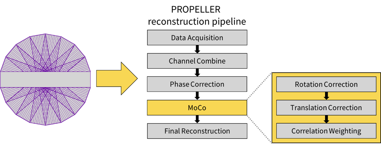
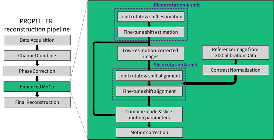
PROPELLER reconstruction with motion correction (MoCo) is illustrated in Figure 1. PROPELLER data is acquired blade-by-blade, then channel combination and phase correction are performed to generate single channel and phase aligned k-space. Conventional MoCo uses the overlapped center k-space to estimate rotation and translation then corrects the blade data. Blades with very low correlation in k-space will be discarded to avoid artifacts. Finally, data from all blades will be interpolated and combined to full resolution k-space to generate the final image. The proposed method is shown in Figure 2. Compared to conventional approach, our proposed motion correction uses the 3D calibration data to extract the corresponding reference slice image then use this reference slice to estimate translation and rotation for later motion correction.Data were acquired using PROPELLER FSE sequence at 3.0T scanners (GE HealthCare, Waukesha, WI). Brain data were acquired with FOV=22-24cm, acquisition matrix size=280x280, echo train length=32, TR=9850-10000ms. Knee data was acquired with FOV=16cm, acquisition matrix size=420x420, echo train length=27, TR=7546ms. For Image quality comparison, same raw datasets are used to reconstruct final images with conventional and proposed MoCo technique.
Results
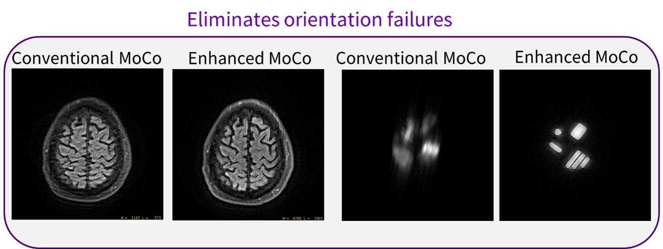
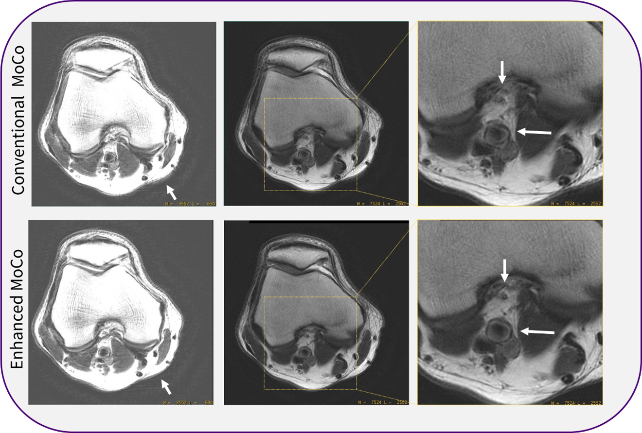
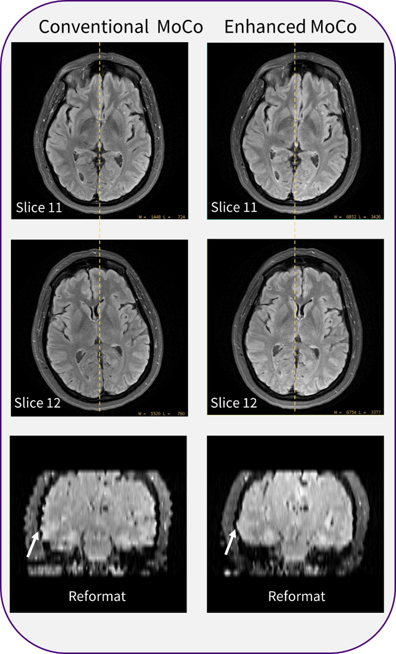
Figure 3 shows the conventional MoCo failed whereas the proposed MoCo removed the motion introduced blurring and streaking. Figrue4 shows a comparison on the knee data, the proposed MoCo approach shows less streaking artifacts and also preserves more detail structures, such as vessels and nerves. Figure5 shows the impact of MoCo on inter-slice alignment. Conventional MoCo doesn’t have a global reference for slice alignment, the reformatted images show discontinuity on slices. The proposed MoCo largely mitigates the misalignment as same fast calibration scan reference volume is used for motion estimation and correction for all slices. This will augment downstream volumetric processes, such as lesion segmentation and measurement and treatment planning.Conclusion
We propose a novel inter-blade and inter-slice motion correction reconstruction technique for PROPELLER MRI by using fast volumetric calibration scan. The results show large improvement brought by the proposed method compared to convention MoCo on both in-plane motion correction and inter-slice alignment.Acknowledgements
No acknowledgement found.References
- Forbes, Kirsten PN, et al. "PROPELLER MRI: clinical testing of a novel technique for quantification and compensation of head motion." Journal of Magnetic Resonance Imaging: An Official Journal of the International Society for Magnetic Resonance in Medicine 14.3 (2001): 215-222.
- Pipe, James G. "Motion correction with PROPELLER MRI: application to head motion and free‐breathing cardiac imaging." Magnetic Resonance in Medicine: An Official Journal of the International Society for Magnetic Resonance in Medicine 42.5 (1999): 963-969.
- Pipe, James G. "Improved in-plane motion correction for PROPELLER MRI." Proc Intl Soc Magn Reson Med. Vol. 9. 2001.
- Pipe, James G., et al. "Revised motion estimation algorithm for PROPELLER MRI." Magnetic resonance in medicine 72.2 (2014): 430-437.
Figures