4641
VAR-NAV: Motion-resolved 4D MRI using vision-assisted reference auto-navigation1Department of Medical Physics, Memorial Sloan Kettering Cancer Center, New York City, NY, United States, 2Department of Radiology, Memorial Sloan Kettering Cancer Center, New York City, NY, United States
Synopsis
Keywords: Motion Correction, Motion Correction
Motivation: Motion navigation with improved data efficiency and robustness is still needed for free-breathing MRI.
Goal(s): To develop VAR-NAV, a new 2D auto-navigation technique with 100% data efficiency for stack-of-stars acquisitions.
Approach: VAR-NAV directly estimates the position of the liver dome using 2D projection images and AM-FM demodulation. Results can be corrected retrospectively, like in soccer video assistant referee (VAR). VAR-NAV is used to sort continuously acquired data and reconstruct motion-resolved 4D images.
Results: VAR-NAV outperforms conventional 1D PCA-based navigation and does not require additional acquisition as in previous 2D navigation techniques, offering an efficient and accurate method for clinical 4D MRI.
Impact: VAR-NAV estimates a motion signal that represents displacements on 2D images, outperforming conventional PCA-based 1D navigation. No extra navigation data are required, and results can be retrospectively analyzed like in soccer video-assisted-referee (VAR). VAR-NAV promises to improve clinical 4D MRI.
INTRODUCTION
Motion-resolved 4D MRI can be used to perform free-breathing imaging and to guide radiotherapy of tumors affected by motion.1,2 The combination of radial acquisition with compressed sensing (XD-GRASP3) and, more recently, with deep learning (Movienet4,5) has enabled to perform 4D MRI with sufficient acquisition and reconstruction speed. A key component in 4D MRI is the navigation technique to sort acquired data into motion states. Most auto-navigation techniques for stack-of-stars acquisition use 1D navigators computed from the central k-space position and do not exploit the 2D structure of each projection. Moreover, they are based on principal component analysis (PCA), which is sensitive to the number of angles and only provides relative rather than absolute displacements. Live-view 4D GRASP6 recently introduced a 2D navigation scheme where a coronal acquisition was performed every two angles, reducing data efficiency to 2/3.This work presents a novel auto-navigation estimation method named Vision-Assisted Reference for auto-NAVigation (VAR-NAV) that does not need extra navigation data and uses 2D images obtained from specific angles from the stack-of-stars acquisition together with AM-FM processing to estimate absolute displacements. The performance of VAR-NAV is tested against the conventional navigation technique based on 1D projection and PCA for motion-resolved 4D MRI of abdominal tumors.
METHODS
Data acquisition: Five patients with abdominal tumors were scanned on different 3T scanners (Signa Premier, GE Healthcare) using a 3D T1-weighted golden-angle stack-of-stars acquisition (Cartesian kz and radial ky-kx) with the following parameters: TR=3-4ms, TE=1.5-2ms, flip angle = 12°, in-plane resolution = 1.25-1.5mm, slice thickness = 4-5mm, number of radial spokes = 1800, and scan time = 4.3-5 min.Quasi-coronal liver-aware image and keypoint estimation: The position of the liver dome was employed for navigation using 2D images from a set of angles that provide adequate liver visualization (Figure 1). The angles were in the range of [-22.5°, 45°] and [157.5°, 225°]. According to the angle, a horizontal image flip processing was applied to have the liver always at the same location. 1D Gaussian filter was applied to each image to enhance the visibility of the liver and smooth the most prominent region (Figure 2). The column with the maximum projected intensity was identified, and the keypoint was calculated using the liver dome location (Figure 1).
Motion signal estimation using AM-FM demodulation: Spline interpolation was used to estimate the locations at each non-used angle. These locations were compared with the images to correct them if needed, like in video-assisted-referee (VAR) for soccer games. A multiscale AM-FM demodulation7,8 was then used to extract the motion signal in the frequency range of interest.
Given r(t) as the interpolated signal, re(t) = a(t)cos(φ(t) was computed, where the instantaneous amplitude, a(t), was calculated using the normalized frequency scales in the range of [0, 1/32], and the instantaneous phase, φ(t), used the normalized frequency scales in the ranges of [1/16, 1/8] and [1/8, 1/4] (Figure 3).
4D reconstruction: Data were sorted accordingly into 10 motion states according to the amplitude of the VAR-NAV motion signal, and 4D reconstruction was performed using the compressed sensing reconstruction from XD-GRASP. For comparison purposes, 4D images were reconstructed with the conventional auto-navigation technique.
VAR-NAV evaluation: The motion in axial, coronal, and sagittal views was compared between images reconstructed with VAR-NAV and the conventional navigation technique using multi-scale SSIM.
RESULTS
Motion signal estimation using VAR-NAV is more stable than conventional PCA, especially when the number of spokes changes (Figure 3). In cases where conventional PCA navigation was successful, VAR-NAV presented similar results (Figure 4). However, in cases where there is no sufficient liver coverage, VAR-NAV outperforms conventional PCA navigation (Figure 5).DISCUSSION
VAR-NAV can estimate liver dome displacement directly from 2D images computed from the imaging data without requiring extra navigation data. Navigation is expected to perform similarly to Live-View GRASP but with 100% data efficiency. Unlike PCA-based methods, VAR-NAV performs a direct location estimation for each time point without a transformation, allowing for stable motion estimation regardless of the number of spokes. Moreover, patient motion can be analyzed since 2D images are available, like using VAR in soccer games. One limitation of VAR-NAV is increased computation time due to 2D analysis, which will be addressed by parallel computing on a GPU.CONCLUSION
This work introduced a new auto-navigation technique for stack-of-starts acquisition called VAR-NAV that does not require additional navigation data and estimates direct displacement from 2D projection images. VAR-NAV provides a more stable and accurate motion estimation than existing PCA-based methods, which can improve 4D MRI reconstruction in a clinical setting.Acknowledgements
The work was supported by NIH grant R01-CA255661.References
1. Otazo R, Lambin P, Pignol JP, et al. MRI-guided Radiation Therapy: An Emerging Paradigm in Adaptive Radiation Oncology. Radiology. 2021;298(2):248-260.2. Siddiq S, Murray V, Tyagi N, Borman P T S, Raaymakers B W, Wu C and Otazo R 2023 MR signature matching (MRSIGMA) for volumetric MRI with less than 300 ms latency on a 1.5T MR-Linac system Proc. Intl. Soc. Mag. Reson Med. 31 1023
3. Feng, Li, Leon Axel, Hersh Chandarana, Kai Tobias Block, Daniel K. Sodickson, and Ricardo Otazo. "XD‐GRASP: golden‐angle radial MRI with reconstruction of extra motion‐state dimensions using compressed sensing." Magnetic resonance in medicine 75, no. 2 (2016): 775-788.
4. Murray V, Siddiq S, Crane C, El Homsi M, Kim T, Wu C, Otazo R. Movienet: Deep space-time-coil reconstruction network without k-space data consistency for fast motion- resolved 4D MRI. Magn Reson Med. 2023;1-15. doi: 10.1002/mrm.29892.
5. Murray V, Siddiq S, Jafari R, Wu C and Otazo R 2023 MR-movienet: fast motion-resolved 4D MRI reconstruction exploiting space-time-coil correlations without k-space data consistency Proc. Intl. Soc. Mag. Reson Med. 31 5485.
6. Feng L. Live‐view 4D GRASP MRI: A framework for robust real‐time respiratory motion tracking with a sub‐second imaging latency. Magnetic Resonance in Medicine. 2023;90(3):1053-1068.
7. Agurto C, Murray V, Barriga E, et al. Multiscale AM-FM Methods for Diabetic Retinopathy Lesion Detection. IEEE Transactions on Medical Imaging. 2010;29(2):502-512.
8. Murray V, Rodriguez P, Pattichis MS. Multiscale AM-FM Demodulation and Image Reconstruction Methods With Improved Accuracy. IEEE Transactions on Image Processing. 2010;19(5):1138-1152.
Figures
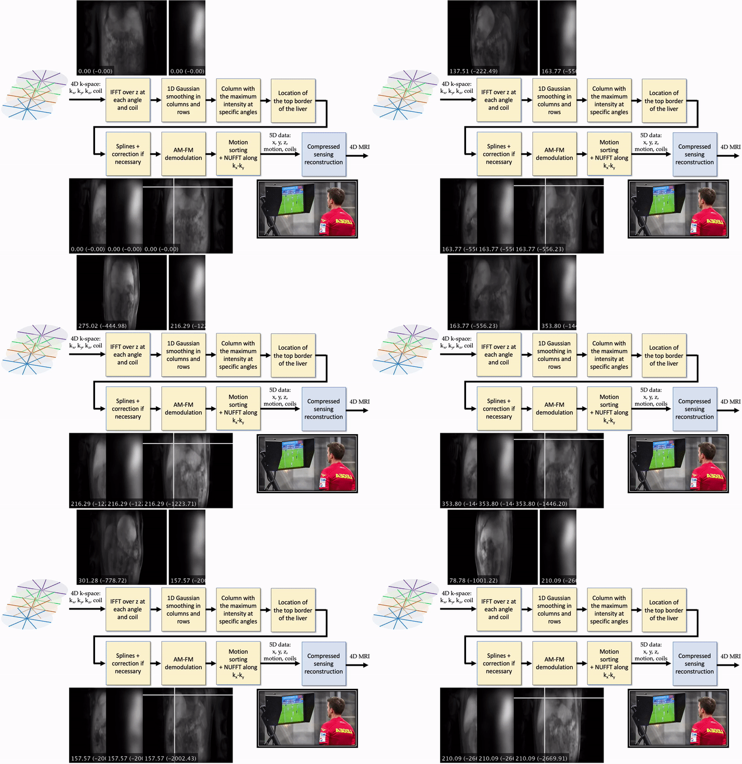
Figure 1. VAR-NAV estimates motion signal from stack-of-stars data without extra navigation data. The liver dome is located using a 1D analysis of rows and columns at specific angles. Splines and AM-FM demodulation process data to produce the motion signal. (Top) Images at every angle and the part of the image smoothed. (Bottom) Location estimated of the top border of the liver. The locations could be corrected if necessary using the vision-assisted reference (VAR). Angles are displayed as the normalized angle from 0° to 360° and the corresponding angle value from the scanner.
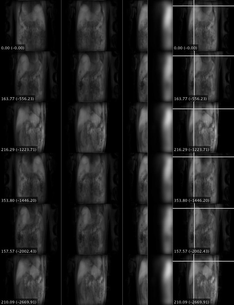
Figure 2. Selected angles to analyze the location of the liver dome. The format of the angles is similar to Figure 1. The original input image obtained at each selected angle using an IFFT through z is shown in the first column. The image is horizontally flipped when necessary to have the liver on the left side of the image (second column). Only half of the image (third column) is used for smoothness (fourth column) with separable 1D Gaussian filters. The final estimation is shown in the fifth column.
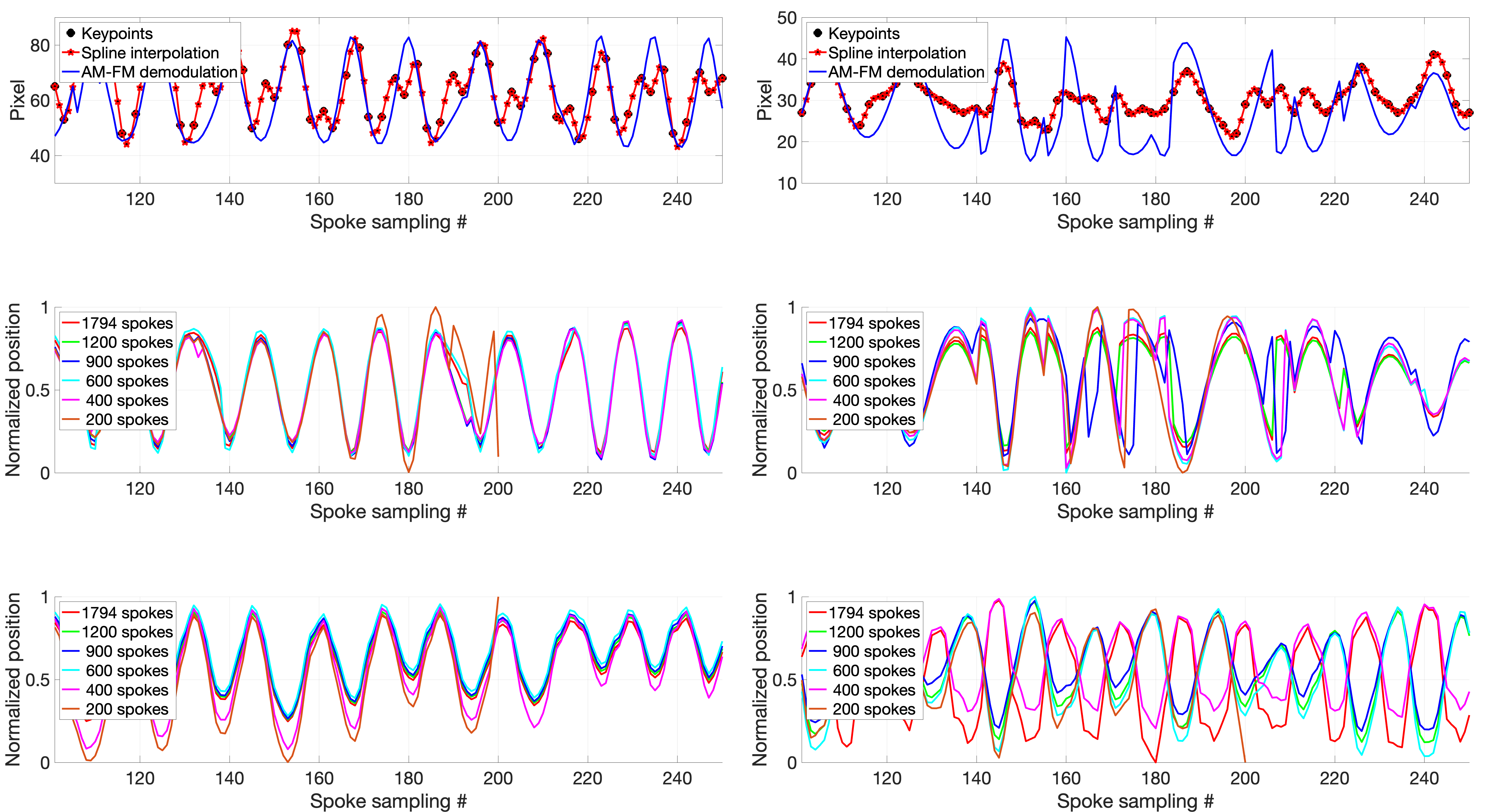
Figure 3. Estimated motion signals. Left: comparison between the motion estimations from a case where the conventional auto-navigation was successful (see Figure 4). Right: comparison between the motion estimations from a case where conventional navigation failed (see Figure 5). (A) Keypoints (marked with a black circle) are used as a reference for the interpolation using splines (marked in red). The AM-FM demodulation of the latter is shown in blue. (B) VAR-NAV estimation using a different number of spokes. (C) Conventional navigation using different numbers of spokes.
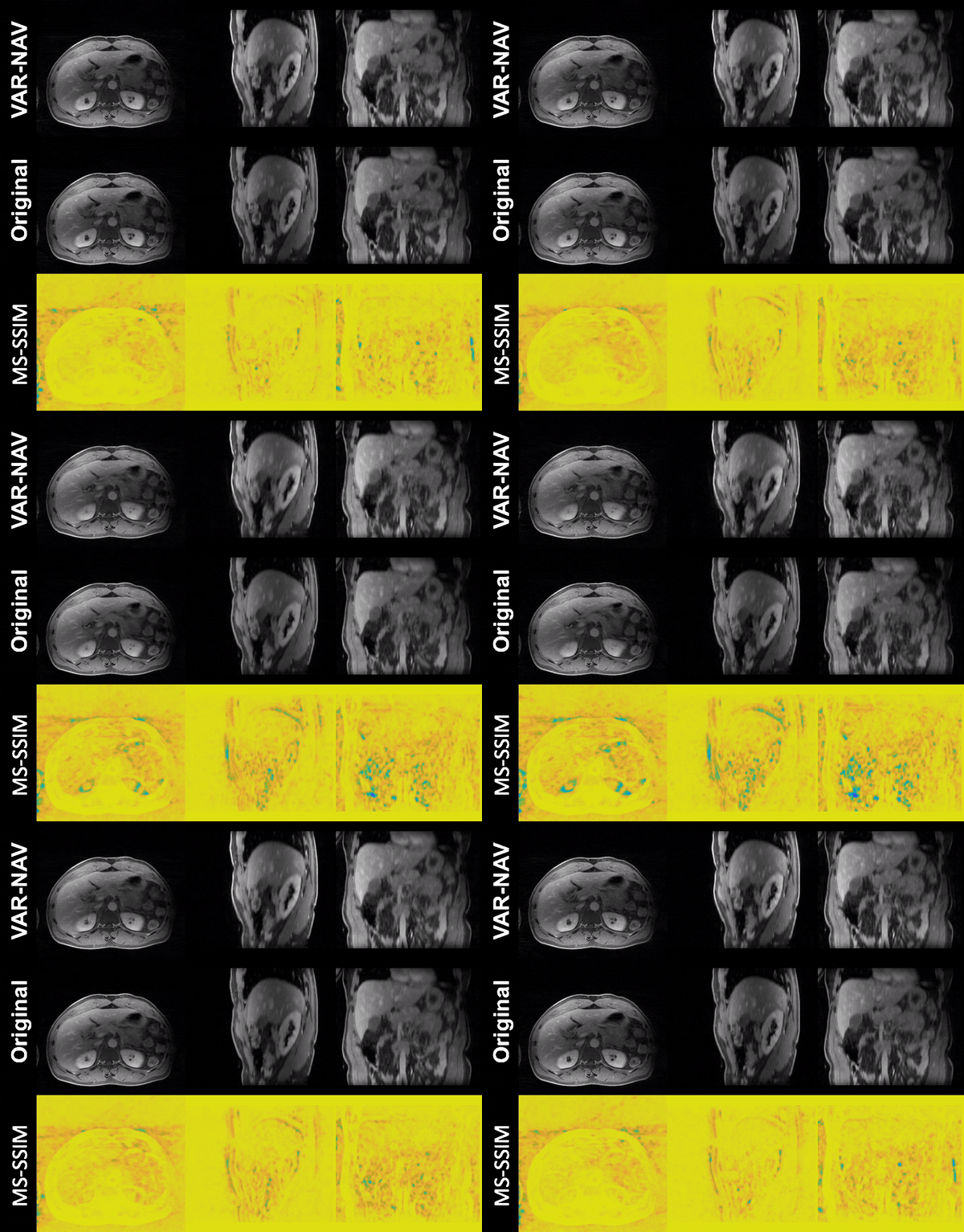
Figure 4. 4D MRI reconstruction using VAR-NAV (top) and conventional navigation (middle). In this case, both navigation methods presented similar results. The MS-SSIM (bottom) is used to analyze the differences described in 4.
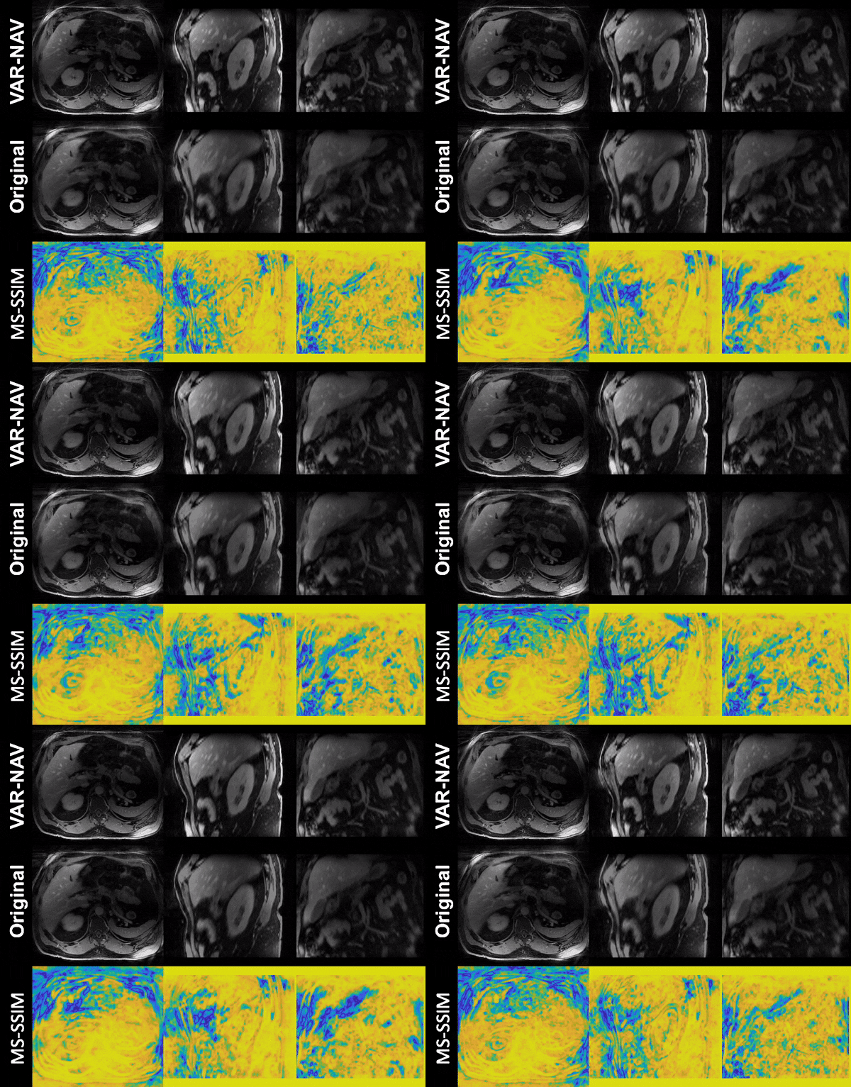
Figure 5. 4D MRI reconstruction using the new VAR-NAV method (top) and the conventional navigation (middle). In this case, the conventional navigation method could not estimate the motion due to reduced liver coverage. However, VAR-NAV successfully estimated the motion signal. The MS-SSIM (bottom) is used to analyze the differences described in 4.