4638
Diffusion and Perfusion Measurements of Uterine Corpus Fluctuate During Menstrual Cycle: an IVIM based study1The Second Affiliated Hospital of Xi'an Jiaotong University, Xi'an, China, 2Xian Yang Central Hospital, Xian Yang, China, 3GE HealthCare MR Research, Beijing, China
Synopsis
Keywords: Uterus, Perfusion
Motivation: Correct identification of the typical appearance of the distinct uterine corpus layers during the menstrual cycle is crucial for evaluating uterine lesions.
Goal(s): To investigate the diffusion and perfusion of uterine corpus fluctuate during menstrual cycle.
Approach: Thirty-one volunteers underwent standard pelvic IVIM-DWI in menstrual period, mid-proliferative period and mid-secretory period. The differences of D, D*, f and f ×D* among three-layer structure or different menstrual period were compared.
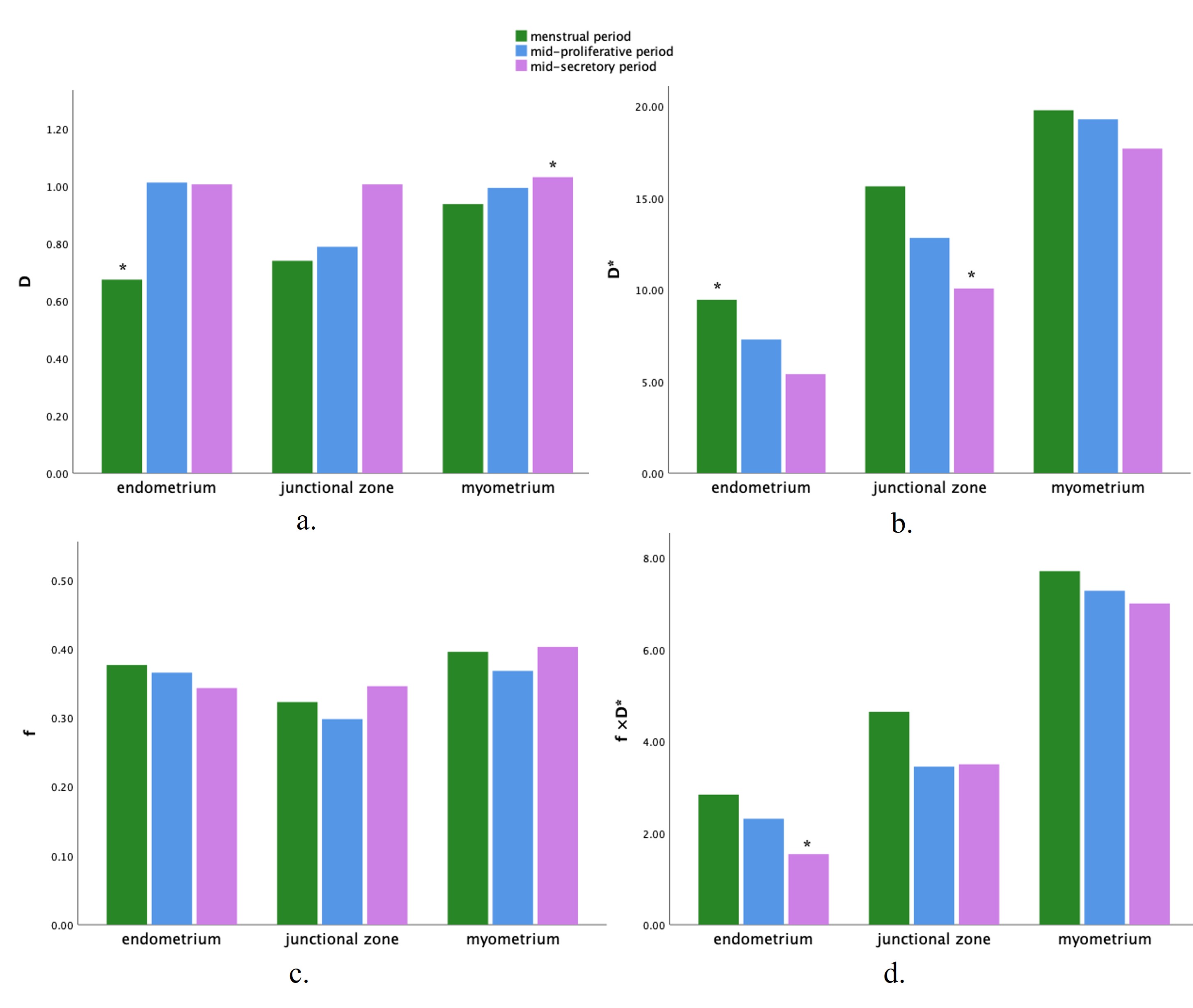
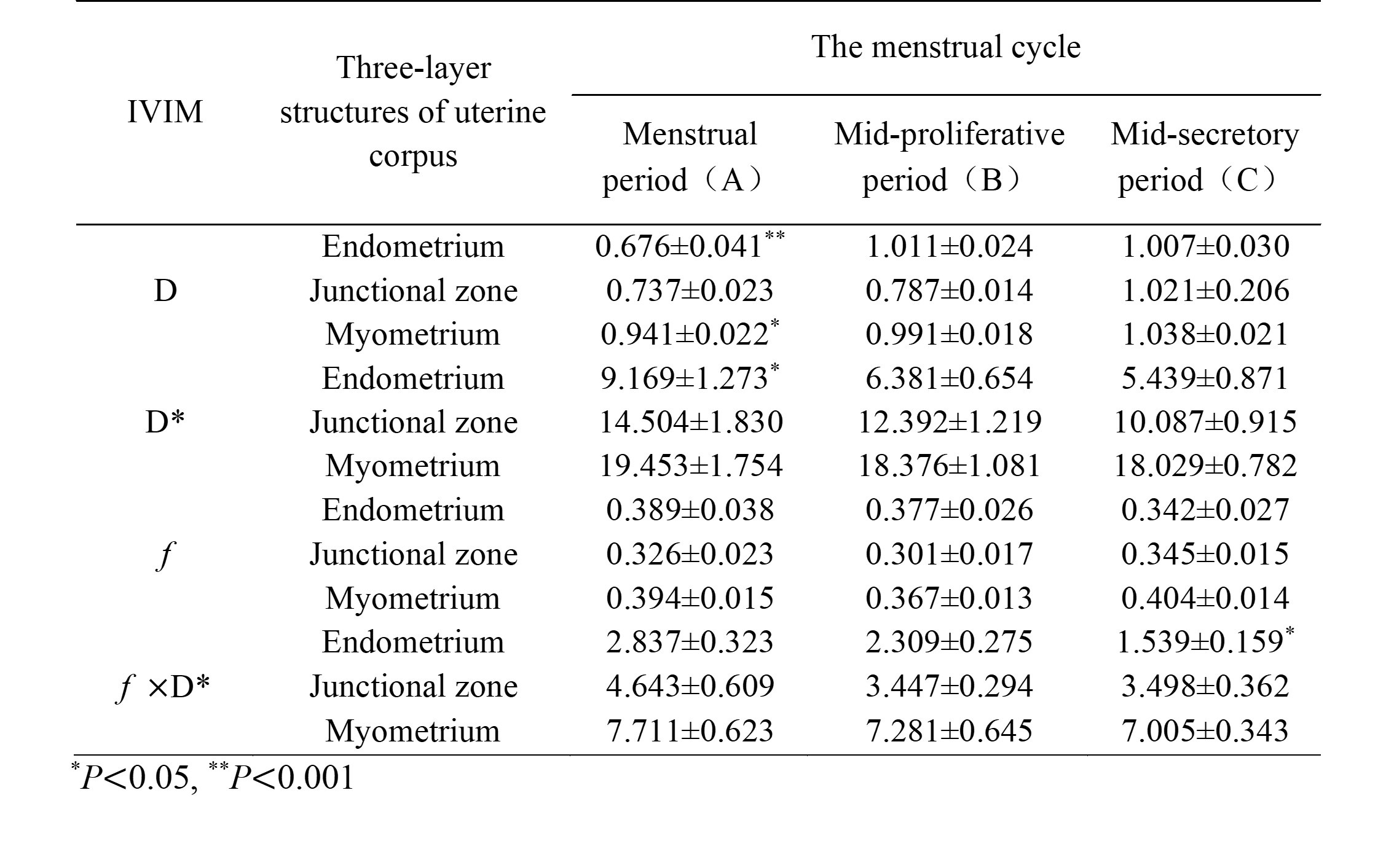
Results: It was found that an exacerbation of diffusion restriction, occurring from the menstrual period to the mid-secretory period, and perfusion driven by IVIM of uterine corpus was generally decreased, especially in endometrium.
Impact: Considering the menstrual status is necessary on assessment of uterine lesions. It is recommended to study uterine lesions within the same menstrual cycle.
Introduction
Uterine conditions are prevalent among women of all age groups. Magnetic Resonance Imaging (MRI) has emerged as the optimal modality for the assessment and management of gynecologic disorders [1,2]. Diffusion-Weighted Imaging (DWI), a quantitative tool in the realm of MRI, has demonstrated its value in characterizing and distinguishing lesions. Notably, some pathological conditions can exhibit a semblance of normality in uterine imaging, potentially leading to missed or incorrect diagnoses. Therefore, it becomes imperative to discern the typical appearance of the distinct uterine corpus layers during the menstrual cycle. Previously, Apparent Diffusion Coefficient (ADC) values derived from DWI showcased periodic fluctuations in the three uterine corpus layers during the menstrual cycle in pre-menopausal women [3-5]. However, it is important to note that ADC, which stems from the mono-exponential DWI model, is unable to disentangle pure molecular diffusion from micro-circulation perfusion. Differing from conventional DWI, Intravoxel Incoherent Motion (IVIM) leverages a biexponential model, enabling the separation of pure water diffusion (Tissue-Diffusion: D) from micro-circulation perfusion (Pseudo-Diffusion: D* and Perfusion-Fraction: f) within the tissue [6]. This study aims to independently investigate the alterations in diffusion and perfusion within the normal uterine corpus across the menstrual cycle using IVIM as a tool.Methods
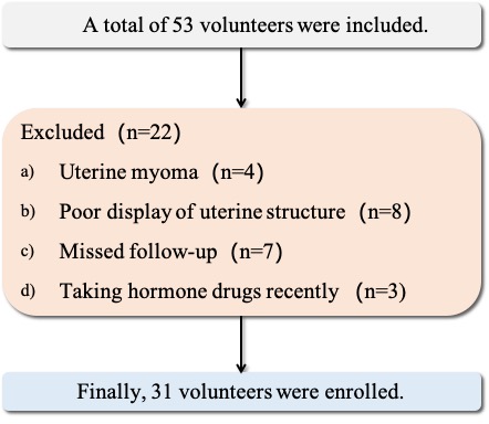
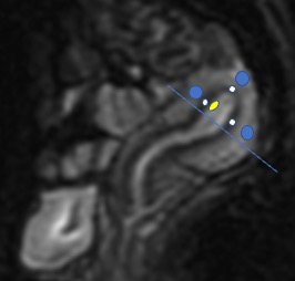
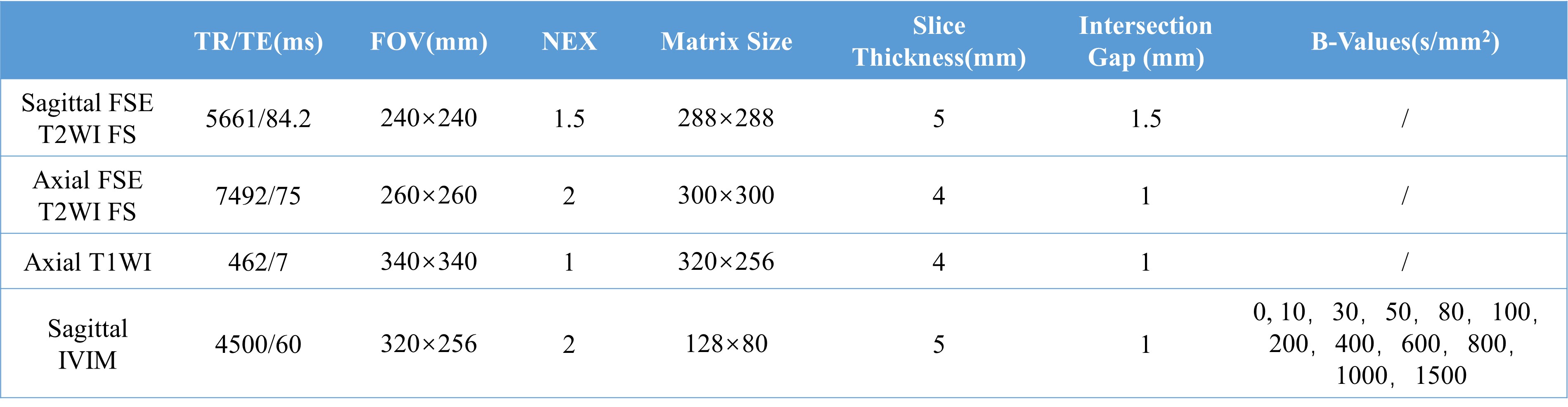
This study was approved by the institutional review board and the written informed consent was obtained from all subjects. From April 2022 to December 2022, thirty-one healthy volunteers (mean age, 24.2±0.49 years; range, 23-49 years) underwent pelvic MRI in the menstrual period ( the 2nd to 4th day of menstrual cycle), mid-proliferative period (the 10th day of menstrual cycle) and mid-secretory period (the 20th day of menstrual cycle). Detailed information about the inclusion criteria were shown in Figure 1. All the volunteers had regular menstrual cycles (mean, 29.2 days; range, 20-31 days). MR imaging was conducted on a 3.0T MR systems (Architect, GE Healthcare) with an 32-channel body coil. The standard pelvic MR protocols and IVIM-DWI were applied. The scan parameters were shown in Table 1. Two radiologists with 8 and 10 years of experience in gynecologic radiology reviewed the T2 and IVIM images. Using midsagittal-section of the b0 image, elliptical ROIs were then manually drawn on endometrium, junctional zone and myometrium of uterus, respectively (Figure 2). The differences of D, D*, f and f ×D* among three-layer structure or different menstrual period were compared using Student’s t-test or Kruskal-Wallis test as appropriate. A two-tailed P-value of < 0.05 was considered statistically significant.Results
In the context of the endometrium, D exhibited a consistent upward trajectory across the menstrual cycle. Conversely, D*, and f, showcased a gradual descent over this timeframe. As for the junctional zone and the myometrial region, D experienced a progressive augmentation throughout the menstrual cycle, while D* exhibited a corresponding reduction. Notably, the parameter f demonstrated an initial decline followed by an elevation, observed in both the junctional zone and myometrium, spanning from the menstrual phase to the mid-secretory phase. Furthermore, f×D*, for both the endometrium and myometrium, displayed a steady decrease throughout the menstrual cycle, as detailed in Table 2 and Figure 3.Acknowledgements
No acknowledgement found.References
1. Taylor, H. S., Kotlyar, A. M. & Flores, V. A. Endometriosis is a chronic systemic disease: clinical challenges and novel innovations. Lancet397, 839-852, doi:10.1016/s0140-6736(21)00389-5 (2021).
2. Nougaret, S., Cunha, T. M., Benadla, N., Neron, M. & Robbins, J. B. Benign Uterine Disease: The Added Role of Imaging. Obstet Gynecol Clin North Am 48, 193-214, doi:10.1016/j.ogc.2020.12.002 (2021).
3. Kara Bozkurt, D., Bozkurt, M., Nazli, M. A., Mutlu, I. N. & Kilickesmez, O. Diffusion-weighted and diffusion-tensor imaging of normal and diseased uterus. World J Radiol 7, 149-156, doi:10.4329/wjr.v7.i7.149 (2015).
4. Kuang, F., Chen, Z., Zhong, Q., Fu, L. & Ma, M. Apparent diffusion coefficients of normal uterus in premenopausal women with 3 T MRI. Clin Radiol 68, 455-460, doi:10.1016/j.crad.2012.09.011 (2013).
5. Tsili, A. C. et al. Apparent diffusion coefficient values of the normal uterus: Interindividual variations during menstrual cycle. Eur J Radiol 81, 1951-1956, doi:10.1016/j.ejrad.2011.04.057 (2012).
6. Iima, M. Perfusion-driven Intravoxel Incoherent Motion (IVIM) MRI in Oncology: Applications, Challenges, and Future Trends. Magn Reson Med Sci 20, 125-138, doi:10.2463/mrms.rev.2019-0124 (2021).
7. Li, X. et al. Field-of-view optimized and constrained undistorted single-shot study of intravoxel incoherent motion and diffusion-weighted imaging of the uterus during the menstrual cycle: a prospective study. Diagn Interv Radiol 29, 656-663, doi:10.4274/dir.2023.232188 (2023).
8. Liubing, C. et al. Magnetic resonance imaging thicknesses and apparent diffusion coefficient values of the endometrium and junction zone in women of reproductive age. Acta Radiol 64, 1263-1271, doi:10.1177/02841851221117559 (2023).
9. Joo, I., Lee, J. M., Han, J. K. & Choi, B. I. Intravoxel incoherent motion diffusion-weighted MR imaging for monitoring the therapeutic efficacy of the vascular disrupting agent CKD-516 in rabbit VX2 liver tumors. Radiology 272, 417-426, doi:10.1148/radiol.14131165 (2014).
Figures