4637
Accelerated Abdominal 3D T1rho Mapping using Diamond Radial Sampling1Department of Medical Physics, Memorial Sloan Kettering Cancer Center, New York, NY, United States, 2Department of Radiology, Albert Einstein College of Medicine and Montefiore Medical Center, Bronx, NY, United States, 3MR Clinical Science, Philips Healthcare, Cambridge, MA, United States, 4Department of Radiology, Memorial Sloan Kettering Cancer Center, New York, NY, United States
Synopsis
Keywords: Liver, Quantitative Imaging, T1rho, Abdomen, Radial, Free-breathing, Motion
Motivation: Current T1rho mapping of the abdomen is performed using breath-hold, respiratory triggering, or stack-of-stars acquisitions, which have limited spatial resolution and coverage or require a long scan time.
Goal(s): To develop accelerated free-breathing 3D T1rho mapping technique for the abdomen using efficient diamond sampling.
Approach: Free-breathing 3D T1rho imaging was performed on six volunteers using the MAPSS sequence. T1rho values were compared between stack-of-stars and diamond sampling.
Results: The scan time was reduced from 5:24min per TSL for stack-of-stars to 2:26min per TSL for diamond sampling. The T1rho values obtained with both methods were comparable.
Impact: The proposed free-breathing 3D T1rho mapping of the abdomen with accelerated diamond sampling has the potential to provide a quantitative assessment of abdominal lesions for improved diagnosis and treatment response evaluation.
INTRODUCTION
T1rho relaxation is known for its sensitivity to low-frequency water-macromolecular interactions1, which can be helpful for detecting and monitoring abdomen lesions through quantitative imaging. Previous reports have highlighted the potential of quantitative T1rho imaging for the liver and pancreas2-7. However, abdominal T1rho imaging faces challenges from respiratory motion, field inhomogeneities on a large field-of-view (FOV), and the absence of a specialized transmitter and receiver coil. Previous studies mainly relied on respiratory-triggered acquisition, resulting in limited spatial resolution and volumetric coverage. T1rho imaging using a 3D radial stack-of-stars sampling (SoS) with golden-angle acquisitions has shown advantages to mitigate motion artifacts as compared to Cartesian sampling8, at the expense of long scan times.This study compares the performance of accelerated radial diamond sampling against stack-of-stars for free-breathing T1rho imaging using a state-of-the-art magnetization-prepared (MP) angle-modulated partitioned k-space spoiled gradient echo snapshots (MAPSS) sequence1,9.
METHODS
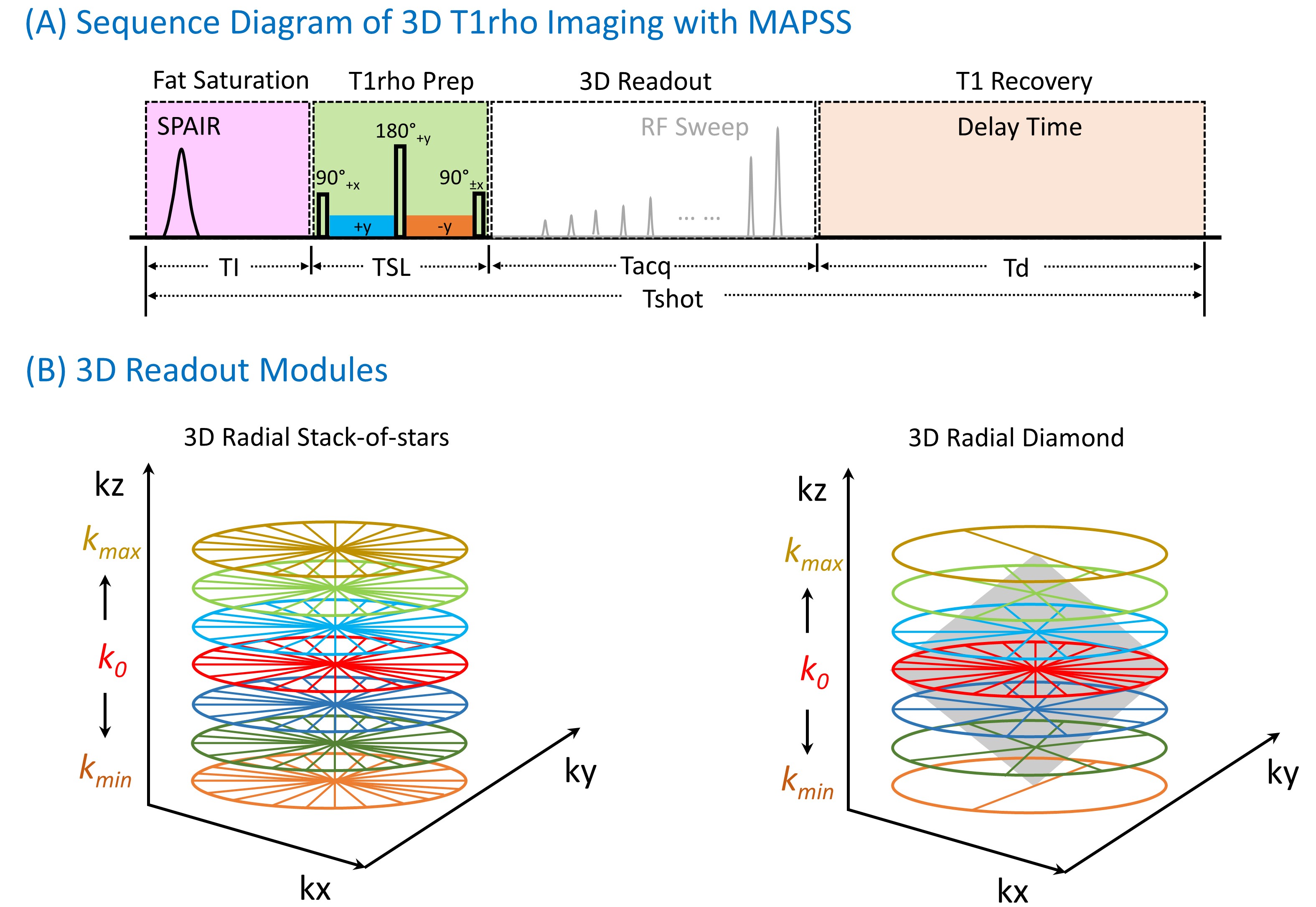
The 3D MAPSS T1rho sequence9 includes four modules: fat suppression (SPAIR), composite spin-locking T1rho MP10, 3D readout, and T1 recovery, as shown in Figure 1A. Two 3D readout modules (Figure 1B) were compared: a) SoS with full sampling in the kz direction, whereas b) diamond sampling with under-sampling in the kz direction, where the sampling density decreases from the center (k0) to peripheral k-space (kmax, kmin) forming a diamond shape11. Variable flip angle and phase cycling with positive (M+) and negative (M-) magnetizations are used for 3D MAPSS T1rho imaging9.Six volunteers were scanned for free-breathing abdominal 3D T1rho-imaging using a 3T-MRI scanner (Ingenia Elition X, Philips Healthcare) with anterior and posterior coils. The scan parameters were the same for both SoS and diamond acquisitions: FOV=360×360×200mm3, voxel size=2.0×2.0×4.0mm3, TR/TE =6.6/2.8ms, bandwidth=382 Hz/pixel, shot duration=1800ms, and delay time=1200ms. Further, spin-locking frequency (FSL)=300 Hz, and spin-locking times (TSLs)=±0, ±30, ±40ms were used for T1rho imaging.
The 3D volumes from all TSLs were motion-corrected using Elastix12. T1rho maps were generated using mono-exponential curve-fitting of the complex-valued data9 for conventional MAPSS (6 TSLs) and fast MAPSS (3 TSLs)9. Interleaved phase cycling (e.g., +0, -30, +40ms) was used for the fast MAPSS T1rho mapping9. Five regions of interest (ROIs), including liver, pancreas, spleen, and bilateral posterior muscles, were manually drawn on a selected slice to evaluate the T1rho values. The difference between diamond and SoS radial T1rho-mapping was computed for the ROIs. Besides, the Pearson correlation coefficients were evaluated for diamond versus SoS and MAPSS versus fast MAPSS.
RESULTS
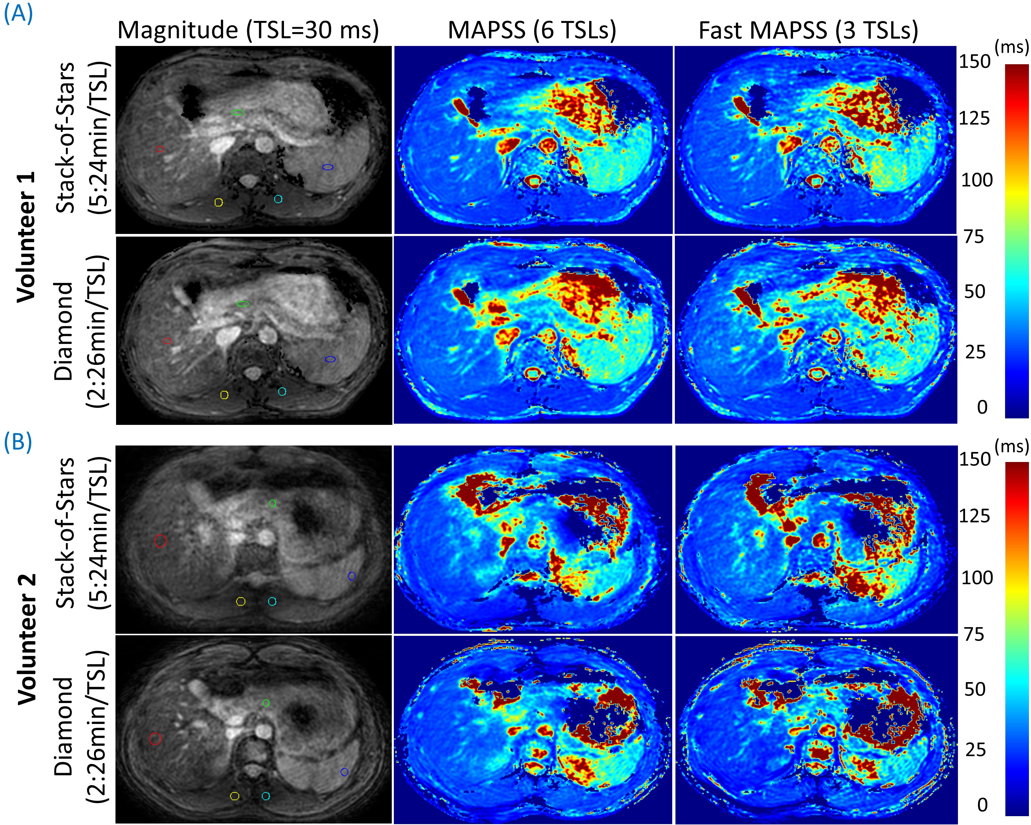
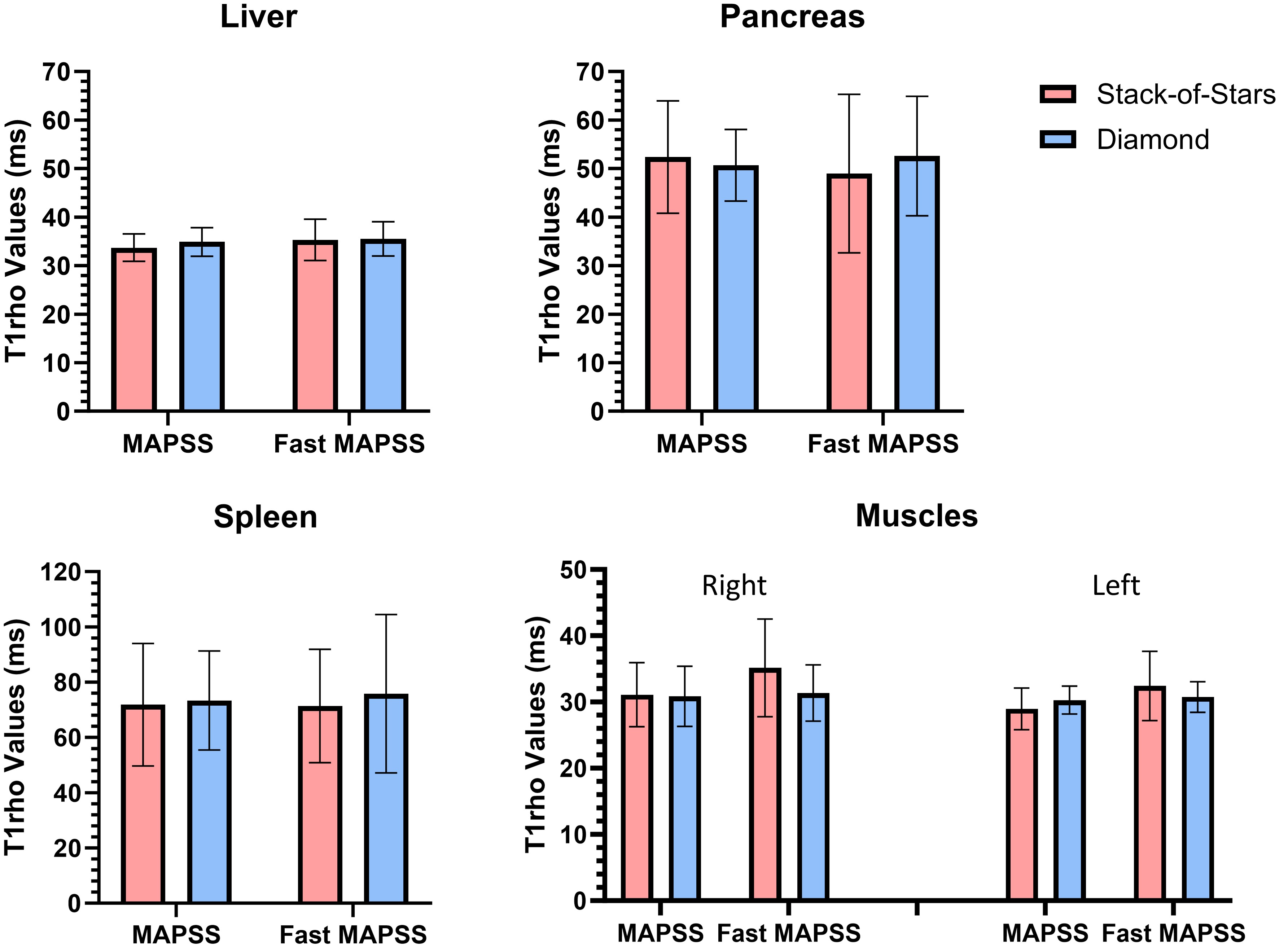
The scan time for SoS and diamond sampling in 3D MAPSS T1rho-imaging was 5:24min per TSL and 2:26min per TSL, respectively. When fast MAPSS T1rho imaging with 3 TSLs was used, the total scan time decreased from 16:12min for SoS to 7:17min for diamond sampling. Figure 2 presents the quantitative T1rho maps from two of the volunteers. It is noticed that the vessel structures of the liver appear less sharp using SoS compared to diamond sampling.There is no statistical difference in the T1rho measurements between SoS and diamond sampling, or between MAPSS with 6TSLs and fast MAPSS with 3TSLs. Figure 3 compares the T1rho values in the liver, pancreas, spleen and muscles. In 3D MAPSS T1rho imaging, the difference in T1rho-values between SoS and diamond sampling was <5% for the liver, pancreas, and spleen. In 3D fast MAPSS T1rho imaging, the difference was <5% for the liver, and <8% for the pancreas and spleen.
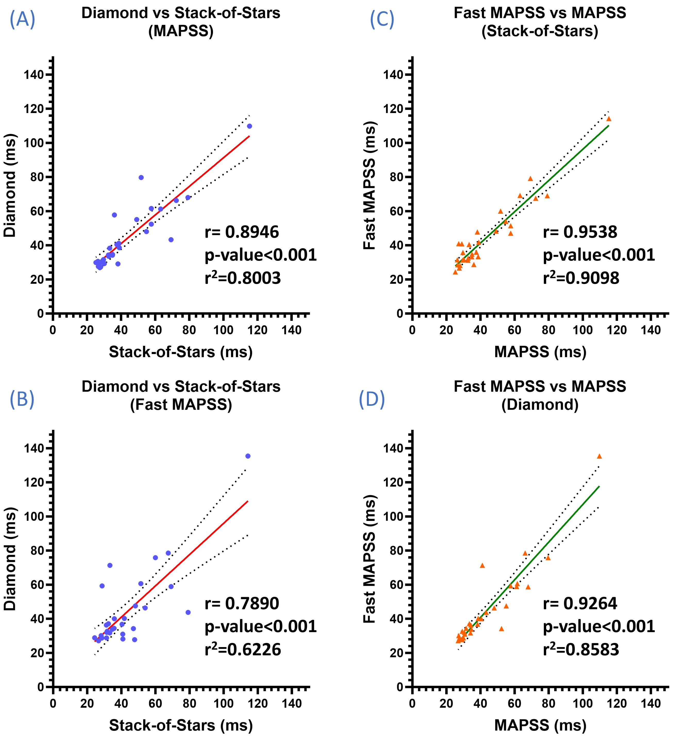
Furthermore, the Pearson correlations between the T1rho measurements were found to be statistically significant for both SoS and diamond sampling, as well as for MAPSS and fast MAPSS (Figure 4).
DISCUSSION
Diamond sampling reduced the acquisition time for 3D T1rho imaging by 2.2-fold, improved image quality, and preserved quantitative T1rho values compared to SoS. The higher standard deviation observed in SoS (Figure 3) may be attributed to increased motion during scanning due to the longer acquisition.CONCLUSION
The study demonstrated that radial diamond sampling efficiently accelerated data acquisition and enabled free-breathing 3D T1rho mapping of the abdomen in under eight minutes. This technique has the potential to provide a quantitative assessment of abdominal lesions.Acknowledgements
The work was supported by NIH Grant R01-AR076328.References
- Li X, Han ET, Busse RF, et al. In vivo T1ρ mapping in cartilage using 3D magnetization‐prepared angle‐modulated partitioned k‐space spoiled gradient echo snapshots (3D MAPSS). Magn Reson Med 2008;59(2):298-307.
- Hou J, Wong VW, Qian Y, Jiang B, Chan AW, Leung HH, Wong GL, Yu SC, Chu WC, Chen W. Detecting Early-Stage Liver Fibrosis Using Macromolecular Proton Fraction Mapping Based on Spin-Lock MRI: Preliminary Observations. J Magn Reson Imaging. 2023;57(2):485-492
- Xie S, Li Q, Cheng Y, Zhang Y, Zhuo Z, Zhao G, Shen W. Impact of Liver Fibrosis and Fatty Liver on T1rho Measurements: A Prospective Study. Korean J Radiol. 2017;18(6):898-905.
- Chen W, Chan Q, Wang Y. Breath-hold black blood quantitative T1rho imaging of liver using single shot fast spin echo acquisition. Quant Imaging Med Surg 2016; 6(2): 168-177.
- Wang Y, Deng M, Lin J, Kwok, Liu E, Chen W. Age- and gender-associated liver physiological T1rho dynamics demonstrated with a clinically applicable single-breathold acquisition. SLAS Technol 2018; 23(2): 179-187.
- Wang Y, Deng M, Lo GG, Liang D, Yuan J, Chen W. Breath-hold black-blood T1rho mapping improves liver T1rho quantification in healthy volunteers. Acta Radiol 2018; 59(3): 257-265.
- Sun S, Zhou N, Feng Y, Lv Y, He J, Liu S, Chen W, Kong W, Zhou Z. Evaluation of Chronic Pancreatitis With T1 ρ MRI: A Preliminary Study. J Magn Reson Imaging. 2021 ;53(2):577-584.
- Sharafi A, Baboli R, Zibetti M, Shanbhogue K, Olsen S, Block T, Chandarana H, Regatte R. Volumetric multicomponent T1ρ relaxation mapping of the human liver under free breathing at 3T. Magn Reson Med. 2020 ;83(6):2042-2050.
- Peng Q, Wu C, Kim J, et al. Efficient phase‐cycling strategy for high‐resolution 3D gradient‐echo quantitative parameter mapping. NMR in Biomedicine. 2022 ;35(7):e4700.
- Witschey II WR, Borthakur A, Elliott MA, et al. Artifacts in T1ρ-weighted imaging: Compensation for B1 and B0 field imperfections. J Magn Reson 2007; 186(1); 75-85.
- Can Wu, Qi Peng, Ramin Jafari, et al. Fast Free-Breathing 3D T1ρ Abdominal Imaging Using an Efficient Diamond Radial Sampling Strategy at 3T. Proc. Intl. Soc. Mag. Reson. Med. 2023; 1848
- Klein S, Staring M, Murphy K, et al. Elastix: a toolbox for intensity-based medical image registration. IEEE Trans Med Imaging. 2009 ;29(1):196-205. (https://elastix.lumc.nl/)
Figures