4635
Multiparametric MRI based assessment of kidney injury in a mouse model of ischemia reperfusion injury1Symbiosis Centre for Medical Image Analysis, Symbiosis International University, Pune, India, 2Institute for Advancing Intelligence, TCG Crest, Kolkata, India, 3Molecular & Clinical Cancer Medicine, University of Liverpool, Liverpool, United Kingdom, 4University of Liverpool, Liverpool, United Kingdom, 5Department of Women’s and Children’s health, Institute of Systems, Molecular and Integrative Biology, University of Liverpool, University of Liverpool, Liverpool, United Kingdom, 6Department of Women’s and Children’s health, University of Liverpool, Liverpool, United Kingdom, 7Mathematical Sciences, University of Liverpool, Liverpool, United Kingdom
Synopsis
Keywords: Kidney, Kidney, Multiparametric MRI, IRI
Motivation: Assessment of kidney health is paramount for early diagnosis of impairment and subsequent methods of intervention.
Goal(s): This study enhances our understanding of kidney pathophysiology using multiparametric MRI analysis
Approach: Parsimonious pharmacokinetic modelling of the DCE-MRI data along with ASL and DWI was used to assess longitudinal changes in glomerular filtration rate (GFR) and kidney function in a mouse model of ischemia reperfusion injury.
Results: Findings suggest that Renal Blood Flow (RBF), parsimonious filtration quotient (Ft), GFR, kidney volume, and Apparent Diffusion Coefficient (ADC) are important in comprehending the renal effects induced by ischemia reperfusion injury.
Impact: This study provides an improved understanding of renal pathophysiology and underscores the value of multi-parametric MR imaging for assessing early changes in kidney function due to IRI, which may aid in early diagnosis of kidney injury and monitoring treatment response.
Authorship Information
* Denotes equal contributionIntroduction
Accurate assessment of glomerular filtration rate (GFR) is crucial for timely diagnosis of renal disorder1. Conventional techniques used for measuring GFR suffer from limited adaptability and reliability in the assessment of renal 2 This study utilizes Dynamic Contrast-Enhanced MRI (DCE-MRI), Arterial Spin Labelling (ASL) and Intravoxel Incoherent Motion Diffusion-Weighted Imaging (IVIM-DWI) along with different pharmacokinetic modelling approaches for multiparametric analysis to identify the most reliable indicators of longitudinal changes in a mouse model of ischemia reperfusion injury (IRI).Methods
Nine C57BL/6 mice (8-10 weeks old, 25-30 g) were used for ischemia reperfusion injury (IRI). Unilateral IRI was induced by clamping the right renal pedicle for 40 minutes. Longitudinal imaging studies were performed at baseline, one day post-IRI surgery, and 15 days post-IRI surgery. A 9.4 T scanner with an 86-mm birdcage transmit coil and a 4-channel receive array coil was used. DCE-MRI was performed using a multi-gradient-echo (MGE) sequence. DCE-MRI data were analysed using renal-specific pharmacokinetic (PK) models like the Patlak3, 2 Compartment Filtration (2CFM)4 and modified 2 Compartment Filtration with outflow5 models and a population-based arterial input function. The Akaike information criterion6 based parsimonious model selection was used to estimate filtration coefficient (Ft) and consequently measure GFR by multiplying it with kidney4 Renal blood flow (RBF) was quantified with a FAIR-RARE based ASL sequence, and IVIM-DWI was performed using a spin-echo echo planar imaging (EPI) sequence with 13 b values. Data analysis was performed in MATLAB. Statistical analysis was done using Origin Pro. Univariate analysis was done using two-way ANOVA with Bonferroni correction at p < 0.05. For multivariate analysis to distinguish between injured and contralateral kidney, Principal Component Analysis (PCA) was done with data from the baseline classified as healthy, day 1 and day 15 data from the IRI kidney classified as injured.Results
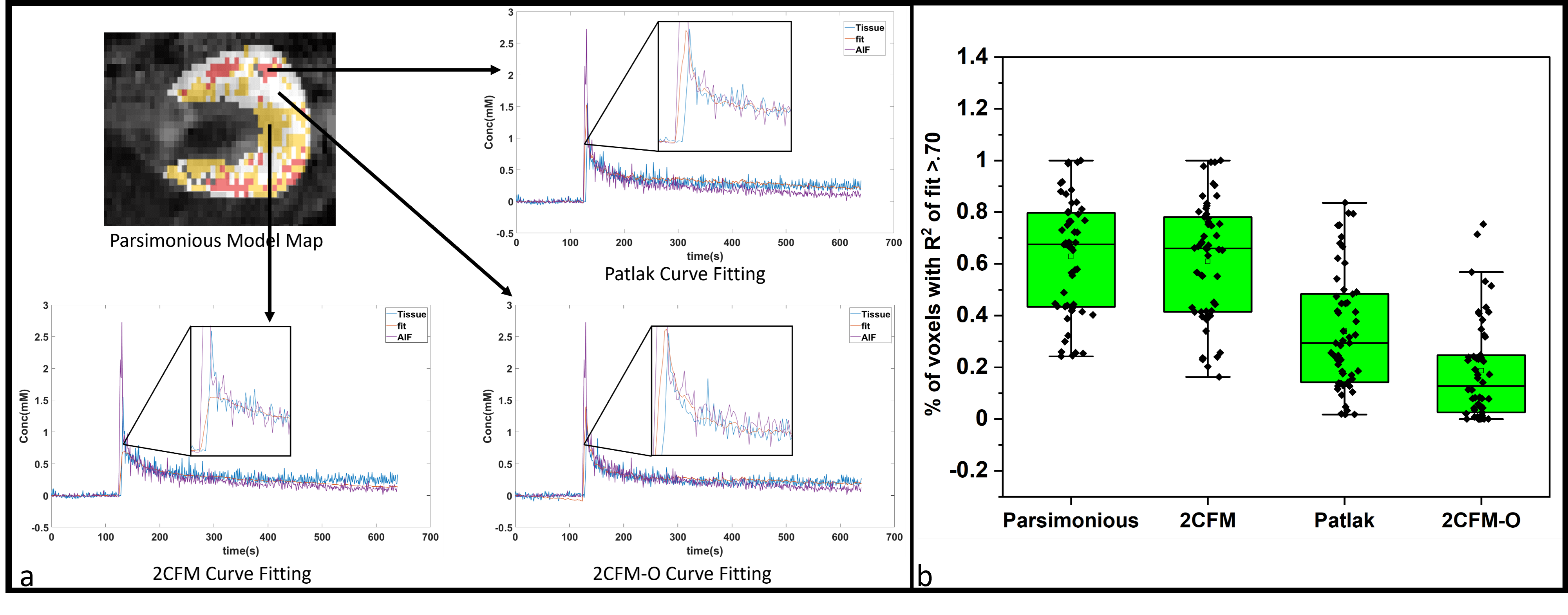
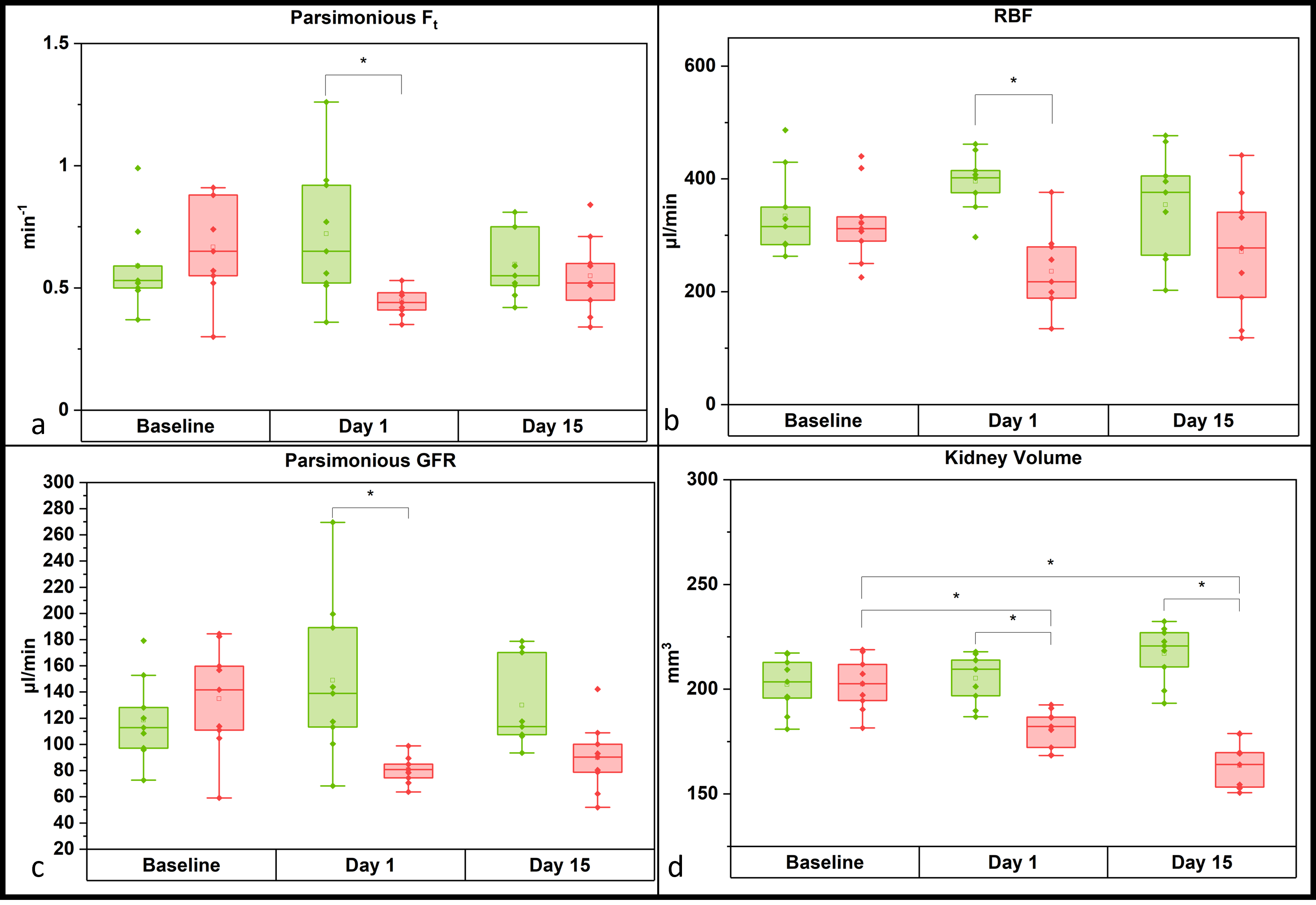
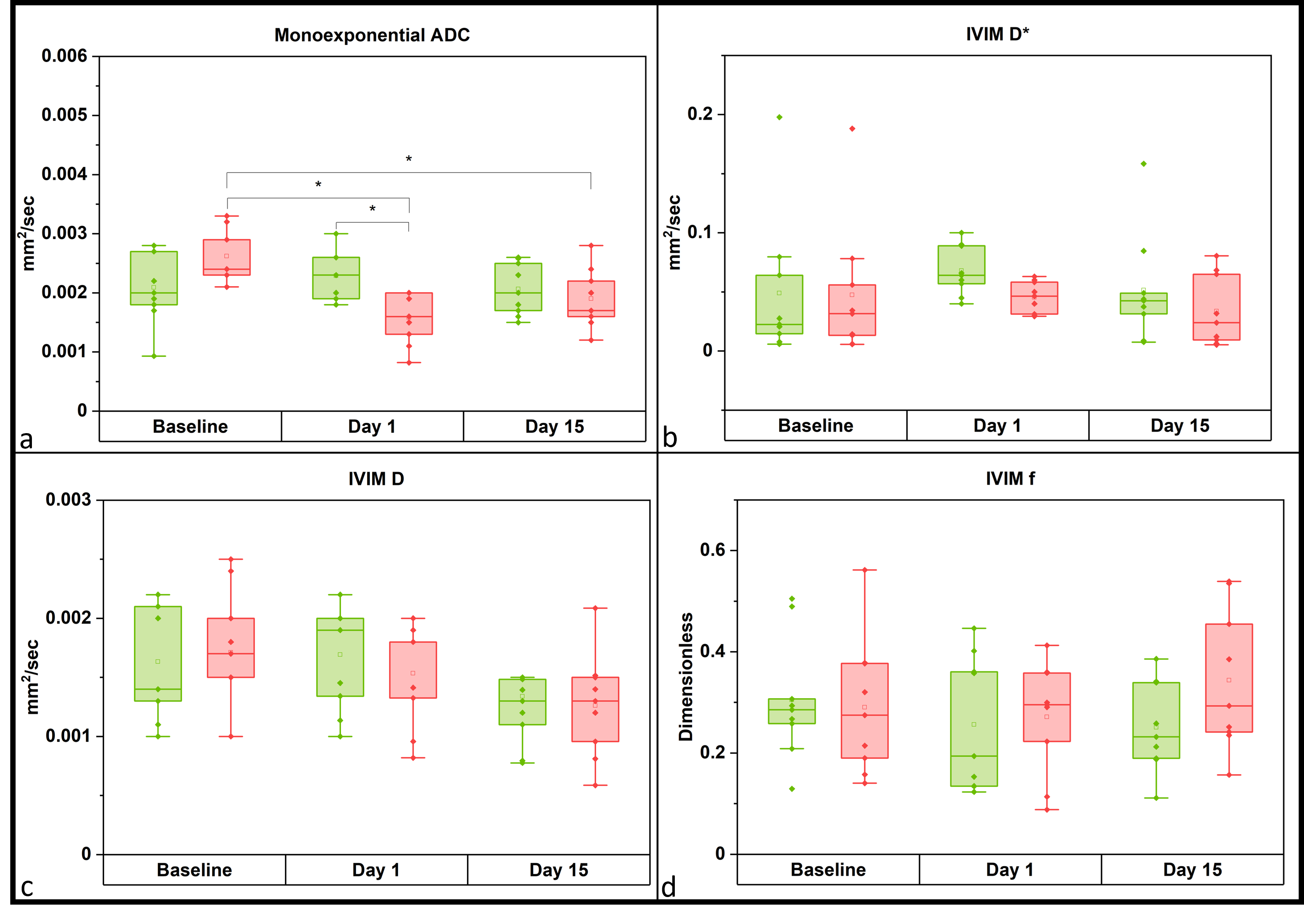
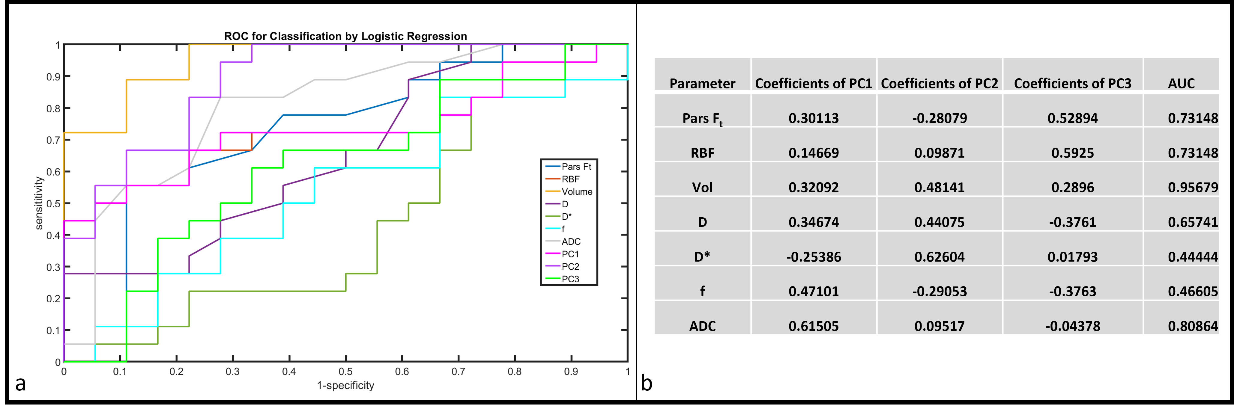
Figure 1 shows the efficacy of the parsimonious PK model with regard to goodness of fit and is compared against individual models. Following IRI surgery, a significant reduction in Ft (p=0.038) and GFR (p=0.005) (from DCE-MRI using parsimonious PK model), RBF (p=0.002), kidney volume (p=0.001) and apparent diffusion coefficient (p=0.048) (ADC, measured through mono-exponential modeling with the higher b values of DWI data) was observed in the injured kidney compared to the contralateral kidney on day 1 post-surgery as shown in Figures 2a-d and 3a. Kidney volume also exhibited significant differences between contralateral and injured kidney on day 15 (p<0.005), as well as a significant reduction of injured kidney volume on day 1 (p=0.006) and day 15 (p<0.005) from baseline. ADC also exhibited a significant difference in ADC on day 1 (p<0.005) and day 15 (p= 0.041) from baseline. Other DWI-based parameters derived from IVIM-DWI data did not yield significant results (Fig 3b-d). Using univariate analysis, figure 4a. shows the receiver operating characteristic (ROC) and corresponding area under the curve (AUC) values (Figure 4b.). Parameters that showed a statistically significant difference in two-way ANOVA between the injured and contralateral kidney provided AUC > 0.7. Multivariate logistic regression was also performed along with Principal Component Analysis (PCA) and coefficients from the three principal components are shown in Figure 4b. Principal Component 2 was identified as the most discriminative component, with an AUC of 0.89.Discussion
The parsimonious PK model-based GFR estimation correlated with transcutaneous GFR measurement, showing promise for non-invasive renal injury assessment and detecting significant differences between the injured and contralateral kidney post-IRI surgery along with a few other parameters that align with previous research7, 8. GFR, RBF, and Ft showed no significant difference on day 15. This may be due to variability in how individual kidneys normalize - some injured kidneys recover while others progress to acute kidney injury. The IVIM-DWI results such as true diffusion (D) and perfusion fraction (f) are at variance with earlier studies9 which might be due to differing injury and imaging timelines. Multivariate analysis with PCA demonstrates the potential for combining parameters to detect renal pathology.Conclusion
These studies highlight the potential of multiparametric MRI approaches for improving diagnostic accuracy in kidney health.Acknowledgements
This project has received funding from the European Union’s Horizon2020 research and innovation programme under the Marie Skłodowska-Curie grant agreement No 813839.References
1. Soveri, I., et al., Measuring GFR: a systematic review. American Journal of Kidney Diseases, 2014. 64(3): p. 411-424.
2. Granerus, G. and M. Aurell, Reference values for 51Cr-EDTA clearance as a measure of glomerular filtration rate. Scandinavian journal of clinical and laboratory investigation, 1981. 41(6): p. 611-616.
3. Sourbron, S.P., et al., MRI-measurement of perfusion and glomerular filtration in the human kidney with a separable compartment model. Investigative radiology, 2008. 43(1): p. 40-48.
4. Annet, L., et al., Glomerular filtration rate: assessment with dynamic contrast-enhanced MRI and a cortical-compartment model in the rabbit kidney. J Magn Reson Imaging, 2004. 20(5): p. 843-9.
5. Jiang, K., et al., Measurement of Murine Single-Kidney Glomerular Filtration Rate Using Dynamic Contrast-Enhanced MRI. Magn Reson Med, 2018. 79(6): p. 2935-2943.
6. Ingrisch, M., et al. Model selection in dynamic contrast enhanced MRI: the Akaike information criterion. in World Congress on Medical Physics and Biomedical Engineering, September 7-12, 2009, Munich, Germany: Vol. 25/4 Image Processing, Biosignal Processing, Modelling and Simulation, Biomechanics. 2009. Springer.
7. Hueper, K., et al., Acute kidney injury: arterial spin labeling to monitor renal perfusion impairment in mice-comparison with histopathologic results and renal function. Radiology, 2014. 270(1): p. 117-24. 8. Hueper, K., et al., Functional MRI detects perfusion impairment in renal allografts with delayed graft function. Am J Physiol Renal Physiol, 2015. 308(12): p. F1444-51.
9. Cheung, J.S., et al., Diffusion tensor imaging of renal ischemia reperfusion injury in an experimental model. NMR in Biomedicine, 2010. 23(5): p. 496-502.
Figures