4633
Preliminary feasibility study of localized 31P spectroscopy in normal human pancreas at 3 Tesla1Philips Healthcare, Shanghai, China, 2Department of Radiology, Ruijin Hospital, Shanghai Jiaotong University School of Medicine, Shanghai, China
Synopsis
Keywords: Pancreas, Pancreas, Multi-nuclei, Phosphorus, Feasibility, Phosphomonoester, Phosphodiester
Motivation: Phosphorus (31P) MR spectroscopy is quite challenging in deeply lying pancreas due to limited coil sensitivity. To our knowledge, its feasibility at 3T has not been reported yet.
Goal(s): This study aims to investigate the feasibility of localized 31P magnetic resonance spectroscopy of normal human pancreas at 3T.
Approach: 2D CSI 31P spectroscopy sequence were applied on pancreas of 4 healthy volunteers and phosphate metabolite ratios were calculated.
Results: 31P spectrum acquired from one normal pancreas achieved comparable quality and similar PME/PDE ratio to a previous pancreas 31P spectroscopy report at 7T. Others failed due to severe contamination from neighboring muscle or gallbladder.
Impact: In one among the four attempts, we obtained good 31P spectrum and PME/PDE ratio in normal pancreas. Despite low success rate, this preliminary exploration showed that localized 31P spectroscopy of human pancreas at 3T is promising although challenging.
Introduction
Phosphorus (31P) MR spectroscopy is a promising technique to reflect energy metabolism and membrane synthesis non-invasively in vivo. Localized 31P MR spectroscopy could detect important phosphate metabolites in pancreatic cancer to explore its pathology. Although there are few pancreas 31P MRS studies at 7 Tesla [1], it is extremely challenging to perform 31P MRS in human pancreas at 3 Tesla due to limited coil sensitivity and lower SNR. To our knowledge, there are no reports of 31P spectroscopy in human pancreas at 3 Tesla yet. This study aimed to preliminarily investigate the feasibility of a localized 31P spectroscopy in normal human pancreas at 3 Tesla.Methods
A 2D 31P MRS sequence with Image-Selected In vivo Spectroscopy(ISIS) localization and Chemical Shift Imaging(CSI) phase encoding was applied in 11min. 20sec. With local IRB approval and written informed consents, four healthy volunteers were scanned on a clinical 3T MR scanner (Elition X, Philips Healthcare, Best, the Netherlands) using a vendor-supplied transmit-receive 31P flex coil with a 14cm diameter. Additional parameters of the 2D 31P CSI sequence included: TR/TE = 4000/0.22 ms, acquired voxel size = 35x35 mm2, acquired matrix = 4x4, reconstructed voxel size = 28x28 mm2, FOV = 140 mm/140 mm, slice thickness = 50 mm, adiabatic excitation pulse, NSA = 14, number of samples = 2048, spectral bandwidth = 3000 Hz, broadband NOE, and broadband proton decoupling. In addition, a T2 weighted multi-slice multivane sequence was scanned in 3min. 45sec. to provide anatomical reference for the 31P spectra, and the sequence parameters were: FOV = 350x350x89 mm3, TR/TE = 945/72 ms, acquired voxel size = 1.5x1.5x4 mm3, reconstructed voxel size = 0.55x0.55x4 mm3. All 31P spectra were processed using the scanner software (SpectroView, Philips Healthcare), with gaussian (24Hz) and exponential (-3Hz) apodization. Peak fitting was performed in a range of -25 ppm to 15 ppm for a variety of metabolites including PCr, α-ATP, β-ATP, γ-ATP, Pi, PC, PE, GPC, and GPE, and the peak areas of these metabolites were obtained for each of the voxels. PE and PC was conventionally combined to represent phosphomonoester(PME), while the addition of GPE and GPC means phosphomonoester(PDE). Various ratios of these phosphate metabolites were calculated and compared to previous findings at 7 Tesla[1].Results
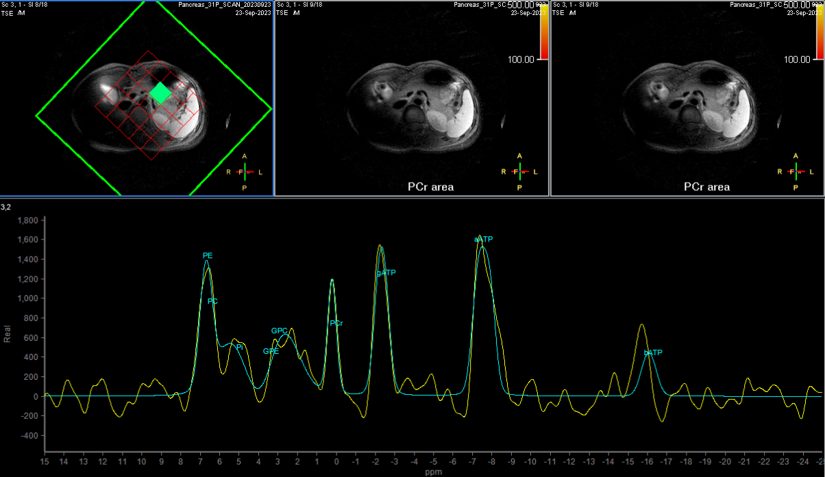
Three voxels were included in each pancreas and analyzed individually. 31P spectra of three normal pancreases were excluded as the PCr peaks were higher than corresponding α-ATP peaks. 31P spectrum of the other pancreas at the central voxel was considered as reasonable with lower-than-αATP PCr peak, as shown in Figure 1. Corresponding fitted peak areas under the curve of β-ATP, α-ATP, γ-ATP, PCr, PDE(GPE+GPC), Pi, PME (PE+PC) were 414, 1179, 1331, 713, 597, 1657 and 987, respectively, as demonstrated in Figure 2. Furthermore, Figure 2 also listed relevant phosphate ratios PME/PDE=1.65, PME/Pi=0.6, PDE/Pi=0.36, PME/α-ATP=0.84, PDE/α-ATP=0.84, PME/β-ATP=2.39 and γ-ATP/α-ATP=1.13, respectively.Discussion
Despite small cohort size, we successfully obtained a similar localized 31P spectrum in the central pancreas body of a healthy volunteer at 3 Tesla, with reference to a previous 7 Tesla finding [1]. Our PME/PDE ratio 1.65 was comparable to the reported ratio 1.9±0.8 at 7 Telsa. However, the PCr peak of pancreas body was relatively higher than the example spectrum at 7 Tesla, and only one spectrum of the total 12 analyzed pancreas voxels was valid for comparison in our study. This indicates that localized 31P spectroscopy at 3 Tesla are more vulnerable to PCr contamination from neighboring muscle or gallbladder than 31P MRS at 7 Telsa. Thanks to the high SNR in ultrahigh magnetic field, 31P spectroscopy at 7 Tesla could utilize smaller voxel size (20x20x36mm3 vs. 28x28x50mm3 , 4 voxels vs. 3 voxels) than 31P MRS acquisition at 3 Tesla. Hence, it suffered less from PCr contamination. Nevertheless, considering our acquisition time was only half of the 7 Tesla acquisition(11min.20sec. vs. 22min.37sec.), we could also reduce the acquisition voxel size and double number of signal average (NSA) to increase success rate and SNR at 3 Tesla in the future investigation.Conclusion
This preliminary study demonstrated that in vivo localized 31P spectroscopy of human pancreas on clinical 3T scanners is possible and promising although challenging. It could potentially detect various phosphate metabolites including PME and PDE as early biomarkers of abnormal energy metabolism and membrane synthesis in pancreatic cancer.Acknowledgements
No acknowledgement found.References
1. Comparison of 31P MRS in human pancreas and liver at 7 Tesla, Leonard W.F. Seelen et. al., ID0513, ISMRM 2023 Proceedings.Figures