4632
Free-breathing 3D Radial DIXON Look-Locker sequence for whole-liver simultaneous quantification of water/fat separated T1 and PDFF1Philips Japan, Tokyo, Japan, 2Department of Radiology, Kawasaki Medical School, Okayama, Japan, 3Philips Healthcare, Tokyo, Japan
Synopsis
Keywords: Liver, Liver
Motivation: T1 and PDFF mapping, which could serve as imaging biomarkers for liver disease, require breath-holds; T1 mapping commonly used in clinical practice, such as MOLLI, has limited spatial coverage. In addition, the presence of liver fat reduces T1 accuracy.
Goal(s): Our goal is to enable simultaneous whole liver water/fat separated T1 and PDFF quantification with clinically acceptable accuracy without breath-holds.
Approach: We introduced the 3D Radial DIXON LL sequence and validated in phantom and in vivo.
Results: Free-breathing 3D Radial Dixon LL sequence results in a proportional bias for T1 measurement and the fixed bias for PDFF measurement compared to current standard techniques.
Impact: The 3D Radial Dixon LL sequence did not result in sufficiently good agreement with reference methods for water/fat separated T1 and PDFF, suggesting that further technical validation and the optimization of imaging parameters will be needed.
INTRODUCTION
T1 mapping, which has the potential to assess inflammation, fibrosis, and function of the liver1-3, gains attention. However, T1 in liver is not yet an established biomarker. It is known that one of the confounders for T1 measurement in liver is fat4-6. Therefore, T1 mapping without the influence of the fat is clinically required for accurate evaluation. Recently, the water/fat separated T1 mapping of the liver to exclude the influence of fat in the T1 measurement has been proposed7-9. The water T1 (W-T1) mapping obtained from the Dixon based sequence could be useful for accurately evaluating the T1 value of the liver without the impact of fat9.Proton density fat fraction (PDFF), which can quantify tissue fat concentration, is increasingly used in liver in clinical practice10-12. However, breath-holding is basically necessary for the measurement of PDFF as well as T1. Therefore, these two measurements are often difficult or not feasible in patients with severe illness or young and old age. Moreover, whereas PDFF is measured in one 3D acquisition, T1 usually needs to be measured with a few breath-holds to cover multiple slices, which can cause extra burden on patients. To address these problems, whole liver free-breathing 3D acquisition has been proposed in a few articles8,13. Inspired by several recent works, we extend 3D pseudo golden-angle radial trajectory with Look-Locker (LL) sampling14 to 6-point multi-echo acquisition, which enables simultaneous whole liver water/fat separated T1 and PDFF quantification under free-breathing. Our goal is to validate the 3D Radial DIXON LL technique in phantom and in vivo.
METHODS
The following five experiments were performed: (1) validating 3D Radial DIXON LL for T1 against inversion recovery based spin echo (IR-SE) and 2D Modified Look-Locker Imaging (MOLLI) in a home-made T1 mapping phantom, (2) validating 3D Radial DIXON LL for PDFF in a PDFF phantom (Fat Fraction Phantom, Model 600; Calimetrix), (3) comparing 3D Radial DIXON LL with reference 2D MOLLI for T1 of the liver, (4) comparing 3D Radial DIXON LL with reference 3D mDIXON Quant for PDFF of the liver, and (5) comparing the difference between IP-T1 and W-T1 and between OP-T1 and W-T1 with PDFF of the liver. A total of 6 subjects (6 males, age = 38 ± 3 years) were recruited for liver imaging. Multiple imaging protocols were prepared for phantom and liver imaging as listed in Figure 1. Bland-Altman analysis and Pearson's correlation analysis were performed. A P value of < 0.05 was considered to indicate statistical significance.RESULTS and DISCUSSION
Figure 2A shows comparisons of T1 maps obtained from IR-SE and 3D Radial DIXON LL and from 2D MOLLI and 3D Radial DIXON LL in a phantom. T1 values from different phantom tubes are highly correlated between IR-SE and 3D Radial DIXON LL (y=0.95x+31.2, R2 = 0.998) and between 2D MOLLI and 3D Radial DIXON LL (y=0.97x+27.0, R2=0.998). The Bland-Altman plot shows a proportional bias for T1 values between 200 and 1350 ms in both studies. 3D Radial DIXON LL shows higher value compared to the true value in a fat fraction phantom (Figure 2B). PDFF was calculated from the multi echo images acquired with the longest TI delay. Therefore, at the longest TI delay water may not have relaxed to its equilibrium state, while fat has. Figure 3 shows IP-T1, OP-T1, W-T1, and PDFF maps obtained with 3D Radial DIXON LL in three volunteers with various PDFF. Figure 4 shows comparisons of T1 and PDFF obtained from 2D MOLLI and 3D Radial DIXON LL in volunteers. T1 shows no significant fixed and proportional bias. PDFF shows significant fixed and no significant proportional bias. As in the PDFF phantom study, this may be related to the fact that water at the longest TI delay has not reached its equilibrium state. Moreover, as PDFF increases, the difference between IP-T1 and W-T1 tends to decrease, but the difference between OP-T1 and W-T1 tends to increase. This indicates that W-T1 can assess T1 value without the influence of fat. Finally, one subject with liver steatosis is shown in Figure 5. In relation to the difference in PDFF between the right and left lobes of the liver, IP-T1 and OP-T1 also differ in both lobes (especially OP-T1). However, the difference in W-T1 is small, indicating that the effect of fat has been reduced.CONCLUSION
Compared to the reference standard, T1 and PDFF measurements using free-breathing 3D Radial Dixon LL show a proportional bias for T1 in a phantom study and a fixed bias for PDFF. Future optimization is needed.Acknowledgements
No acknowledgement found.References
- Banerjee R, Pavlides M, Tunnicliffe EM, et al. Multiparametric magnetic resonance for the non-invasive diagnosis of liver disease. J Hepatol. 2014;60(1):69-77.
- Horowitz JM, Venkatesh SK, Ehman RL, et al. Evaluation of hepatic fibrosis: a review from the society of abdominal radiology disease focus panel. Abdom Radiol (NY). 2017;42(8):2037-2053.
- Katsube T, Okada M, Kumano S, et al. Estimation of liver function using T1 mapping on Gd-EOB-DTPA-enhanced magnetic resonance imaging. Invest Radiol. 2011;46(4):277-83.
- Mozes FE, Tunnicliffe EM, Pavlides M, et al. Influence of fat on liver T1 measurements using modified Look-Locker inversion recovery (MOLLI) methods at 3T. J Magn Reson Imaging. 2016;44(1):105-11.
- Erden A, Kuru Öz D, Peker E, et al. MRI quantification techniques in fatty liver: the diagnostic performance of hepatic T1, T2, and stiffness measurements in relation to the proton density fat fraction. Diagn Interv Radiol. 2021;27(1):7-14.
- Ahn JH, Yu JS, Park KS, et al. Effect of hepatic steatosis on native T1 mapping of 3T magnetic resonance imaging in the assessment of T1 values for patients with non-alcoholic fatty liver disease. Magn Reson Imaging. 2021;80:1-8.
- Jaubert O, Arrieta C, Cruz G, et al. Multi-parametric liver tissue characterization using MR fingerprinting: Simultaneous T1, T2, T2*, and fat fraction mapping. Magn Reson Med. 2020;84(5):2625-2635.
- Feng L, Liu F, Soultanidis G, et al. Magnetization-prepared GRASP MRI for rapid 3D T1 mapping and fat/water-separated T1 mapping. Magn Reson Med. 2021;86(1):97-114.
- Higashi M, Tanabe M, Yamane M, et al. Impact of fat on the apparent T1 value of the liver: assessment by water-only derived T1 mapping. Eur Radiol. 2023;33(10):6844-6851.
- Reeder SB, Hu HH, Sirlin CB. Proton density fat-fraction: a standardized MR-based biomarker of tissue fat concentration. J Magn Reson Imaging. 2012;36(5):1011-1014.
- Zhong X, Nickel MD, Kannengiesser SA, et al. Liver fat quantification using a multi-step adaptive fitting approach with multi-echo GRE imaging. Magn Reson Med. 2014;72(5):1353-1365.
- Yokoo T, Bydder M, Hamilton G, et al. Nonalcoholic fatty liver disease: diagnostic and fat-grading accuracy of low-flip-angle multiecho gradient-recalled-echo MR imaging at 1.5 T. Radiology. 2009;251(1):67-76.
- Wang N, Cao T, Han F, et al. Free-breathing multitasking multi-echo MRI for whole-liver water-specific T1, proton density fat fraction, and R2∗ quantification. Magn Reson Med. 2022;87(1):120-137.
- Noda T, Sofue K, Shimada R, et al. Whole Upper Abdominal T1 Mapping on Free-Breathing 3D Look-Looker Sequence using Radial Sampling Acquisition: A Phantom and Volunteer Study. Proceedings of the 31th Annual Meeting of ISMRM, 2023 #2040.
Figures
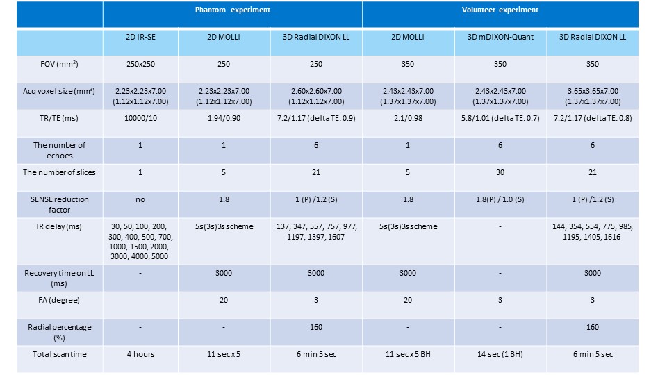
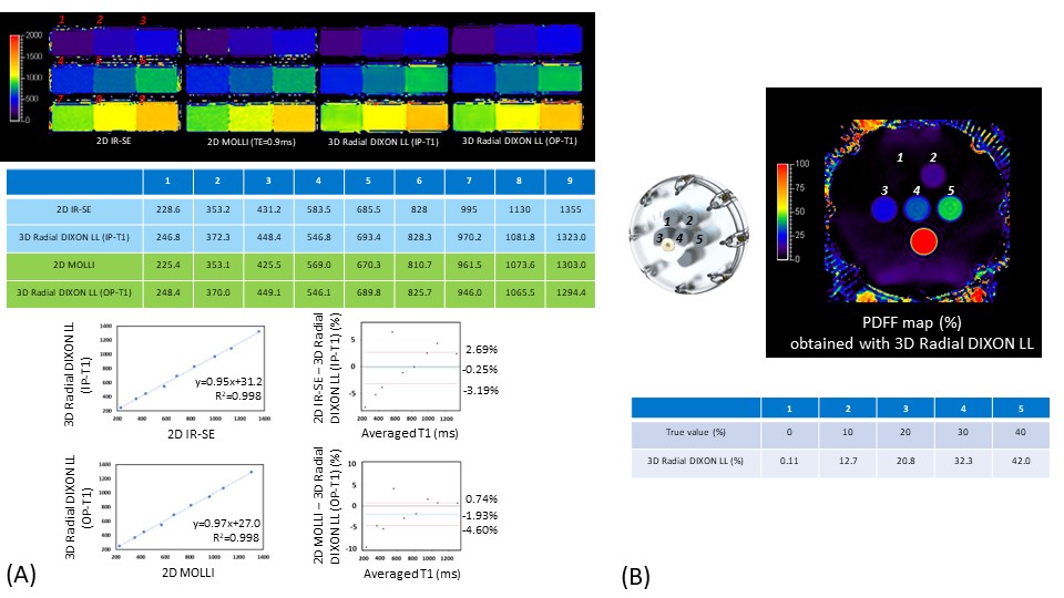
Figure 2. (A) Comparisons of T1 maps obtained from 2D IR-SE and 3D Radial DIXON LL, and from 2D MOLLI and 3D Radial DIXON LL in a phantom. T1 values are highly correlated between 2D IR-SE and 3D Radial DIXON LL (y=0.95x+31.2, R2 = 0.998) and between 2D MOLLI and 3D Radial DIXON LL (y=0.97x+27.0, R2 = 0.998). The Bland-Altman plot shows a proportional bias for T1 values in both studies. (B) Comparisons of PDFF obtained from 3D Radial DIXON and the true value in a fat fraction phantom. 3D Radial DIXON LL shows higher value compared to the true value.
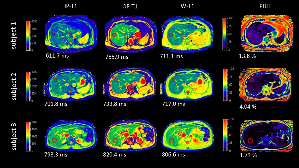
Figure 3. Comparison of IP-T1, OP-T1, W-T1, and PDFF acquired with 3D Radial DIXON LL in three volunteers. Subject 1 is confirmed to have liver steatosis. W-T1 is higher than IP-T1 and lower than OP-T1 in the liver. The differences are found to be larger in subject 1 with liver steatosis compared to subject 2 and 3.
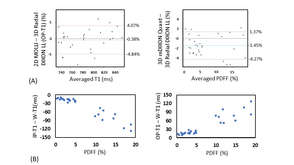
Figure 4. Comparisons of T1 and proton density fat fraction (PDFF) obtained from 2D MOLLI and 3D Radial DIXON LL in volunteers. T1 shows no significant fixed and proportional bias. PDFF shows significant fixed and no significant proportional bias. As PDFF increases, the difference between IP-T1 and W-T1 tends to decrease, but the difference between OP-T1 and W-T1 tends to increase.
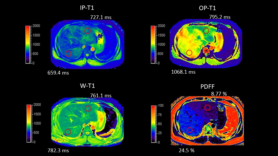
Figure 5. One subject with liver steatosis is shown. In relation to the difference in PDFF between the right and left lobes of the liver, IP-T1 and OP-T1 also differ in both lobes (especially OP-T1). However, the difference in W-T1 is small, indicating that the effect of fat has been reduced.