4627
Optimization of Parallel Imaging in Flip Angle Modulated 2D Sequential CSE-MRI for SNR-Efficient and Motion-Robust Liver Fat Quantification1Medical Physics, University of Wisconsin-Madison, Madison, WI, United States, 2Radiology, University of Wisconsin-Madison, Madison, WI, United States, 3Calimetrix, LLC, Madison, WI, United States, 4Biomedical Engineering, University of Wisconsin-Madison, Madison, WI, United States, 5Medicine, University of Wisconsin-Madison, Madison, WI, United States, 6Emergency Medicine, University of Wisconsin-Madison, Madison, WI, United States
Synopsis
Keywords: Liver, Fat, data acquisition, liver, pulse sequence design, parallel imaging
Motivation: Flip-angle modulation (FAM) in a 2D sequential acquisition is one potential means to improve the motion-robustness and SNR of CSE-MRI for liver fat quantification, and the motion-robustness and SNR-efficiency of FAM can be further improved through parallel imaging and capturing respiratory motion in-plane.
Goal(s): To determine the feasibility and performance (with respect to SNR, repeatability, and bias) of FAM-based free-breathing liver fat quantification acquired across imaging orientations and accelerations.
Approach: Trials of various parameters of FAM in volunteers and in a phantom with controlled PDFF and T1 values.
Results: Accelerated FAM demonstrates few artifacts, low bias, and increased SNR-efficiency compared to unaccelerated FAM.
Impact: Increasing the motion-robustness and SNR-efficiency of FAM may enable and enhance its use for improved detection, staging, and monitoring of steatotic liver disease. Non-axial acquisitions may mitigate through-plane respiratory motion compared to axial acquisitions.
Background and Theory
2D sequential chemical-shift-encoded (CSE)-MRI with flip-angle modulation (FAM) is a motion-robust, non-invasive liver fat quantification method1. FAM enables motion-robust PDFF mapping through its short temporal footprint: 1-2 seconds per slice in its original implementation.The temporal footprint of FAM can be further shortened through parallel imaging (PI) acceleration. The cost function for optimizing FAM flip angles1 was generalized to PI by matching the desired Gaussian k-space profile to PI’s nonuniform k-space sampling.
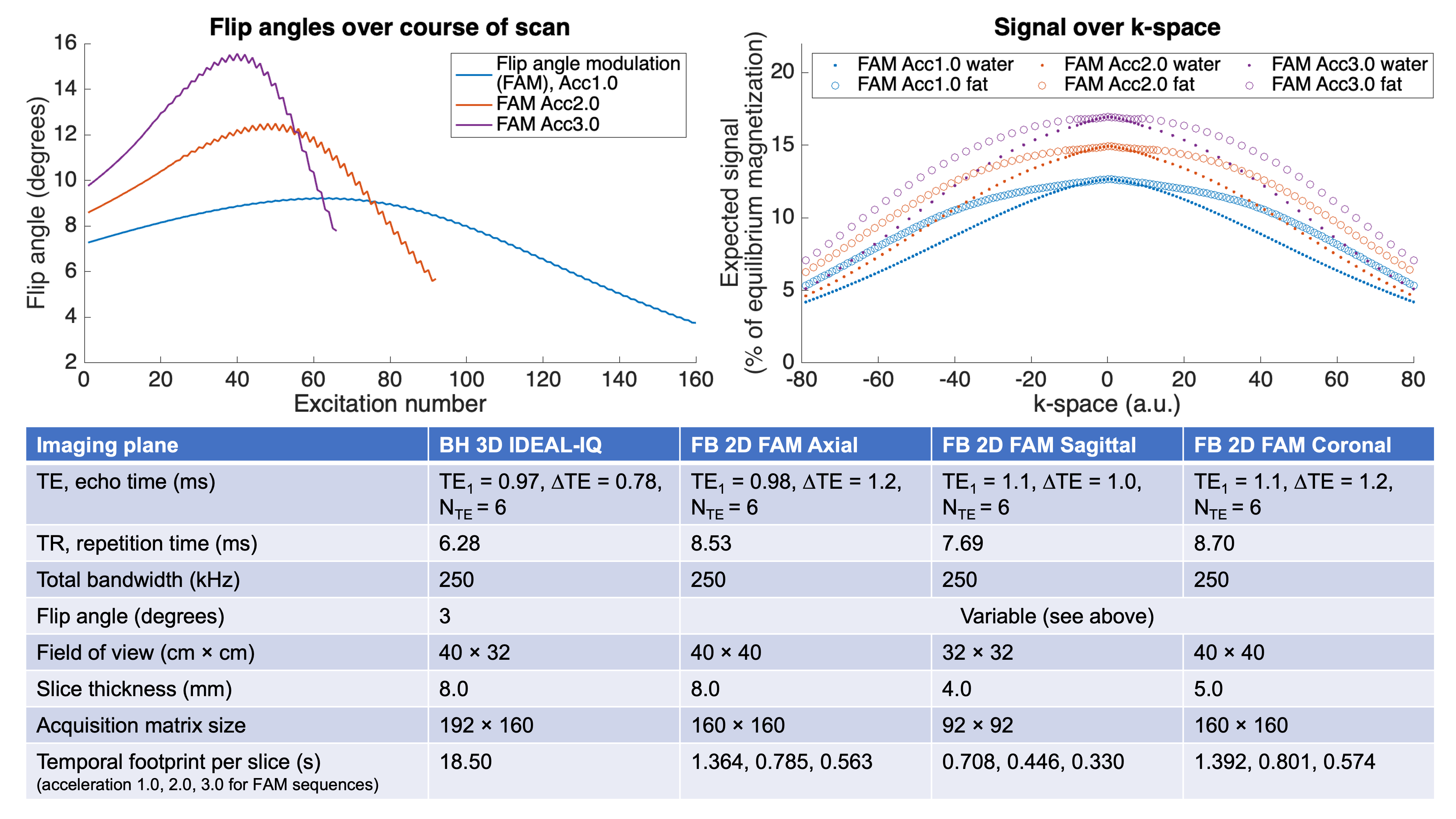
Further, due to the non-steady-state, centric-encoded FAM acquisition, it may be possible to acquire accelerated FAM with reduced SNR penalty by using higher flip angles (FAs). Generally, there is a tradeoff between increasing FAs and T1-bias1. However, in a non-steady-state method like FAM, T1-bias accumulates over multiple excitations. Therefore, if the number of excitations is reduced through PI acceleration, FAs can be increased, utilizing the magnetization more efficiently and potentially increasing SNR. Overall, this may offset the SNR penalty of PI, without the corresponding T1-bias penalty (Figure 1). Further, imaging in coronal or sagittal orientations may mitigate through-plane respiratory motion that can lead to missed tissue in axial free-breathing 2D acquisitions. Therefore, the purpose of this work was to evaluate SNR loss mitigation and enhanced motion-robustness in accelerated FAM in three imaging planes.
Methods
Pulse SequencesFAM sequences were created for axial, coronal, and sagittal imaging planes, and nominal 1D PI phase-encoding acceleration factors set to 1.0, 2.0, and 3.0. The acquisition of autocalibration lines meant real acceleration factors were lower than nominal. A commercially-available 3D-CSE-MRI method (IDEAL-IQ, GE HealthCare), designed for breath-held acquisitions, was also acquired as the reference. These sequences were evaluated in phantom and in vivo at 3.0T (GE Signa Premier, Waukesha, WI).
Phantom Acquisitions & Analysis
A commercial PDFF-T1 phantom (Calimetrix LLC, Madison, WI), containing vials with controlled PDFF values (0,10,20,30%) and T1water (200,600,1000,1400ms), was imaged using the above sequences. The phantom was rotated as imaging planes changed so acquired slices were always orthogonal to the vials’ axes. 10 repetitions of each sequence were acquired without repositioning to evaluate voxel-wise PDFF standard deviations (SDs) as a surrogate of SNR. Circular ROIs were drawn at each vial’s center, and mean voxel-wise SDs and mean PDFF values in each ROI recorded.
Volunteer Acquisitions & Analysis
Volunteers were recruited to represent a wide range of BMIs as a proxy for liver PDFF, and imaged in an IRB-approved study with informed written consent. Volunteers breath-held for the 3D-CSE-MRI acquisition and breathed normally for FAM acquisitions. Test-retest repeatability was evaluated by repeating the 3D-CSE-MRI and FAM acquisitions after volunteer repositioning. From the test and retest acquisitions, analysts drew ROIs on the nine liver segments. ROI means were compared between each FAM acquisition and the reference 3D-CSE-MRI acquisition. Test and retest ROI means were also compared. SDs within each ROI were also measured as a surrogate of SNR. This is different from the phantom SD methodology, where voxel-wise SDs were calculated across repetitions; in-vivo SDs were calculated across the ROI. Although the PDFF heterogeneity of the liver contributes to this SD measurement, noise is also expected to be a major contributor with small ROIs.
Results
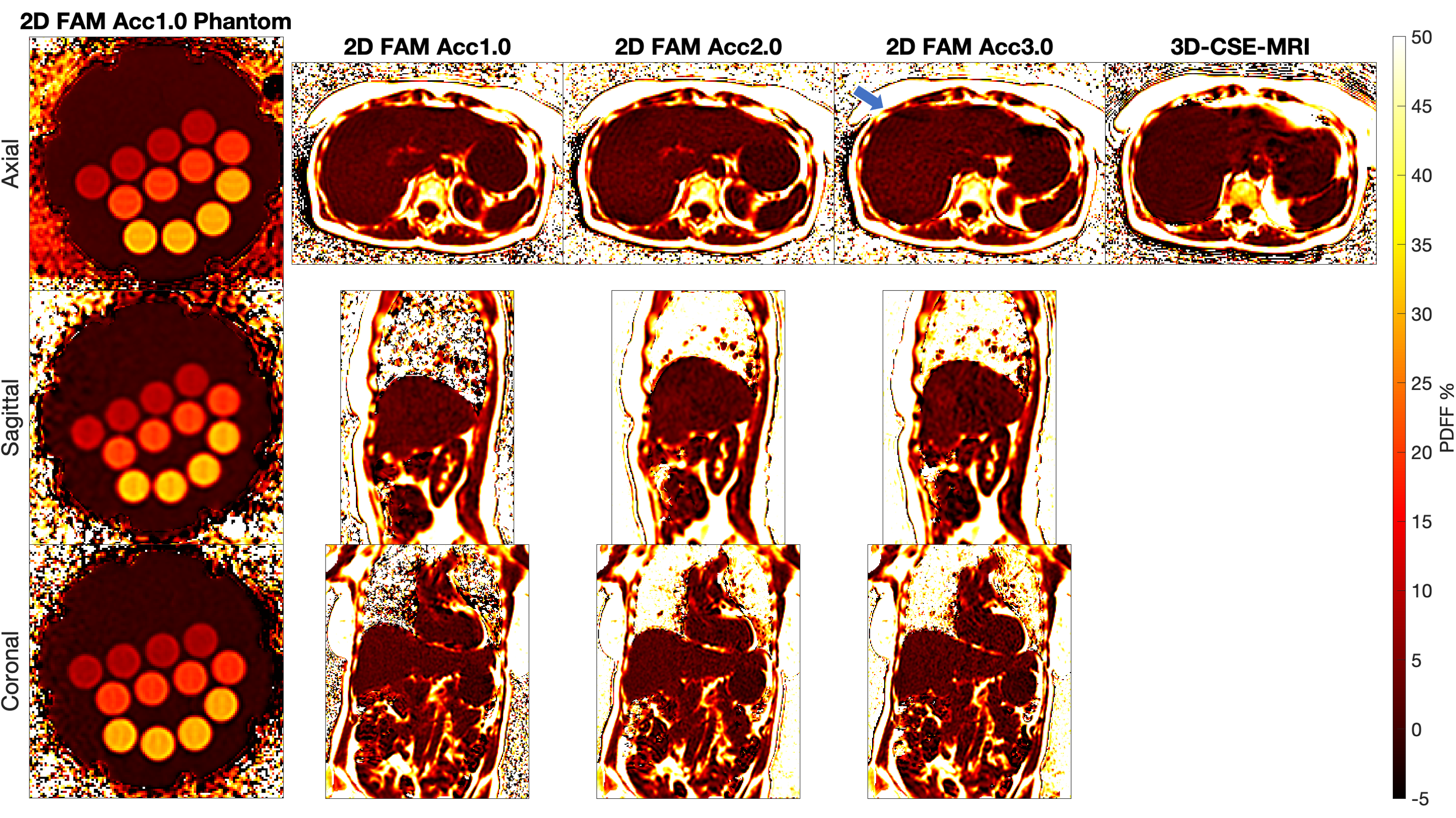
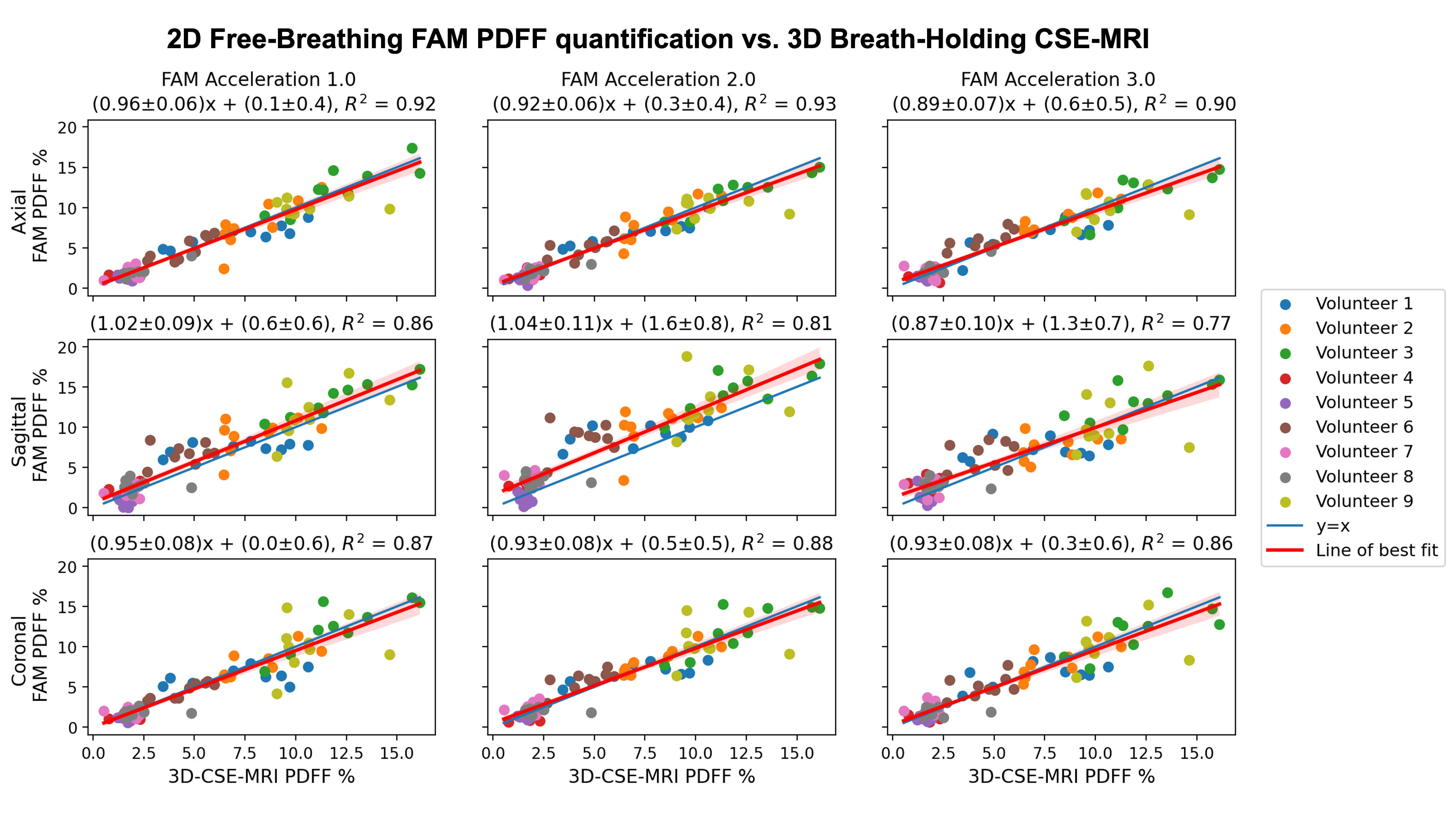
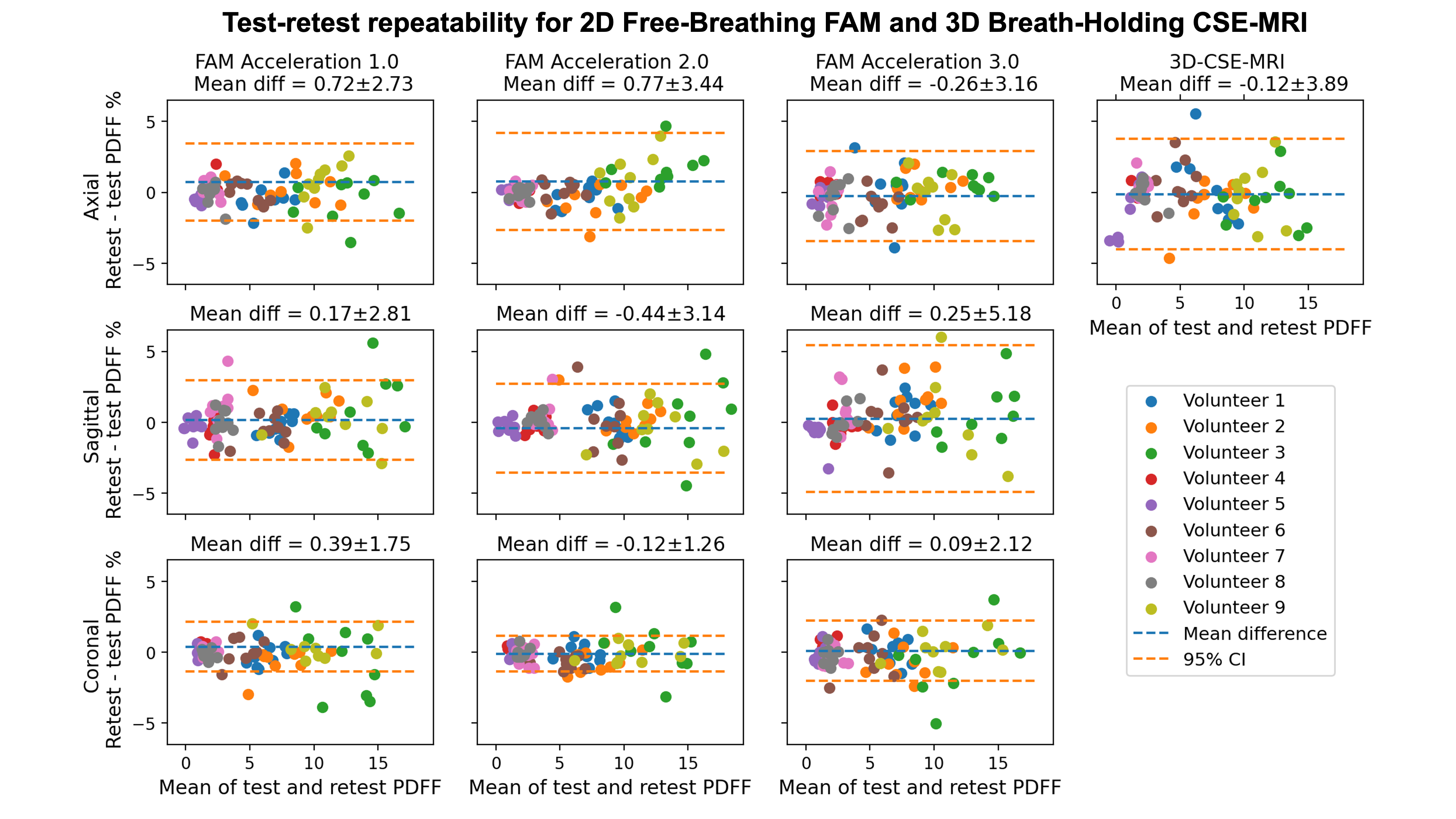
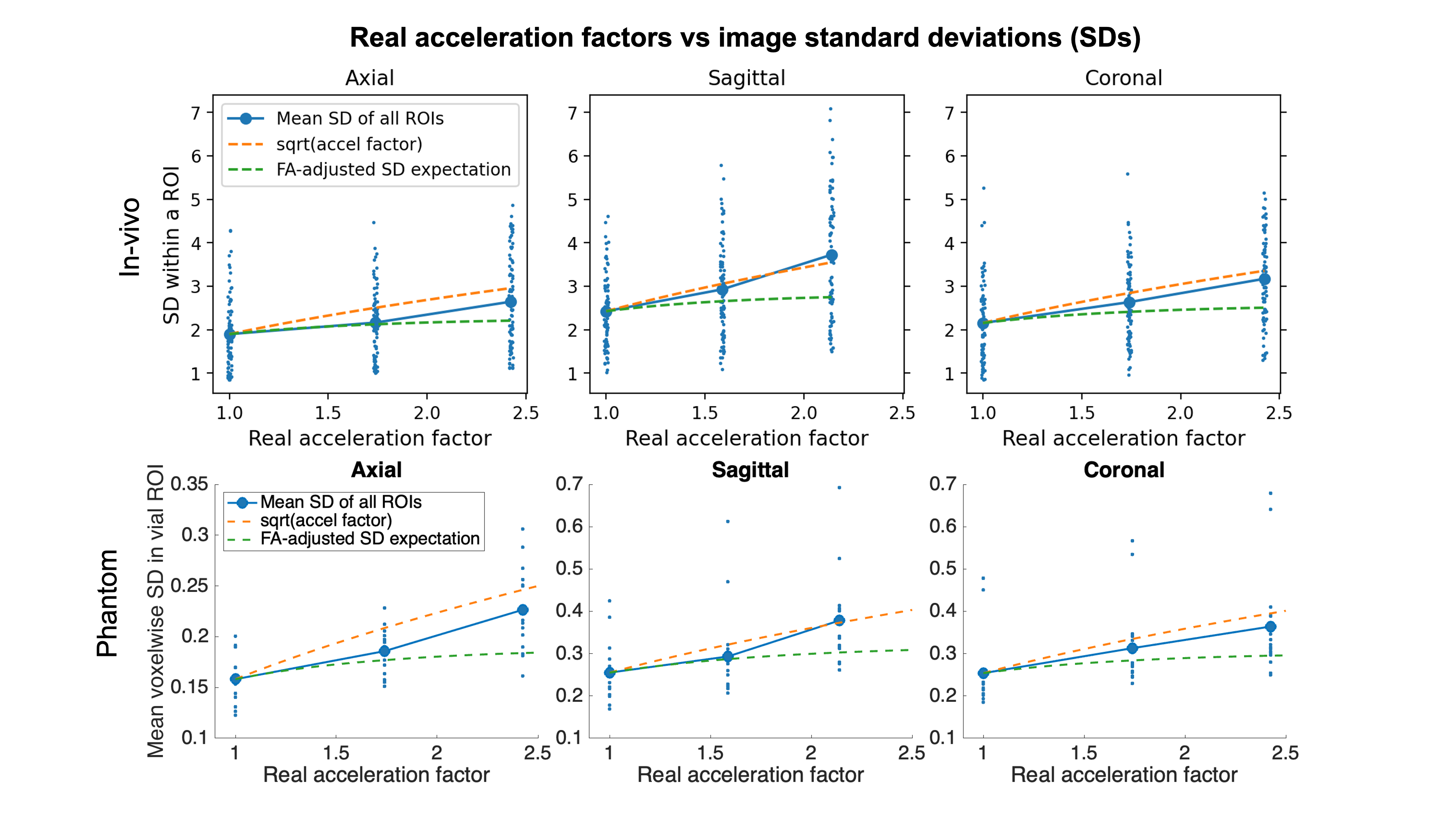
Nine volunteers (ages 28-63, 44.4% female, BMI 19.3-41.7) were recruited and imaged as above.Images were relatively free of artifacts in all three imaging planes up to acceleration factor 2.0. Residual aliasing from high acceleration was observed in some acceleration factor 3.0 images (Figure 2). In vivo, all FAM methods show good agreement with 3D-CSE-MRI (Figure 3), and Bland-Altman analysis shows good test-retest repeatability (Figure 4). In phantoms and in vivo, FAM with PI acceleration showed slower SNR degradation than predicted with conventional PI acceleration, where SNR degrades with $$$\sqrt{\text{acceleration factor}}$$$ plus g-factor effects2,3, due to offsetting increases in the FAs used for higher acceleration FAM sequences (Figure 5).
Discussion
This work extends FAM-based PDFF mapping to include PI acceleration. We evaluated this method in a PDFF-T1 phantom and in volunteers across acceleration factors and imaging planes. We demonstrated that accelerated FAM provides a shorter acquisition temporal footprint (improved motion-robustness), with better SNR-efficiency from higher FAs, while avoiding T1-bias. Specifically, these results show good potential for FAM with acceleration factor 2.0 for motion-robust, free-breathing fat quantification. Further, accelerated non-axial FAM acquisitions show excellent quantitative performance and could enable free-breathing PDFF mapping with whole-liver coverage without missing any tissue.Limitations of this work include evaluation at a single center, vendor, and field strength, and lack of evaluation in patients.
Altogether, accelerated FAM CSE-MRI obtained with an optimized imaging orientation may enable error-proof, free-breathing PDFF mapping across broad patient populations.
Acknowledgements
The authors acknowledge support from NIH grants R44EB025729 and R01EB031886.The authors also gratefully acknowledge the assistance of the study coordination and MRI technologist teams at the Wisconsin Institutes for Medical Research.
Dr. Hernando and Dr. Reeder are co-founders of Calimetrix, LLC, which manufactured and loaned to the authors the phantom used in this study.
Dr. Reeder is the John H. Juhl Endowed Chair of Radiology.
Jiayi Tang is a shareholder of GE HealthCare.
GE HealthCare provides research support to the University of Wisconsin.
References
- Zhao R, Zhang Y, Wang X, et al. Motion-robust, high-SNR liver fat quantification using a 2D sequential acquisition with a variable flip angle approach. Magn Reson Med. 2020;84(4):2004-2017. doi:10.1002/mrm.28263
- Rabanillo I, Aja-Fernández S, Alberola-López C, Hernando D. Exact Calculation of Noise Maps and g -Factor in GRAPPA Using a k -Space Analysis. IEEE Trans Med Imaging. 2018;37(2):480-490. doi:10.1109/TMI.2017.2760921
- Robson PM, Grant AK, Madhuranthakam AJ, Lattanzi R, Sodickson DK, McKenzie CA. Comprehensive Quantification of Signal-to-Noise Ratio and g-Factor for Image-Based and k-Space-Based Parallel Imaging Reconstructions. Magn Reson Med. 2008;60(4):895-907. doi:10.1002/mrm.21728
Figures