4625
Lipid composition mapping of the breast in BRCA1/2 genetic mutation carriers using chemical shift-encoded imaging1Institute of Medical Sciences, University of Aberdeen, Aberdeen, United Kingdom, 2Massachusetts General Hospital, Boston, MA, United States, 3Breast Unit, Broomfield Hospital, Mid and South Essex NHS Trust, Chelmsford, United Kingdom, 4Radiology Department, Aberdeen Royal Infirmary, Aberdeen, United Kingdom, 5Newcastle Magnetic Resonance Centre, Newcastle University, Newcastle, United Kingdom
Synopsis
Keywords: Screening, Fat, BRCA1/2, chemical shift-encoded imaging, early detection
Motivation: Deregulation of lipid composition in the breast of BRCA1/2 carriers has been observed using single voxel spectral edited MRS, however the spatial distribution may further distinguish the disease state.
Goal(s): We aimed to determine the power and repeatability of CSEI in detection of lipid deregulation in BRCA1/2.
Approach: Lipid composition maps were acquired from 20 BRCA1/2 and 20 patients, to derive monounsaturated, polyunsaturated and saturated fatty acids in the peri-tumoural region in patients and the whole breast in all subjects.
Results: Lipid composition in BRCA1/2 showed similarity to positive control. CSEI has excellent repeatability in lipid composition mapping of the breast.
Impact: Deregulation of lipid composition in the breast of BRCA1/2 genetic mutation carriers resembles the diseased group, serving as potential precursor of breast cancer. CSEI yields highly repeatable quantitative maps of lipid composition critical for effective disease monitoring.
Introduction
Breast cancer is a major and expanding health challenge, despite significant improvement in survival rate1. Genetic mutation carriers of BRCA1/2 have over 30% increased risk of developing breast cancer and receive annual DCE-MRI surveillance2. MRI contrast is sensitive to tumour angiogenesis, however detects malignancies that are well under development. Deregulation of lipid composition, including monounsaturated, polyunsaturated and saturated fatty acids (MUFA, PUFA, SFA), has been shown in the breast of BRCA1/2 carriers using single voxel spectral edited MR spectroscopy3, and in the peri-tumoural region of the breast4. Novel chemical shift-encoded imaging (CSEI) allows rapid lipid composition mapping of the whole breast, and the spatial distribution may further distinguish the disease state. We therefore hypothesise that lipid composition in the breast of BRCA1/2 carriers show no difference from patients with breast cancer, and determine the repeatability of CSEI.Methods
Twenty patients with invasive ductal carcinoma (age 31 – 55 years) and 20 BRCA1/2 genetic mutation carriers (age 32 – 51 years) participated in the study. Patients with a tumour size larger than 1 cm on mammography and have not had hormonal therapy or chemotherapy were eligible. Participants with diabetes or on long-term medications that might alter lipid metabolism were excluded. The study was approved by the North West – Greater Manchester West Research Ethics Committee (ID: 19/NW/0225), and written informed consents were obtained from all the participants (Figure 1).CSEI
All images were acquired on a 3 T whole-body clinical MRI scanner (Achieva dStream, Philips Healthcare, Best, Netherlands). Lipid composition images were acquired from all participants using a 2D fast field echo sequence5,6 with 16 echoes, initial echo time of 1.14 ms, echo spacing of 1.29 ms, repetition time of 60 ms, reconstruction pixel size of 2.0 × 2.0 mm2 and slice thickness of 3.0 mm, with subsequent repeated acquisition.
Data Processing
Image analysis was conducted in MATLAB (R2020a, MathWorks Inc., Natick, MA, USA). The maps of the number of double bonds in triglycerides were computed from raw data, before subsequent quantification of MUFA, PUFA and SFA as a fraction of total lipids5,6. The delineation of tumour was conducted on the first echo of magnitude image, with reference to dynamic contrast enhanced images. The whole breast in BRCA1/2, and the whole breast and the peri-tumoural region in patients were the three regions-of-interest. The whole breast was defined to contain only adipose and fibroglandular tissue in BRCA1/2, and excluding the tumour in patients. The peri-tumoural region was defined as an annular ring of 16 mm (8 voxels) around the tumour. Adipose voxels with lipid signal below 60% of total signal were excluded from further analysis. The mean lipid composition from the regions-of-interest was subsequently computed.
Statistical Analysis
All statistical analysis was performed in the R software (v4.3.1, R Foundation for Statistical Computing, Vienna, Austria). Wilcoxon signed rank paired tests were performed for comparison of lipid composition in the whole breast and the peri-tumoural region in patients, with Wilcoxon rank sum tests performed between the whole breast of BRCA1/2 and patients. The within-subject coefficient of variance (%wCoV) was calculated as [standard deviation / mean] × 100%. A p-value < 0.017 was considered to indicate a statistically significant difference for 3-group comparisons, after Bonferroni correction.
Results
The participant demographics of the entire cohort are shown in Table 1.BRCA1/2 and Patients
There was no significant difference in mean MUFA, PUFA and SFA in the whole breast of BRCA1/2 against the whole breast nor the peri-tumoural region of patients (Figure 2, Table 2).
Patients
There was a significantly lower mean MUFA (p<0.01) and higher mean SFA (p<0.01) in the peri-tumoural region compared to the whole breast. There was no significant difference in mean PUFA (p=0.02) between the peri-tumoural region and the whole breast (Figure 2, Table 2).
Within-subject CoV
The %wCoV in all the three regions-of-interest were below 10.0% (Figure 3).
Discussion
Deregulation of lipid composition in the breast of BRCA1/2 genetic mutation carriers resembled the diseased group, serving as potential precursor of breast cancer. There was a decrease in MUFA in the peri-tumoural region to support accelerated membrane synthesis for tumour growth7, while an increase in SFA in the peri-tumoural region to avoid lipotoxicity and enhance chemoresistance7. CSEI has excellent repeatability in lipid composition mapping of the breast critical for effective disease monitoring.Conclusion
Lipid composition in BRCA1/2 showed similarity to positive control, and lipid composition in the peri-tumoral region was altered due to tumour growth. CSEI has excellent repeatability for accurate measurement of lipid composition in the breast.Acknowledgements
The authors would like to thank Dr Matthew Clemence (Philips Healthcare Clinical Science, UK) for clinical scientist support, Ms Resifina Seyara, Ms Farah Muir, Ms Susan Rodwell and Ms Vera Herd for patient recruitment support, Ms Teresa Morris and Ms Dawn Younie for logistics support, and Ms Nichola Crouch, Ms Laura Reid, Ms Michelle Mauchline, Mr Arthur Ginsburg and Mr Mike Hendry for radiographer support. The authors would also like to thank Ms Mairi Fuller, Ms Elizabeth Smyth and Ms Beatrix Elsberger for providing access to the patients. This project was funded by Cancer Research UK, and Sai Man Cheung’s research fellow training is currently funded by Chief Scientist Office.References
1. Pan H, Gray R, Braybrooke J, et al. 20-year risks of breast-cancer recurrence after stopping endocrine therapy at 5 years. N Engl J Med. 2017;377(19):1836-1846.
2. Malone KE, Begg CB, Haile RW, et al. Population-based study of the risk of second primary contralateral breast cancer associated with carrying a mutation in BRCA1 or BRCA2. J Clin Oncol. 2010;28(14):2404-2410.
3. Ramadan S, Arm J, Silcock J, et al. Lipid and metabolite deregulation in the breast tissue of women carrying BRCA1 and BRCA2 genetic mutations. Radiology. 2015;275(3):675-682.
4. Chan KS, Cheung SM, Senn N, et al. Peri-tumoural spatial distribution of lipid composition and tubule formation in breast cancer. BMC Cancer. 2022;22(1):285.
5. Bydder M, Girard O, Hamilton G. Mapping the double bonds in triglycerides. Magn Reson Imaging. 2011;29(8):1041-1046.
6. Peterson P, Månsson S. Simultaneous quantification of fat content and fatty acid composition using MR imaging. Magn Reson Med. 2013;69(3):688-697.
7. Broadfield LA, Pane AA, Talebi A, Swinnen JV, Fendt S. Lipid metabolism in cancer: New perspectives and emerging mechanisms. Dev Cell. 2021;56(10):1363-1393.
Figures
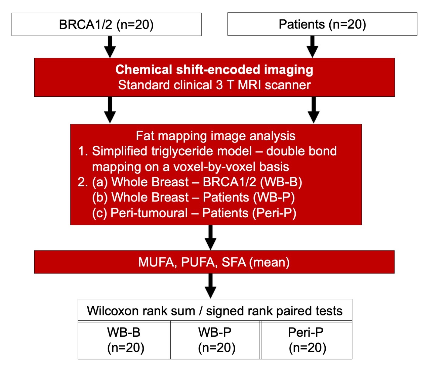
Figure 1. Study design
Twenty genetic mutation carriers of BRCA1/2 and 20 patients with invasive ductal carcinoma of the breast participated in the study. All patients underwent chemical shift-encoded imaging on a clinical 3 T MRI scanner. Fat mapping image analysis was conducted to compute monounsaturated fatty acids (MUFA), polyunsaturated fatty acids (PUFA) and saturated fatty acids (SFA) in three regions-of-interest, with Wilcoxon tests subsequently performed.
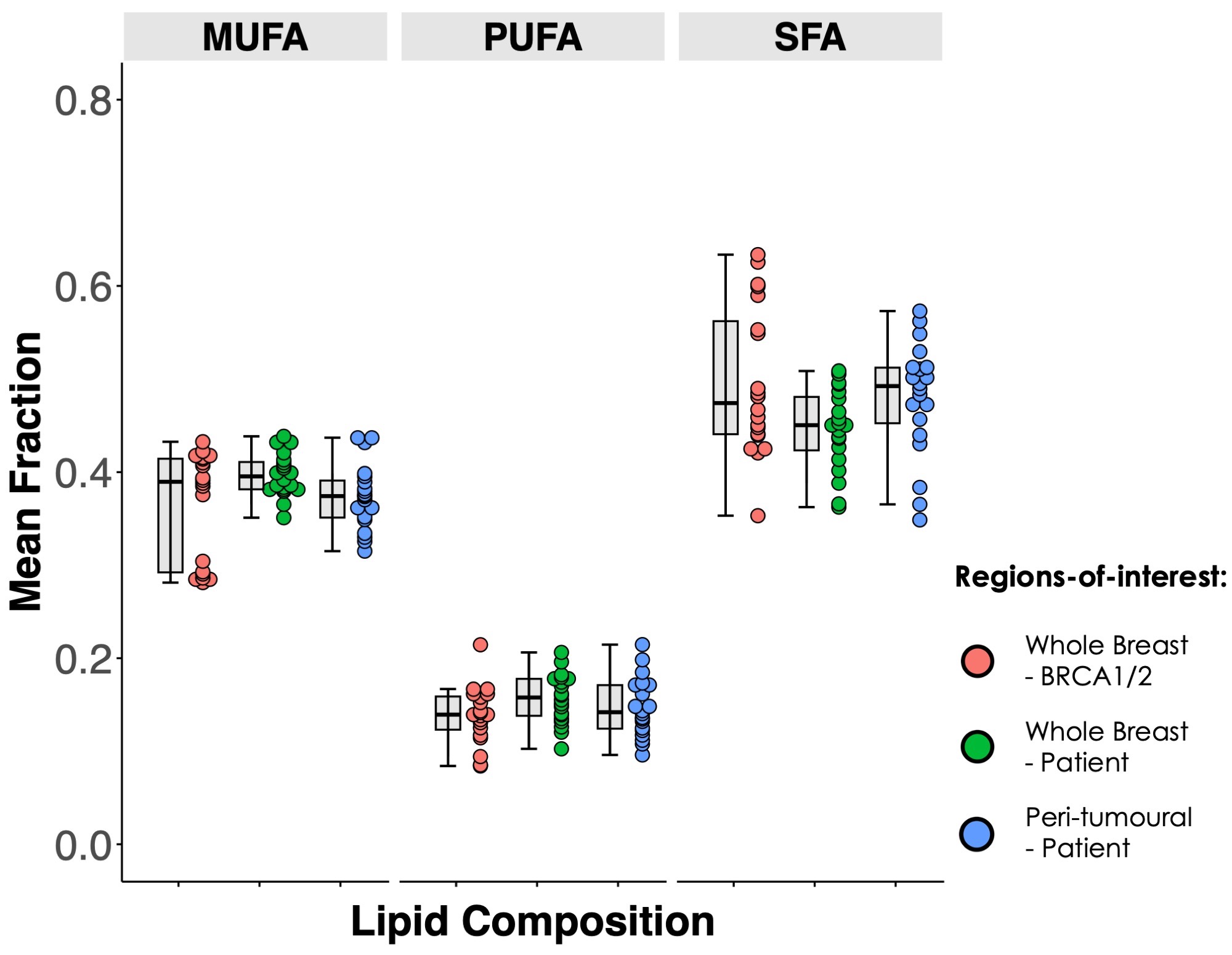
Figure 2. Difference in lipid composition between genetic mutation carriers of BRCA1/2 and patients with breast cancer
Monounsaturated fatty acids (MUFA), polyunsaturated fatty acids (PUFA) and saturated fatty acids (SFA) in the whole breasts of BRCA1/2 and patients, and the peri-tumoural region of patients. Each dot represents a lipid mean fraction, and the dots are organised in three columns corresponding to the three regions-of-interest. Error bars indicate the median and interquartile range.
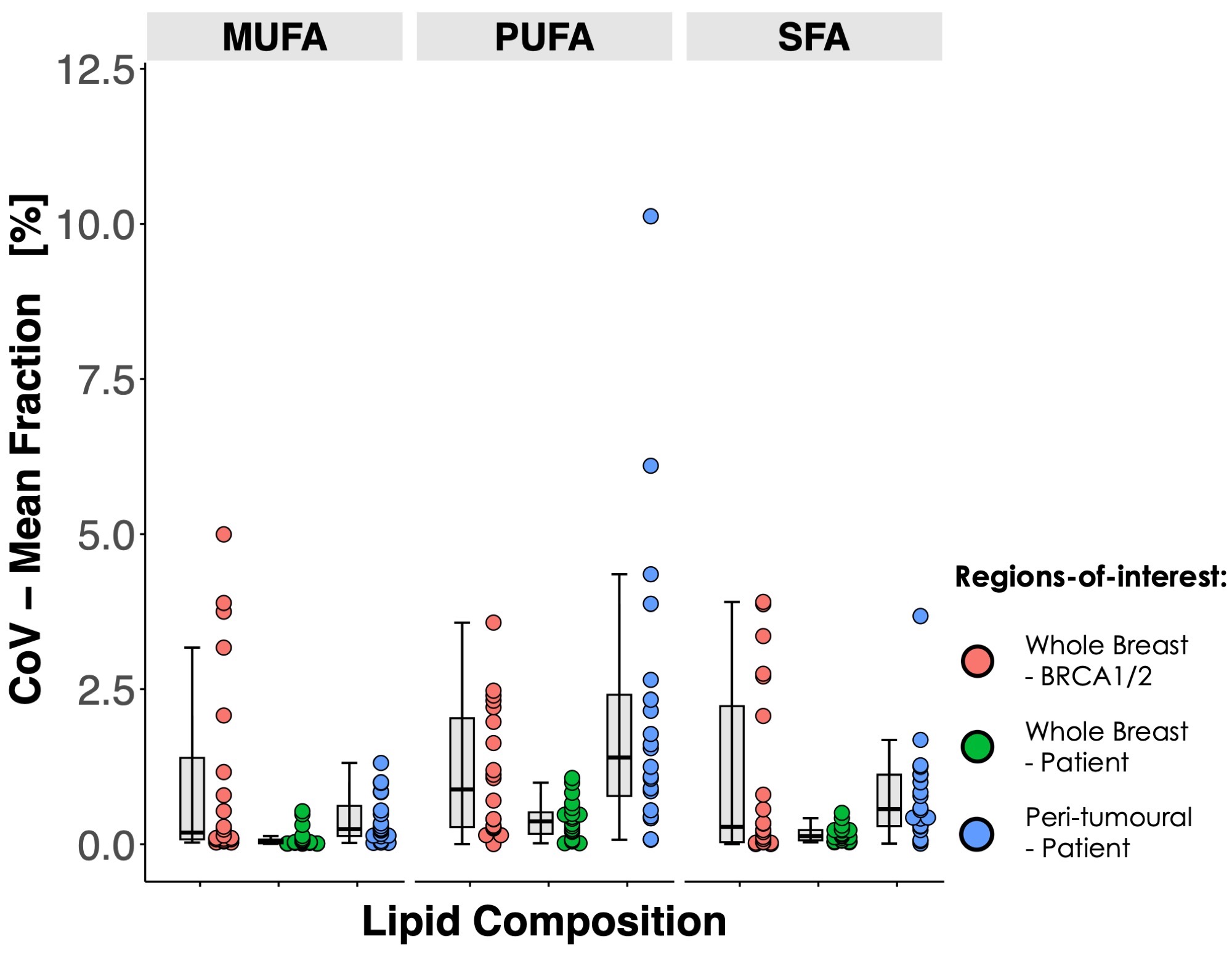
Figure 3. Repeatability of lipid composition in genetic mutation carriers of BRCA1/2 and patients with breast cancer
The within-subject coefficient of variance (%wCoV) from repeated acquisitions of monounsaturated fatty acids (MUFA), polyunsaturated fatty acids (PUFA) and saturated fatty acids (SFA) in the whole breast of BRCA1/2 and patients, and the peri-tumoural region of patients. Error bars indicate the median and interquartile range.
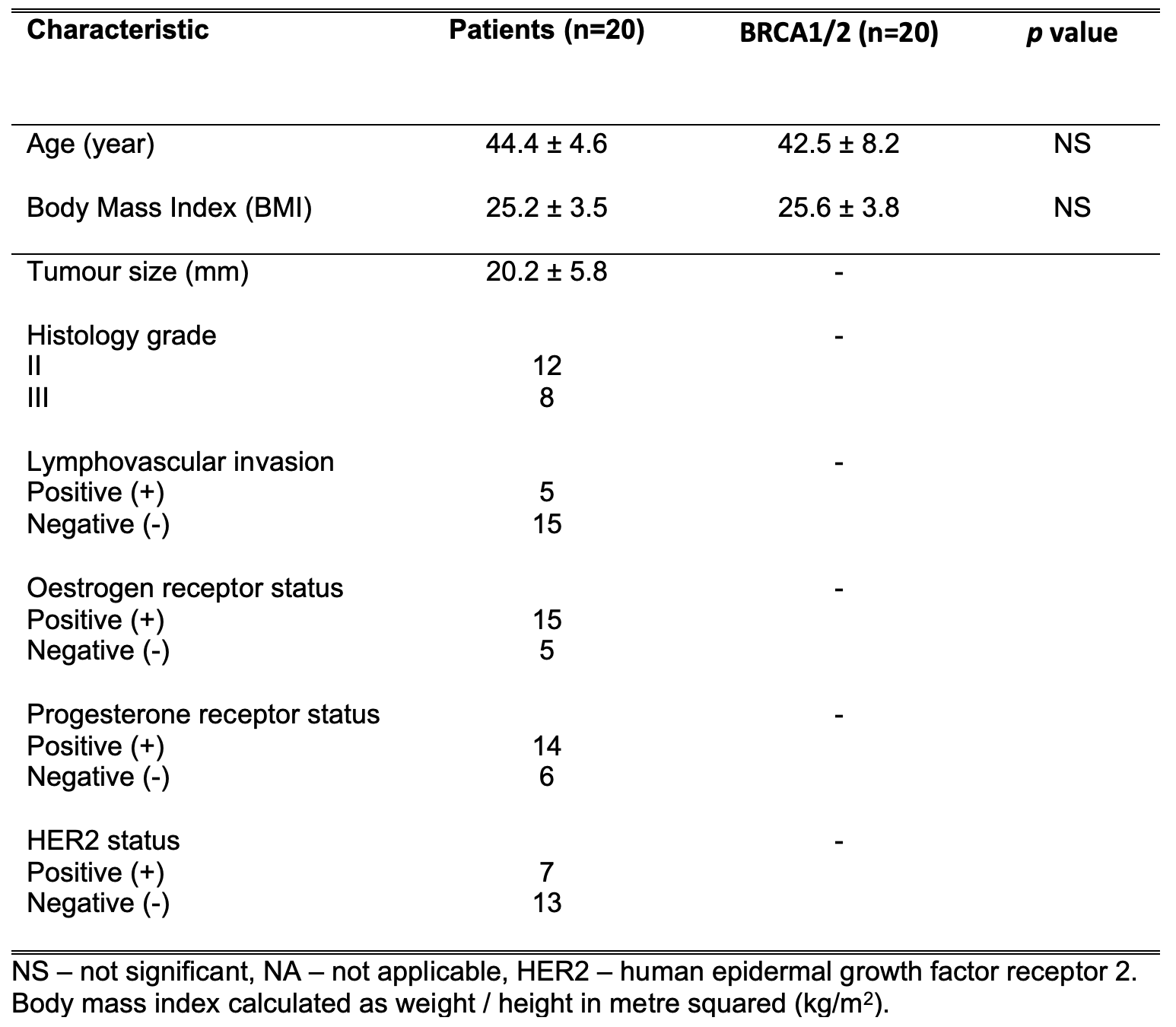
Table 1. Participant demographics
Age and body mass index of all participants are shown. Histopathological findings for patients with breast cancer are also shown. Quantitative entries are expressed as mean and standard deviation (mean ± SD).
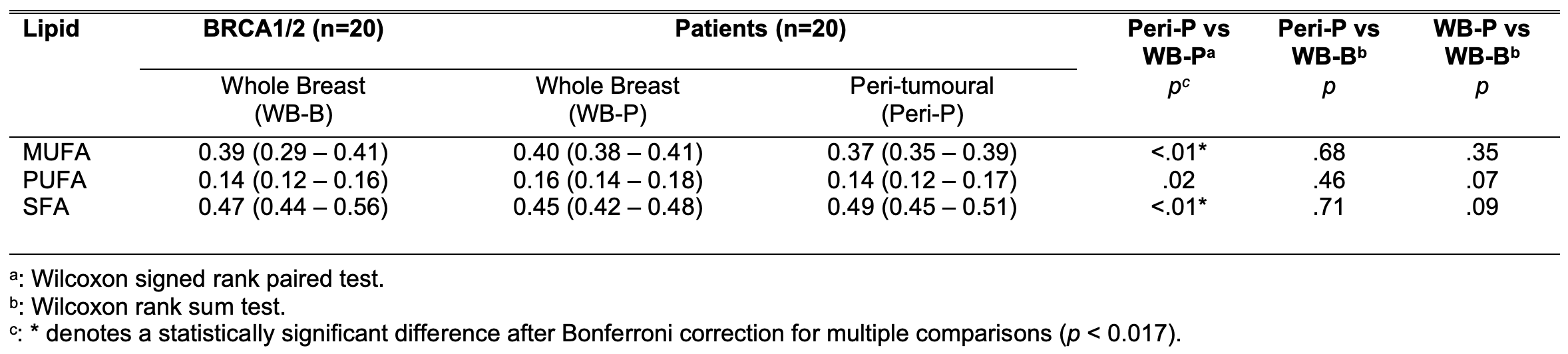
Table 2. Monounsaturated, polyunsaturated and saturated fatty acids (MUFA, PUFA, SRA) in the whole breast of BRCA1/2 mutation carriers, and the whole breast and the peri-tumoural region of patients with breast cancer
The mean MUFA, PUFA, SFA in the three regions-of-interest were compared between BRCA1/2 and patients. Quantitative entries are expressed as median (interquartile range).