4619
The value of quantitative synthetic MRI in differentiating breast cancer patients with HER2 zero-, low- and over-expressing1Department of Radiology, The Second Affiliated Hospital of Nanchang University, Nanchang, China, Nanchang, China, 2MR Research, GE Healthcare, Beijing, China, Beijing, China
Synopsis
Keywords: Breast, Breast, breast cancer, human epidermal growth factor receptor 2 (HER2), magnetic resonance imaging, synthetic imaging
Motivation: Half of breast cancers exhibits HER2 low-expressing and benefits from new antibody-drug conjugates. However, it remains lack of non-invasive technique to identify HER2 expression level. T1, T2, and proton density (PD) are quantitative MRI parameters widely used to characterize tumors.
Goal(s): Investigate if T1, T2, and PD extracted from synthetic MRI (syMRI) can be used to classify HER2 expression level.
Approach: One-hundred patients were enrolled. T1, T2, and PD of tumor were extracted from syMRI and were compared among different HER2 expression level. ROC was used to assess the diagnostic performance.
Results: T1 and PD can be used to diagnose HER2 expression levels.
Impact: Our results suggested quantitative SyMRI can be used as non-invasive biomarker to classify HER2 zero-, low- and over-expressing breast cancer patients. It would be helpful in guiding treatment selection and dynamically monitoring HER2 expressing status during treatment process over time.
Introduction
Breast cancer (BC) had become the most diagnosed cancer in 2020[1]. HER2 is a protein that tightly linked to tumor growth and progression in BC. It is one of the key prognosis factors of BC and has been used to guide the selection of treatment[2]. Clinically, BC can be divided into HER2-overexpressing (HER2-oe) and HER2-negative according to the HER2 expression status[3]. However, recent studies had proposed HER2 low-expressing should be separated from HER2-negative BC and treated as a new subgroup[4]. Recently, the molecular and pathologic differences between HER2-zero, HER2-low, and HER2-oe BC have begun to be explored [4]. HER2-low BC patients might benefit from the HER2-targeted agents[5]. Moreover, the expression status of HER2 in BC can change along the treatment process[6]. Biopsy or surgery is not always appropriate for dynamically monitoring of the HER2 status due to the invasive nature. Thus, it would be highly desirable to find a noninvasive imaging technique to identify HER2 subgroups in BC.Synthetic MRI (SyMRI) is a noninvasive technique to quantitatively evaluate the characteristics of tumors by simultaneously providing longitudinal relaxation time (T1), transverse relaxation time (T2), and proton density (PD) of tissue[7]. Previous studies had reported the value of SyMRI in differentiating HER2-oe from HER2-negative as well as in discriminating other molecular subtypes of BC [8-9], With these promising results, this study aimed at investigating the value of SyMRI in identifying HER2-zero, HER2-low, and HER2-oe BC patients.
Methods and Materials
Study population34 HER2-oe, 48 HER2-low, and 18 HER2-zero BC patients were recruited in our hospital.
MRI experiment
All MRI examinations were performed at 3.0 Tesla scanner (SIGNA Architect; GE Healthcare, USA) using an 8-channel breast coil. SyMRI was scanned with FOV=360mm×360mm, matrix size=256×256, slice thickness=5mm, and number of slices=20. Contrast-enhanced imaging were also carried out.
Imaging analysis
All MRI data were processed using the vendor-provided workstation (GE Healthcare, USA) which was equipped with the syMRI post-processing software (SyMRI 7.2; Linköping, Sweden). The region-of-interest (ROIs) of the tumor was manually delineated. The necrotic or cystic was not included in the ROIs. The average of T1, T2 and PD within each ROI was extracted for further analysis.
Statistical analysis
The quantitative parameters of tumors among HER2-zero, HER2-low, and HER2-oe were compared using ANOVA test. Post hoc comparison was corrected using Bonferroni correction. The receiver operating characteristic (ROC) curve was using to assess the performance for identifying HER2-zero, HER2-low and HER2-oe. P<0.05 was considered statistically significant.
Results
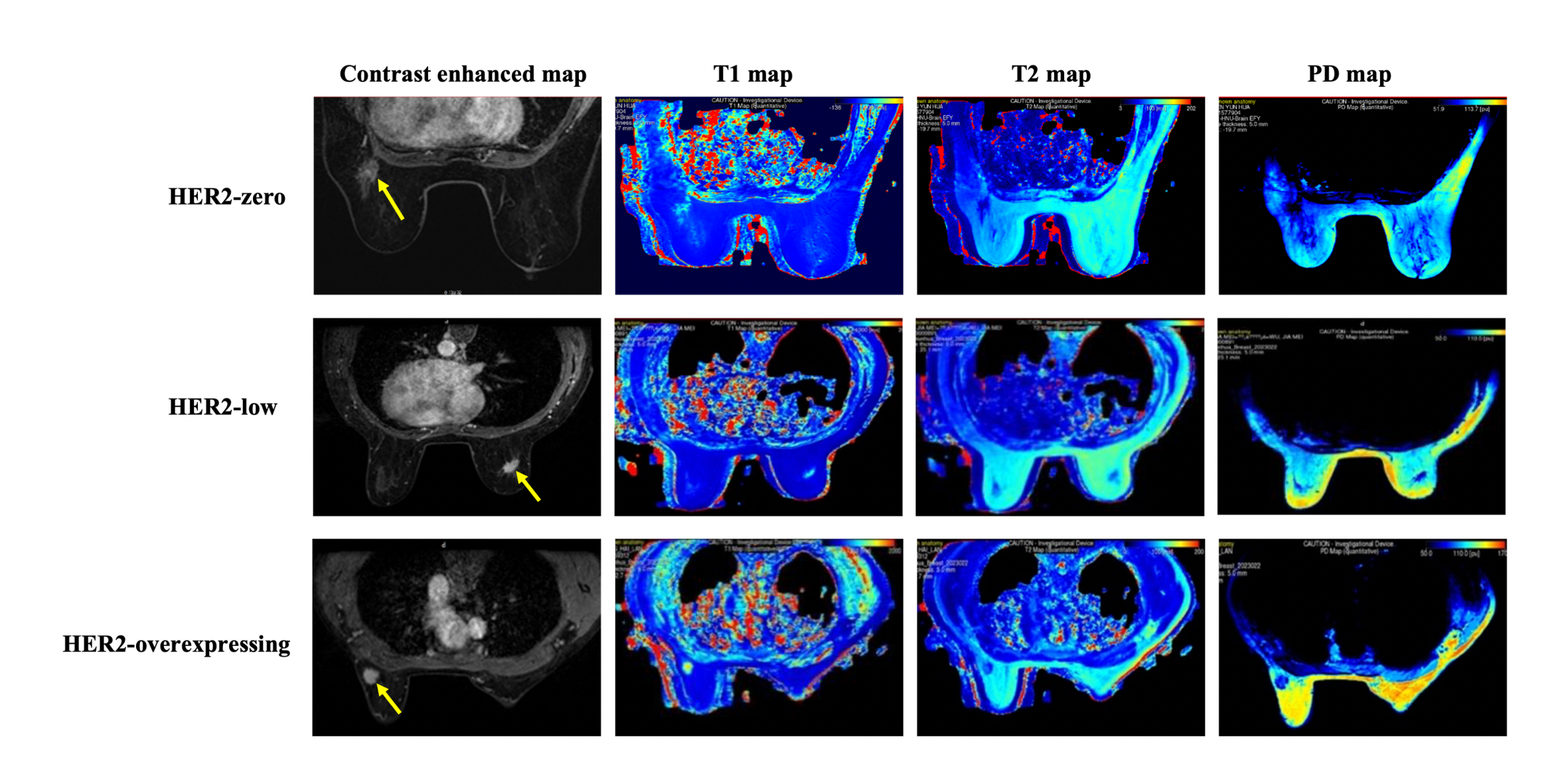
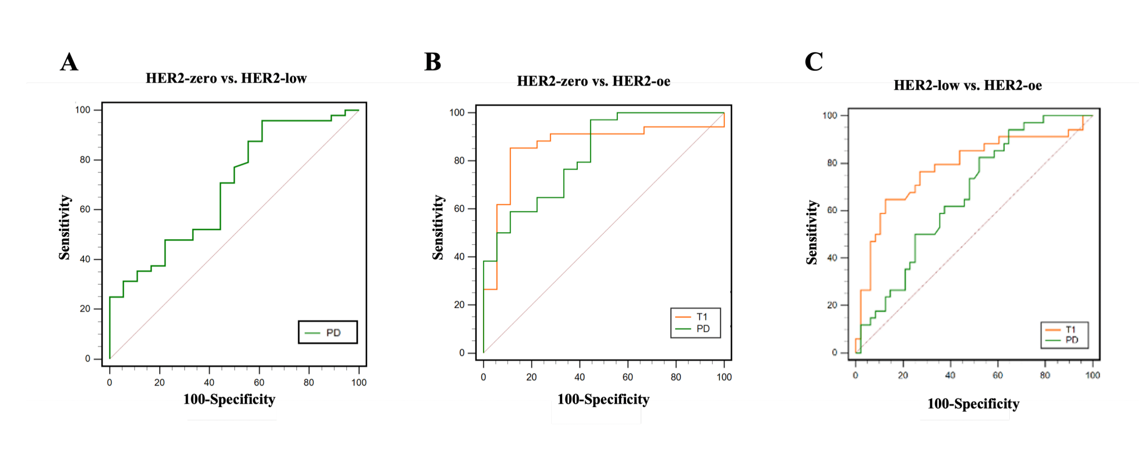
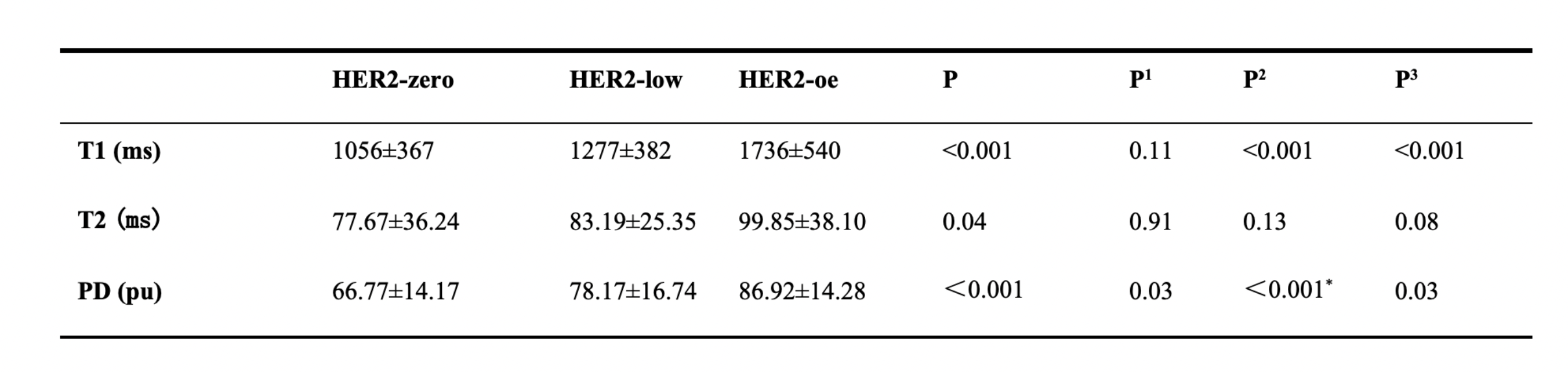
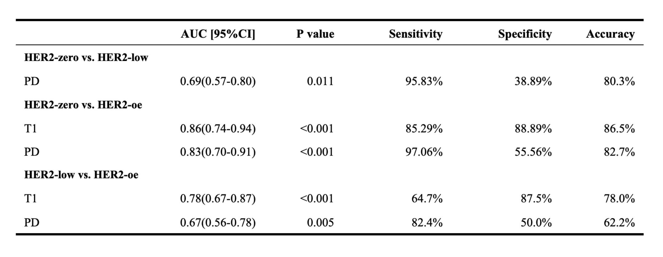
Table 1 and Figure 1 showed the HER2-oe had significantly higher T1 value than HER2-low and HER2-zero (all P<0.05). The T1 value was relatively higher in HER2-low than in HER2-zero but the difference was not significant. The HER2-oe also had significantly higher PD value than HER2-low and HER2-zero (all P<0.05). The PD value was significantly higher in HER2-low than in HER2-zero. Post hoc test showed no significant difference of T2 value among HER2-zero, HER2-low, and HER2-oe.Figure 2 and Table 2 showed PD can significantly differentiating HER2-zero from HER2-low (AUC=0.69). The T1and PD demonstrated good performance in distinguishing HER2-zero from HER2-oe (AUC=0.86 and 0.83, respectively). T1 and PD can significantly discriminate HER2-low from HER2-oe (Table 2 and Figure 2).
Discussion and Conclusion
This study investigated the value of SyMRI in identifying HER2-zero, HER2-low and HER2-oe BC patients. The results showed PD value can classify HER2-zero, HER2-low, and HER2-oe. Although T1 can distinguish HER2-zero from HER2-oe and discriminate HER2-low from HER2-oe, it failed to differentiate HER2-zero from HER2-low. The observed highest T1 and PD in HER2-oe BC patients can be attributed to the high degree of malignancy, poor, differentiation, severe tissue destruction, cell density, and angiogenesis[9].In conclusion, quantitative SyMRI can be used as imaging biomarker to identify HER2-zero, HER2-low and HER2-oe breast cancer patients. It would be beneficial for treatment selection and dynamically monitoring HER2 expressing status during the treatment process.
Acknowledgements
NoneReferences
[1] SUNG H, FERLAY J, SIEGEL R L, et al. Global Cancer Statistics 2020: GLOBOCAN Estimates of Incidence and Mortality Worldwide for 36 Cancers in 185 Countries[J]. CA Cancer J Clin, 2021,71(3): 209-249.
[2] AMOURY M, KOLBERG K, PHAM A T, et al. Granzyme B-based cytolytic fusion protein targeting EpCAM specifically kills triple negative breast cancer cells in vitro and inhibits tumor growth in a subcutaneous mouse tumor model[J]. Cancer Lett, 2016,372(2): 201-209.
[3] SLAMON D J, CLARK G M, WONG S G, et al. Human breast cancer: correlation of relapse and survival with amplification of the HER-2/neu oncogene[J]. Science, 1987,235(4785): 177-182.
[4] DENKERT C, SEITHER F, SCHNEEWEISS A, et al. Clinical and molecular characteristics of HER2-low-positive breast cancer: pooled analysis of individual patient data from four prospective, neoadjuvant clinical trials[J]. Lancet Oncol, 2021,22(8): 1151-1161.
[5] HUANG L, WANG R, XIE K, et al. A HER2 target antibody drug conjugate combined with anti-PD-(L)1 treatment eliminates hHER2+ tumors in hPD-1 transgenic mouse model and contributes immune memory formation[J]. Breast Cancer Res Treat, 2022,191(1): 51-61.
[6] JORDAN N V, BARDIA A, WITTNER B S, et al. HER2 expression identifies dynamic functional states within circulating breast cancer cells[J]. Nature, 2016,537(7618): 102-106.
[7] JI S, YANG D, LEE J, et al. Synthetic MRI: Technologies and Applications in Neuroradiology[J]. J Magn Reson Imaging, 2022,55(4): 1013-1025.
[8] LI Q, XIAO Q, YANG M, et al. Histogram analysis of quantitative parameters from synthetic MRI: Correlations with prognostic factors and molecular subtypes in invasive ductal breast cancer[J]. Eur J Radiol, 2021,139: 109697.
[9] KAZAMA T, TAKAHARA T, KWEE T C, et al. Quantitative Values from Synthetic MRI Correlate with Breast Cancer Subtypes[J]. Life (Basel), 2022,12(9).
Figures