4618
Combination of DCE-MRI and NME-DWI via Deep Neural Network for Predicting Breast Cancer Molecular Subtypes1Imaging Center, Harbin Medical University Cancer Hospital, Harbin, China, 2GE Healthcare, MR Research China, Beijing, China, 3Division of Respiratory Disease, Fourth Affiliated Hospital of Harbin Medical University, Harbin, China
Synopsis
Keywords: Breast, Cancer, Breast Cancer; Molecular subtype; Dynamic contrast-enhanced; Non-mono-exponential model; Deep neural network
Motivation: Breast cancer exhibits diverse molecular subtypes with varying responses to treatment.
Goal(s): This study aims to explore the potential enhancement of breast cancer molecular subtype prediction by combining DCE and NME-DWI through DNNs.
Approach: 475 patients with 480 breast cancers were recruited and classified into molecular subtypes using IHC staining and FISH examination. Manual lesion segmentation and analysis using IVIM, diffusion kurtosis, and stretched exponential models. DNN models for molecular subtype prediction, based on single DCE-MRI or NME-DWI datasets were constructed and compared.
Results: DNN classification accuracy significantly varied among the three imaging datasets (P < 0.05), with MP-MRI outperforming DCE-MRI and NME-DWI.
Impact: This study's integration of DCE-MRI and NME-DWI through DNNs for breast cancer subtype prediction advances non-invasive genotyping, potentially transforming personalized treatment strategies and improving outcomes in breast cancer patients.
Introduction and Purpose
Breast cancer comprises diverse molecular subtypes with varying responses to treatment [1]. Current subtype assessment relies on invasive methods, such as gene expression profiling and immunohistochemical (IHC) analysis of biopsies [2], which may not capture tumor heterogeneity over time [3]. Radiogenomics and deep learning have emerged as powerful tools for non-invasive genotype research [4]. However, there are no reports on predicting molecular subtypes using Deep Neural Networks (DNNs) with dnamic contrast-enhanced (DCE) and non-mono-exponential (NME) model-based diffusion-weighted imaging (DWI). In this study, we aim to investigate whether combining DCE and NME-DWI through DNNs can enhance the prediction of breast cancer molecular subtypes compared to using each imaging technique alone.Materials and Methods
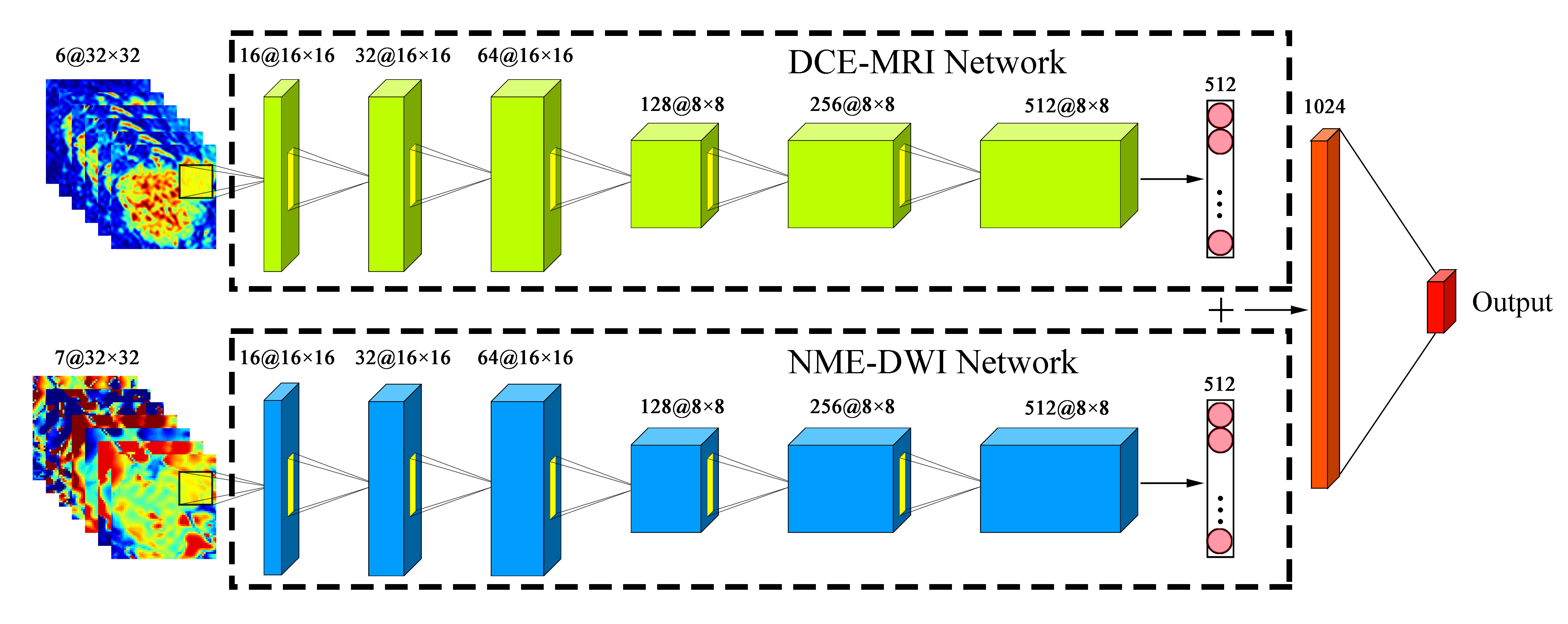
Patients: After obtaining Institutional Review Board approval, we recruited 475 patients with a total of 480 breast cancers. Molecular subtypes were annotated using IHC staining and fluorescence in situ hybridization (FISH) examination results, classified as human epidermal growth factor receptor 2 enriched (HER2-enriched), luminal A, luminal B (HER2–), luminal B (HER2+), and triple-negative (TN) subtypes. Imaging: On a 3.0T MRI scanner (Signa Pioneer, GE Healthcare) with a dedicated seven-channel bilateral breast coil, NME-DWI data was acquired using a single-shot spin-echo echo planar imaging sequence with inversion recovery fat suppression (TR/TE=6443/77 msec, FOV=324×324 mm2, b-values=0, 10, 25, 50, 75, 100, 200, 400, 600, 800, 1000, 1500 and 2000 s/mm2) and DCE-MRI data was acquired using a fat suppressed enhanced T1 high resolution isotropic volume excitation sequence(TR/TE =4.8/2.1 msec, flip angle=12°, FOV=350×350 mm2).Data Processing: Lesions were manually segmented, and multi-b-value DW images were analyzed using the IVIM, diffusion kurtosis, and stretched exponential models. A DNN for predicting molecular subtypes based on single DCE-MRI or NME-DWI datasets was constructed, consisting of an input layer, six convolution layers, and an output layer as shown in Fig.1. We used a training-validation split of 80%-20% with fivefold cross-validation, reserving 20% of cases as an independent test dataset. The predictive performance of the DNN on MP-MRI, DCE-MRI, and NME-DWI testing sets was evaluated using accuracy in 5-way subtype classification, sensitivity, specificity, and the area under the receiver-operating characteristic (ROC) curve (AUC) for binary subtype classification. Statistical Analyses: Group comparisons were conducted using SPSS software (version 25.0, Chicago, IL).Results
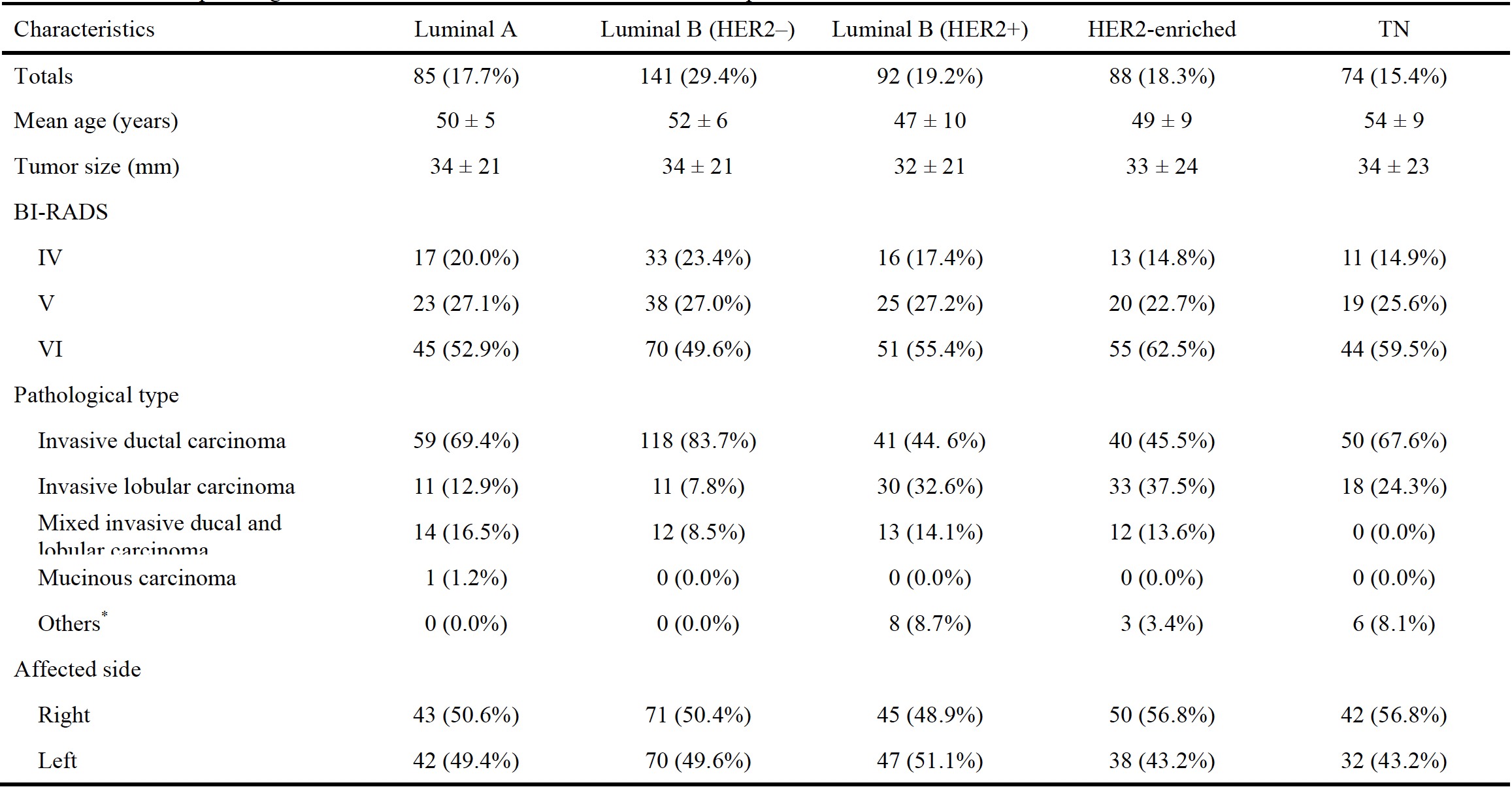
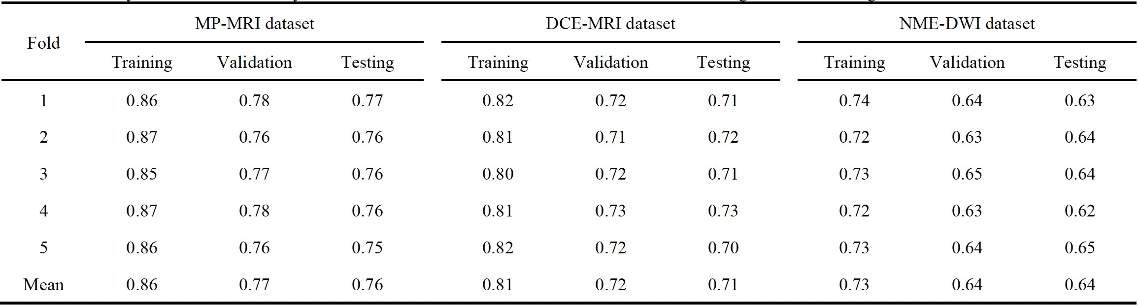
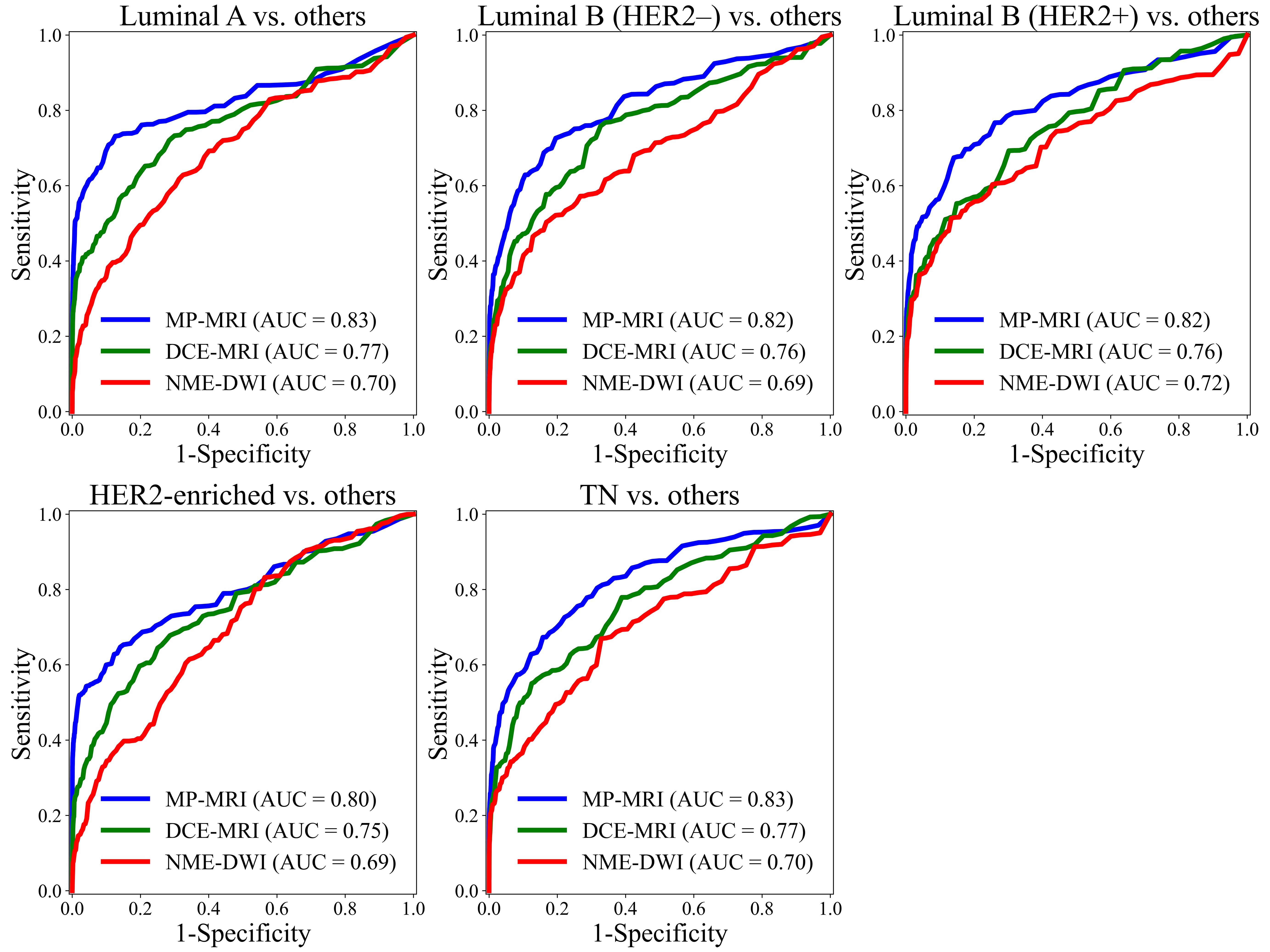
The Kruskal-Wallis H test showed no significant differences between subtypes in terms of age (P = 0.518) and tumor size (P = 0.426) as shown in Fig.2. Categorical characteristics also showed no significant differences between subtypes (P = 0.693 ~ 0.727) by the chi-square or Fisher’s exact test, except for pathological type (P < 0.05). One-way ANOVA with LSD post hoc tests revealed significant differences in DNN classification accuracy among the three imaging datasets (P < 0.05) and between each pair of them (P < 0.05). Specifically, classification accuracies on MP-MRI (0.76) were significantly higher than on both DCE-MRI (0.71) and NME-DWI (0.64) as shown in Fig.3. Additionally, DCE-MRI outperformed NME-DWI, with NME-DWI classification accuracy being significantly lower. The comparative results of binary classification between the three datasets were consistent with the 5-way classification as shown in Fig.4.Discussion and Conclusion
MP-MRI demonstrated a significant improvement in classifying breast cancer molecular subtypes compared to individual imaging techniques. Furthermore, DCE-MRI exhibited superior predictive performance over NME-DWI. The architecture of NME-DWI network was not identical with DCE-MRI network due to more input channels (7 vs. 6 channels), showing the increase of z-axis dimension for filters in the first convolutional layer. This implied that the NME-DWI network had more weights than the DCE-MRI network. But, in practice, the number of added weights was only a very small percentage of all weights in the entire network and thereby had little influence on performance of network. Moreover, despite more weights, the NME-DWI network did not outperform the DCE-MRI network in classifying molecular subtypes. On the other hand, the ensemble network proposed in this study integrated the DCE-MRI and NME-DWI DNNs at the global feature level to differentiate molecular subtypes within the MP-MRI dataset. Comparing this global-feature fusion approach with the input-level ensemble, global-feature fusion enabled a step-by-step training of the network which could effectively mitigate overfitting issues that may arise due to the increased network size resulting from incorporating more input channels. In summary, the combination of DCE-MRI and NME-DWI through DNNs significantly improved breast cancer molecular subtype prediction compared to using each imaging technique alone, with DCE-MRI being the more effective modality for differentiating subtypes.Acknowledgements
Funding: This study was supported by Harbin Medical University Cancer Hospital Haiyan Foundation (No. JJZD2020-02), Harbin Medical University Cancer Hospital Haiyan Foundation (No. JJZD2021-15) and National Natural Science Foundation of China (No. 81701654)References
1.Zardavas D, Irrthum A, Swanton C, Piccart M. Clinical management of breast cancer heterogeneity. Nat. Rev. Clin. Oncol. 2015;12(7):381-394.
2.Perou CM, Sørlie T, Eisen MB, et al. Molecular portraits of human breast tumours. Nature. 2000;406(6797):747-752.
3.Lee JY, Lee K-s, Seo BK, et al. Radiomic machine learning for predicting prognostic biomarkers and molecular subtypes of breast cancer using tumor heterogeneity and angiogenesis properties on MRI. Eur. Radiol. 2022;32(1):650-660.
4.Aerts HJ, Velazquez ER, Leijenaar RT, et al. Decoding tumour phenotype by noninvasive imaging using a quantitative radiomics approach. Nat. Commun. 2014;5:4006.
Figures