4611
Validating the Use of Breast MRE as a Non-Invasive Diagnostic Technique for Breast Cancer1The Ohio State University, Columbus, OH, United States, 2Wexner Medical Center, The Ohio State University, Columbus, OH, United States
Synopsis
Keywords: Breast, Elastography
Motivation: While breast tumors can be identified and characterized through biopsy, MR Elastography (MRE) could provide a method of diagnosis with similar sensitivity and specificity while being non-invasive.
Goal(s): Validate the use of MRE against histological grading in biopsy and contrast enhanced MRI breast tumors.
Approach: MRE was performed on patients with known breast tumors to measure the stiffness non-invasively and validate it against biopsy.
Results: Statistically significant correlation was determined between MRE-derived tumor stiffness and histological grade of biopsy.
Impact: The statistically significant association between breast MRE results, a non-invasive method of breast cancer diagnosis, and histological grading, an invasive method, invites the possibility that physicians could diagnose breast cancer non-invasively through further study.
Introduction
Cancer is the second leading cause of death in the United States and occurs in a multitude of sites in the body, but significant evidence has shown it is more common in some sites than others; for example, female breast cancer1,2. Breast cancer diagnosis is typically done through biopsies, but due to its invasiveness and subjective histological grading, it is not the preferred method, especially if the risk of a malignant tumor is low3. Magnetic Resonance Elastography (MRE) is a noninvasive technique to estimate the stiffness of soft tissues. Earlier studies have demonstrated the use of MRE in diagnosing breast tumors4; however, this study is validating the use of MR Elastography against histological grading of a biopsy.Methods
Fourteen volunteers with known breast tumors were recruited after approval of the institutional review board. All imaging was performed in a 3T MRI scanner (Skyra, Siemens Healthcare, Erlangen, Germany) after obtaining written informed consent from the subjects. Vibrations were introduced at 60 Hz through a soft sternal driver and axial slices of the entire breast volume were collected using a custom-made, single shot, spin-echo echo planar imaging MRE sequence. Imaging parameters included: echo time (TE) = 43.6 ms, repetition time (TR) = 833.33 ms, field of view (FOV) = 360 x 360 mm2, matrix size 128x128, slice thickness = 3 mm, number of slices = 10, MRE phase offsets = 4. Once the images were obtained, the breasts were masked, and custom-made software in Matlab (Mathworks, Natick, MA) was used to apply curl processing to remove longitudinal waves and a bandpass directional filter with cutoff values of 6-20 waves/FOV to remove reflected waves. Finally, Direct Inversion (DI) was performed to obtain stiffness maps. Using contrast-enhanced MRI images that were annotated by a clinician, the tumor was located on the MRE magnitude image and an estimated mean stiffness for the breast tumor was calculated. Through a clinical report, a pathologist confirmed the histological grade for the volunteers’ tumors. A linear correlation was performed between MRE-derived stiffness and histological grade of the tumors.Results
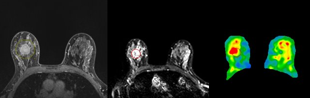
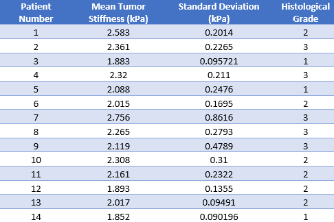
Figure 1 depicts the contrast enhanced breast MRI image used to locate the tumor, the MRE magnitude image, and the corresponding DI stiffness map. Figure 2 shows the mean tumor stiffness (kPa), the standard deviation for each of the fourteen volunteers, and the histological grading. Figure 3 illustrates the significant (p=0.02) correlation between mean tumor stiffness and the histological grading with an R-value of 0.61.Conclusion
The results of this study demonstrate the potential use of breast MRE in diagnosis of breast cancer. The correlation between breast tumor stiffness and histological grading provides the opportunity for a study with a larger sample size to determine the sensitivity and specificity of using MRE for breast tumors.Acknowledgements
This work was supported by NIH R01AR075062References
1. Centers for Disease Control and Prevention. Leading Causes of Death. Centers for Disease Control and Prevention. Published January 18, 2023. https://www.cdc.gov/nchs/fastats/leading-causes-of-death.htm
2. National Cancer Institute. Female Breast Cancer - Cancer Stat Facts. SEER. Published 2018. https://seer.cancer.gov/statfacts/html/breast.html
3. Breast cancer - Diagnosis and treatment - Mayo Clinic. www.mayoclinic.org. https://www.mayoclinic.org/diseases-conditions/breast-cancer/diagnosis-treatment/drc-20352475#:~:text=A%20biopsy%20is%20the%20only
4. Hawley JR, Kalra P, Mo X, Raterman B, Yee LD, Kolipaka A. Quantification of breast stiffness using MR elastography at 3 Tesla with a soft sternal driver: A reproducibility study. Journal of Magnetic Resonance Imaging. 2016;45(5):1379-1384. doi:https://doi.org/10.1002/jmri.25511
Figures
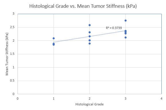
Shows the correlation between mean tumor stiffness and histological grade.