4608
Lipid composition mapping for early response to neoadjuvant chemotherapy in breast cancer using chemical shift-encoded imaging1Institute of Medical Sciences, University of Aberdeen, Aberdeen, United Kingdom, 2Massachusetts General Hospital, Boston, MA, United States, 3Oncology Department, Aberdeen Royal Infirmary, Aberdeen, United Kingdom, 4Royal Marsden Hospital, London, United Kingdom, 5Pathology Department, Aberdeen Royal Infirmary, Aberdeen, United Kingdom, 6Breast Unit, Broomfield Hospital, Mid and South Essex NHS Trust, Chelmsford, United Kingdom, 7Newcastle Magnetic Resonance Centre, Newcastle University, Newcastle, United Kingdom
Synopsis
Keywords: MR-Guided Interventions, Fat, lipid composition, early response, neoadjuvant chemotherapy
Motivation: Deregulation of lipid composition, holding a critical role in breast cancer progression, can be accurately mapped using chemical shift-encoded imaging (CSEI), and might support response monitoring in patients undergoing neoadjuvant chemotherapy (NACT).
Goal(s): We aimed to determine the power of CSEI in differentiation of poor responders after one cycle of NACT.
Approach: Seventeen patients were imaged before and after one cycle of NACT to compute the percentage change in monounsaturated, polyunsaturated and saturated fatty acids.
Results: There was a treatment induced lipid normalisation in all participants, however there was no difference in poor responders in comparison to good responders.
Impact: CSEI is sensitive to the longitudinal change in lipid composition in the peri-tumoural region and the whole breast, however the impact on lipid metabolism is secondary to determining poor responder and other markers of metabolic imaging should be explored.
Introduction
Breast cancer is the most common cancer among women, with age-adjusted annual incidence of 205 per 100,0001. An imbalance of monounsaturated, polyunsaturated and saturated fatty acids (MUFA, PUFA, SFA) has been shown in the peri-tumoural adipose tissue adjacent to breast tumour2. Novel chemical shift-encoded imaging (CSEI) provides a rapid mapping of lipid composition in the breast, and may serve response monitoring of neoadjuvant chemotherapy (NACT) for stratified treatment. We therefore hypothesise that there might be a difference in lipid composition in the peri-tumoural region and the whole breast between good and poor responders after one cycle of NACT.Methods
Seventeen patients (age 46 – 58 years) with invasive ductal carcinoma participated in the longitudinal study to undertake MRI scan at Baseline and after Cycle 1. Patients with a tumour size larger than 2 cm on mammography and have not had hormonal therapy prior to chemotherapy were eligible. Miller-Payne system was used to assess pathologic complete response for good responder3. The study was approved by the London Research Ethics Committee (ID: 17/LO/1777), and written informed consents were obtained from all the participants (Figure 1).Lipid Composition Mapping
All images were acquired on a 3 T whole-body clinical MRI scanner (Achieva TX, Philips Healthcare, Best, Netherlands). Lipid composition images were acquired from the diseased breast in all participants using a 2D CSEI sequence4,5 with 48 echoes, initial echo time of 1.14 ms, echo spacing of 1.14 ms, repetition time of 60 ms, reconstruction matrix of 96 × 96, reconstruction pixel size of 2.5 × 2.5 mm2 and slice thickness of 5.0 mm.
Data Processing
Image analysis was conducted in MATLAB (R2020a, MathWorks Inc., Natick, MA, USA). The maps of the number of double bonds in triglycerides were computed from raw data, before subsequent calculation of quantitative maps of MUFA, PUFA and SFA as a fraction of the total amount of lipids4,5. The boundary of tumour was delineated on the first echo of lipid composition images, with reference to anatomical and diffusion weighted images. The peri-tumoural region was defined as a growth of 15 mm (6 voxels) concentric ring surrounding the tumour boundary. The whole breast was defined to contain only adipose and fibroglandular tissue, and excluding the tumour. Adipose voxels with lipid signal over 60% of total signal were extracted from lipid composition maps. The mean lipid composition from the regions-of-interest was subsequently computed for each lipid constituent. Percentage change in lipid composition was calculated as: [Cycle 1 – Baseline] / Baseline × 100(%).
Statistical Analysis
All statistical analysis was performed in the R software (v3.6.3, R Foundation for Statistical Computing, Vienna, Austria). Wilcoxon signed rank paired tests were performed for comparison of lipid composition in the peri-tumoural region and the whole breast between Baseline and Cycle 1. Wilcoxon rank sum tests were performed for comparison of percentage change in lipid composition between good and poor responders. Statistical significant finding was determined by p < 0.05.
Results
The histopathological findings of the patient cohort are shown in Table 1.Baseline and Cycle 1
In the peri-tumoural region, there was a borderline higher mean MUFA (p=0.055) at Cycle 1 compared to Baseline. There was no significant difference (p=0.073) in mean PUFA between Baseline and Cycle 1. There was a borderline lower mean SFA (p=0.055) at Cycle 1 compared to Baseline (Figure 2).
In the whole breast, there was no significant difference in lipid composition between Baseline and Cycle 1 (Figure 2).
Good and Poor Responders
There was no significant difference in percentage change in lipid composition in the peri-tumoural region and the whole breast between good and poor responders after one cycle of NACT (Figure 3, Table 2).
Discussion
The increase in MUFA after NACT showed treatment induced normalisation, since there was a decrease in MUFA in the peri-tumoural region to sustain elevated membrane synthesis during cancer progression2. The decrease in SFA after NACT showed active tumour regression, since there was an increase in SFA in the peri-tumoural region for enhanced tumour proliferation2. There was no significant difference in the percentage change in lipid composition between good and poor responders, however the cohort size is small and further studies are warranted to unravel lipid regulation during tumour regression and subsequent treatment induced normalisation in patients with breast cancer undergoing NACT.Conclusion
There was a treatment induced lipid normalisation after one cycle of neoadjuvant chemotherapy, although there was no significant difference in percentage change in lipid composition in the breast between good and poor responders.Acknowledgements
The authors would like to thank Dr Matthew Clemence (Philips Healthcare Clinical Science, UK) for clinical scientist support, Ms Erica Banks and Ms Alison McKay for patient recruitment support, Ms Teresa Morris and Ms Dawn Younie for logistics support, and Ms Beverly MacLennan, Ms Nichola Crouch, Ms Laura Reid and Mr Mike Hendry for radiographer support. The authors would also like to thank Ms Mairi Fuller, Mr Dionysios Koufoudakis, Ms Elizabeth Smyth and Ms Beatrix Elsberger for providing access to the patients. This project was funded by Friends of Aberdeen and North Centre for Haematology, Oncology and Radiotherapy (ANCHOR), NHS Grampian Endowment Research Fund and Tenovus Scotland. Sai Man Cheung’s research fellow training is currently funded by Chief Scientist Office.References
1. Smittenaar CR, Petersen KA, Stewart K, Moitt N. Cancer incidence and mortality projections in the UK until 2035. Br J Cancer. 2016;115(9):1147-1155.
2. Chan KS, Cheung SM, Senn N, et al. Peri-tumoural spatial distribution of lipid composition and tubule formation in breast cancer. BMC Cancer. 2022;22(1):285.
3. Ogston KN, Miller ID, Payne S, et al. A new histological grading system to assess response of breast cancers to primary chemotherapy: Prognostic significance and survival. Breast. 2003;12(5):320-327.
4. Bydder M, Girard O, Hamilton G. Mapping the double bonds in triglycerides. Magn Reson Imaging. 2011;29(8):1041-1046.
5. Peterson P, Månsson S. Simultaneous quantification of fat content and fatty acid composition using MR imaging. Magn Reson Med. 2013;69(3):688-697.
Figures
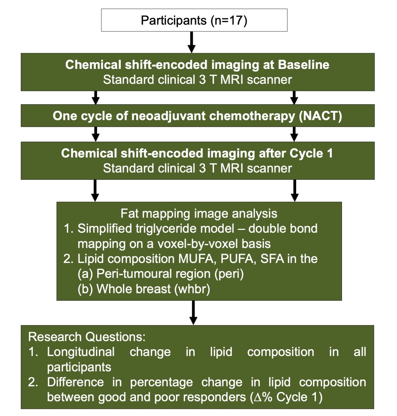
Figure 1. Study design
Seventeen patients with breast cancer participated in the study. All patients underwent chemical shift-encoded imaging on a clinical 3 T MRI scanner at Baseline and after Cycle 1. Fat mapping image analysis was conducted to compute monounsaturated fatty acids (MUFA), polyunsaturated FA (PUFA) and saturated FA (SFA) in the peri-tumoural region (peri) and the whole breast (whbr). Wilcoxon tests were subsequently performed between the time points and responder groups.
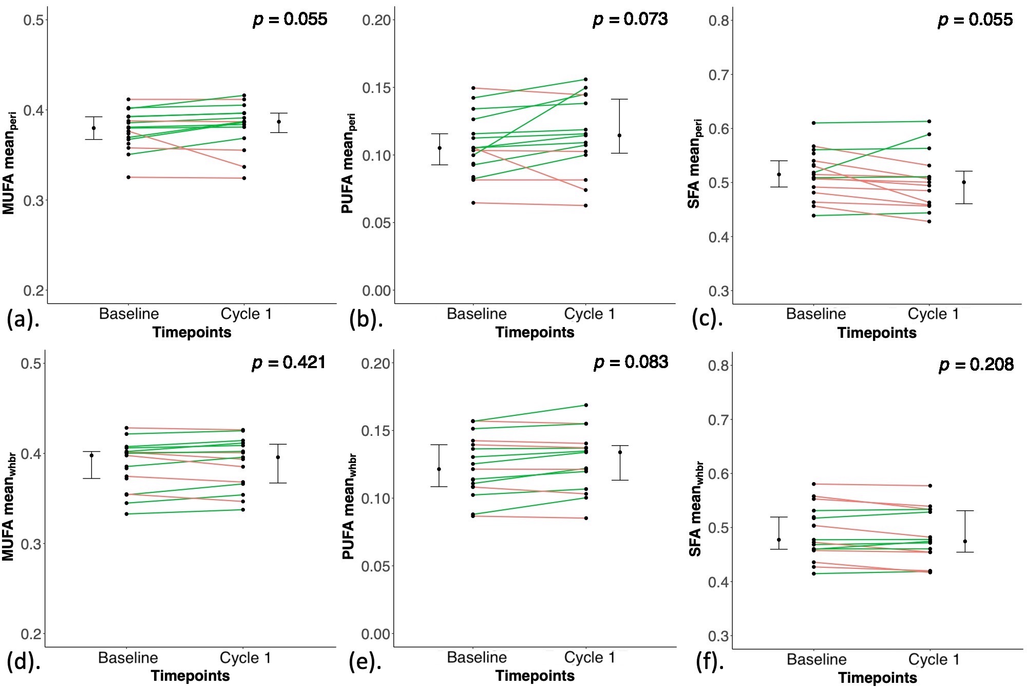
Figure 2. Longitudinal change in lipid composition measurements
The longitudinal change in monounsaturated fatty acids (MUFA), polyunsaturated FA (PUFA) and saturated FA (SFA) in (a-c) the peri-tumoural region (peri) and (d-f) the whole breast at Baseline and after Cycle 1. Each dot represents a peri-tumoural or whole breast mean fraction, and the dots are organised in two columns corresponding to the two time points. Error bars indicate the median (interquartile range).
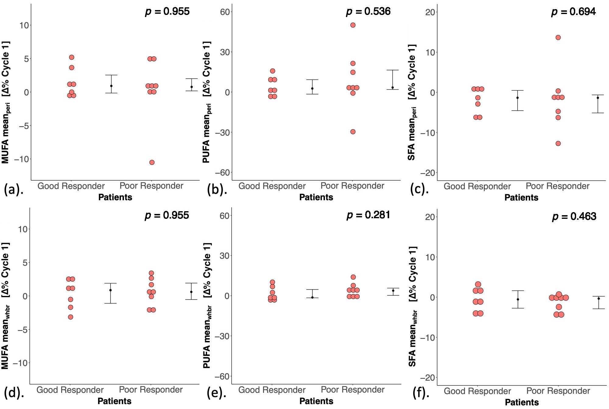
Figure 3. Differences in lipid composition measurements between good and poor responders in the peri-tumoural region and the whole breast
The percentage change after one cycle of NACT (Δ% Cycle 1) in monounsaturated fatty acids (MUFA), polyunsaturated FA (PUFA) and saturated FA (SFA) in (a-c) the peri-tumoural region (peri) and (d-f) the whole breast (whbr) between good and poor responders. Error bars indicate the median (interquartile range).
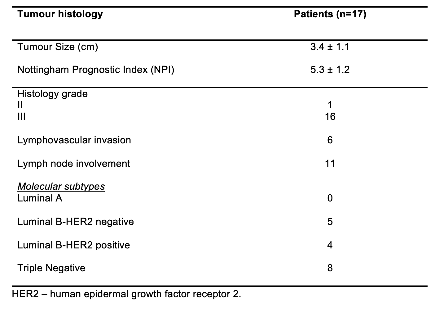
Table 1. Tumour histology in the cancer patient group
Histopathological findings for patients with breast cancer are shown, with quantitative entries expressed as mean and standard deviation (mean ± SD) and qualitative entries expressed as number of positive observations.
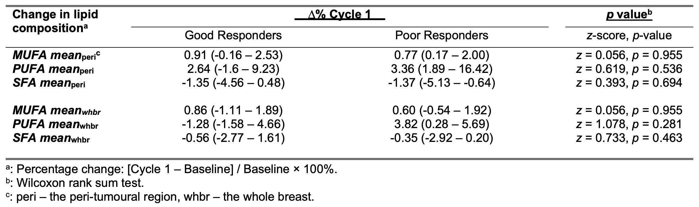
Table 2. Differences in lipid composition measurements between good and poor responders at Cycle 1
Percentage change in the peri-tumoural region and the whole breast monounsaturated, polyunsaturated and saturated fatty acids (MUFA, PUFA, SFA) after one cycle of neoadjuvant chemotherapy (∆% Cycle 1) were compared between good and poor responders.