4598
Preoperative Prediction of Recurrence Risk in Breast Cancer Patients Based on MRI Features1First affiliated hospital of Wenzhou Medical University, Wenzhou, China, 2University of California, Irvine, Irvine, CA, United States, 3Guangxi Normal University, Guilin, China, 4Wenzhou Medical University, Wenzhou, China
Synopsis
Keywords: Breast, Breast
Motivation: Early prediction of recurrence risk is essential to treatment decision-making for breast cancer patients.
Goal(s): To explore potential predictors of recurrence risk based on MRI features and to construct a preoperatively predictive model of risk.
Approach: MRI features of 588 patients were investigated, 397 in training and 191 in testing data. Four machine learning methods were used to construct the predictive model.
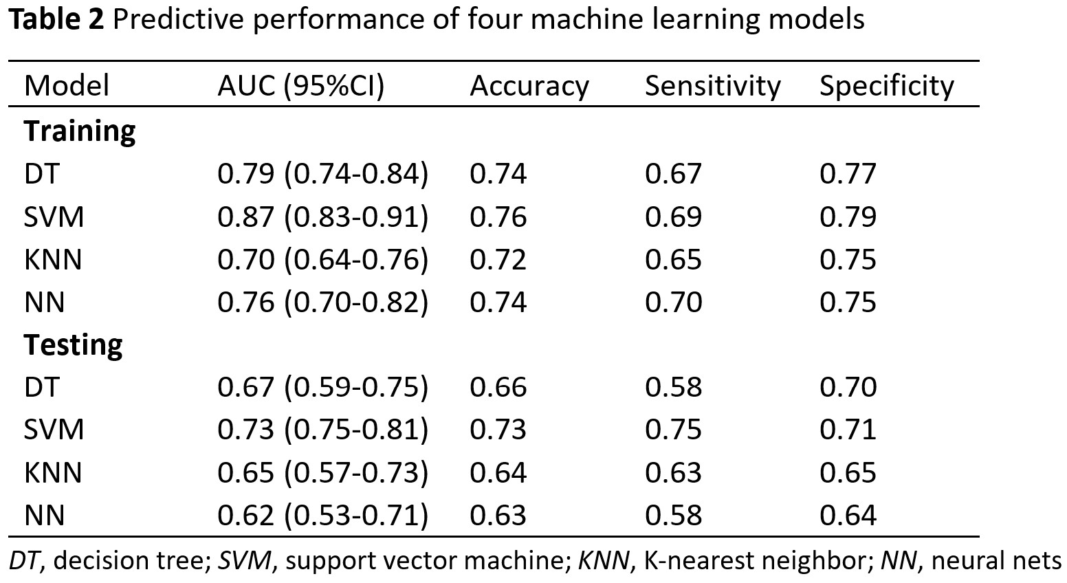
Results: Multiple lesions, irregular shape, spiculated margin, and peritumor edema were identified as predictive factors and used to construct the model. SVM showed the best predictive performance with AUC 0.87 (95%CI 0.83-0.91) and 0.73 (95%CI 0.75-0.81) in training and testing data.
Impact: A preoperative predictive model based on MRI features could be a valuable tool for predicting recurrence risk and assisting in the personalized treatment of breast cancer patients.
Introduction
Breast cancer (BC) is a highly heterogeneous disease, in which patients undergo significant variation in prognosis and survival. Early prediction of recurrence risk and individualized management based on it are highly essential to better prognosis and survival. Multigene test is an accurate method to evaluate risk and prognostic information. However, due to its high cost, the choice of target population, and the lack of a unified interpretation standard, it has yet to be widely used in clinical practice. A more simply used and economic tool is based on comprehensive clinicopathologic parameters. Tumor size, histological grade, and the status of axillary nodes are frequently used for prognosis prediction. Estrogen receptor (ER), progesterone receptor (PR), and human epidermal growth factor receptor-2 (HER2) have been recommended as valuable complement to the risk categories. Ki-67 and age are confirmed to be prognostic factors. However, the main problem is that all of the information can be only obtained by examining tissue samples, which may be confined to the temporal and spatial heterogeneity of cancers. Breast MRI has a unique advantage in obtaining images of the entire tumor and the surrounding tissue. Multi-parametric MRI (mpMRI) with variable functional and morphologic parameters can provide detailed information on the characteristics of BC. Numerous studies have shown that MRI features of BC could be used to assess the prognosis of patients. It is reported that apparent diffusion coefficient (ADC) values correlated with recurrence risk likelihood stratified using Oncotype Dx test recurrence scores and intrinsic imaging phenotypes of BC tumor heterogeneity can predict 10-year recurrence. MpMRI has shown the potential to be an essential tool for prognosis prediction. Therefore, in this study, we aimed to explore potential predictors of recurrence risk based on MRI features and construct a preoperatively predictive model of the risk in BC patients.Methods
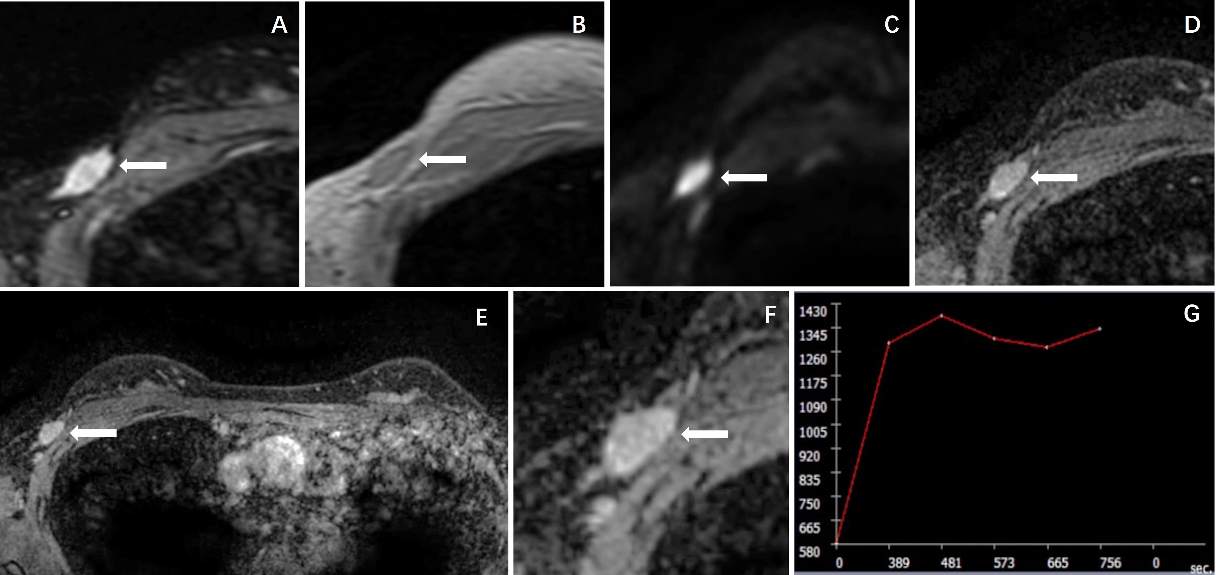
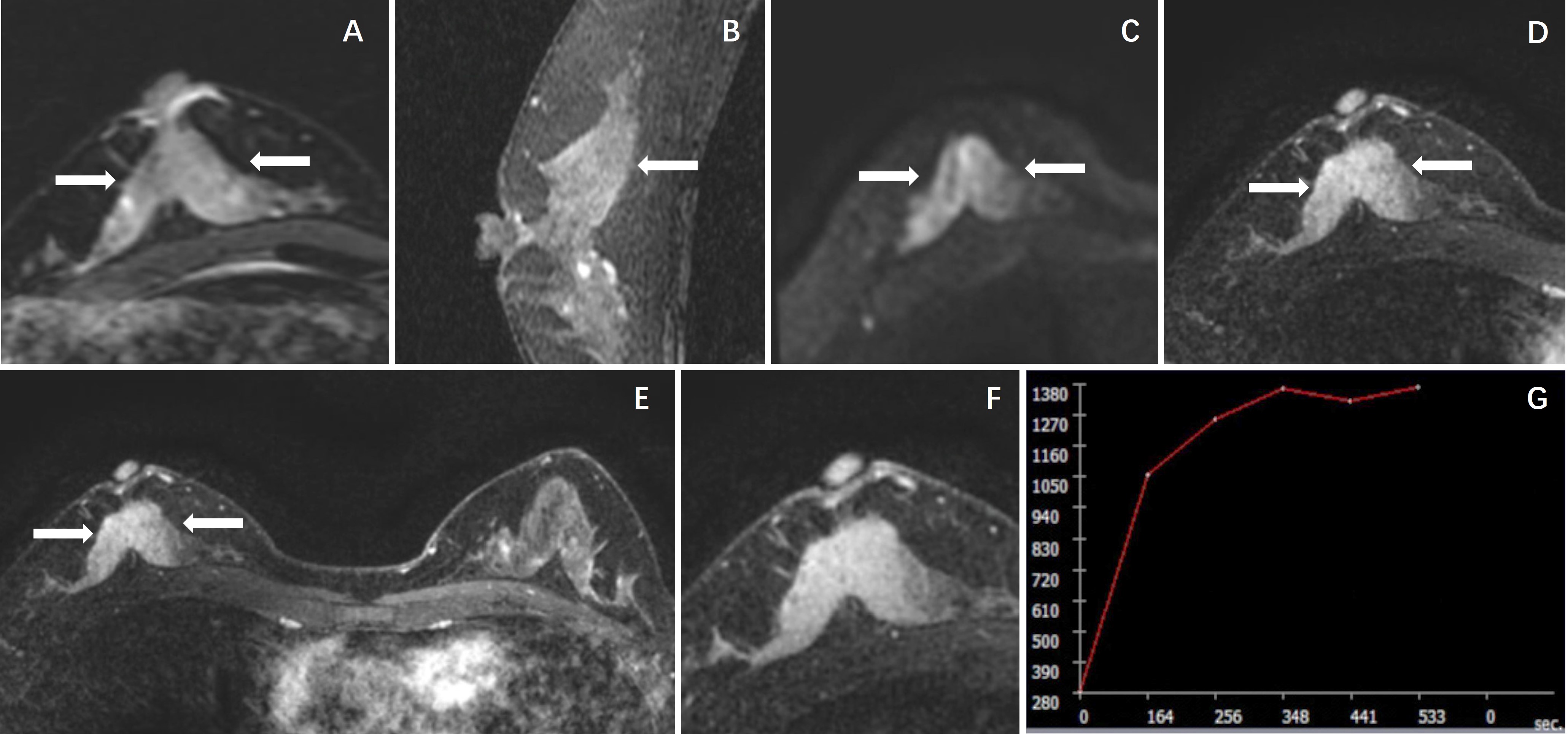
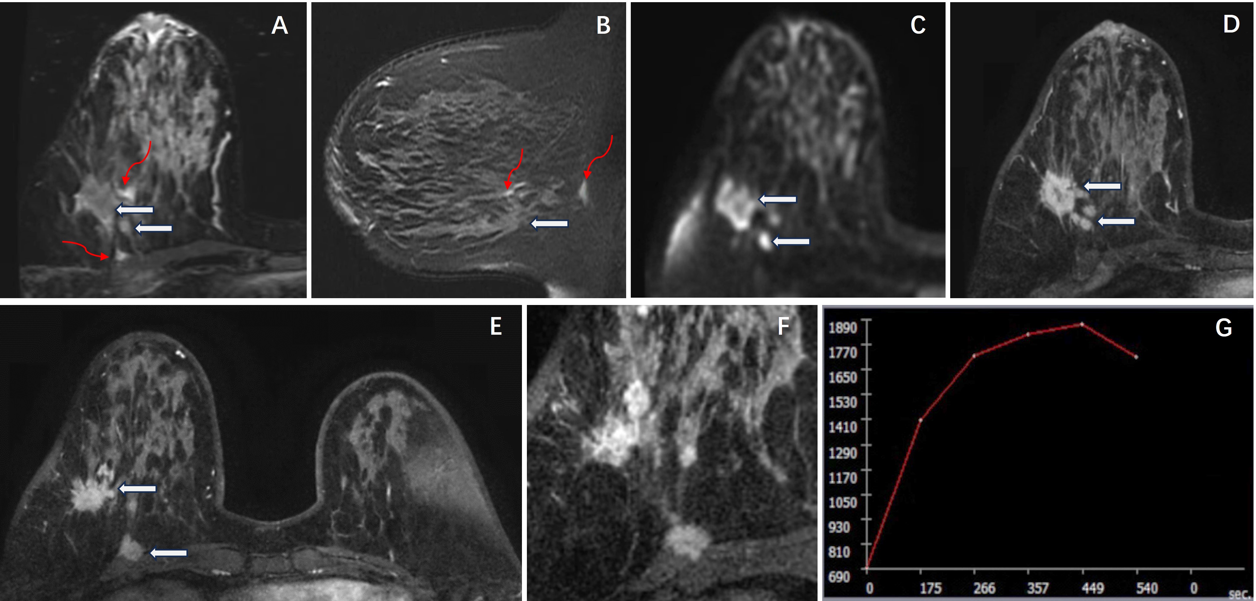
The retrospective study included 588 patients, separating into training (397) and testing (191) datasets. Low-/-intermediate and high-risk were classified based on clinicopathological parameters, including histologic grade, size, lymphovascular invasion (LVI), Ki-67 index, the status of ER, PR, HER2 and axillary node status, and patient age. Patients will be categorized high-risk, if they have 1-3 positive nodes, and the tumor is associated with ER or PR positive and HER2 negative, and Grade 3 or pathological size >5cm; or if they have ≥4 positive nodes. MRI features were reviewed by two radiologists in consensus, including morphology as mass or non-mass enhancement (NME), shape, margin, number of lesions, internal enhancement pattern (IEP), peritumoral edema, ADC value, largest diameter on MRI, and DCE kinetic curve. The predictors of risk were identified by multivariable analysis and used to construct the predictive model by machine learning (ML) algorithms, including Decision Tree (DT), Support Vector Machine (SVM), K-nearest Neighbor (KNN) and Neural Nets (NN).Results
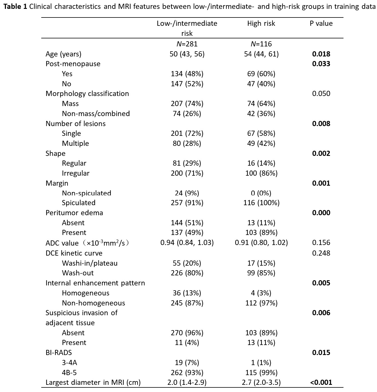
In training data, 281 and 116 patients were classified as low-/intermediate- and high-risk. Age, post-menopause, shape, margin, edema, IEP, suspicious invasion of adjacent tissue, BI-RADS, and largest diameter on MRI showed significant differences in the univariable analysis (Table 1). Four independent factors, multiple lesions, irregular shape, spiculated margin, and peritumor edema, were selected to construct the predictive model. Among four ML models, SVM showed the best predictive performance with AUC 0.87, accuracy 0.76, sensitivity 0.69, and specificity 0.79 in the training data, which was 0.73, 0.73, 0.75, and 0.71, respectively, in testing data (Table 2).Discussion
Although the advancement of adjuvant chemotherapy has reduced the mortality of BC patients, it is still challenging to determine who will truly benefit from such treatments. Accurate assessment of the high risk of recurrence is an essential basis of systemic treatment for patients. Different from invasive and poorly repeatable examination of tissue samples, breast MRI could be a convenient and non-invasive preoperative prediction tool for BC. In this study, multiple lesions, irregular shape, spiculated margin, and peritumor edema were identified as independent predictive factors and included in the machine learning modeling. Four ML models presented decent predictive performance, especially for SVM of AUC 0.87. The results showed a high specificity of above 70%, which means that most of the patients with non-high risk could be predicted by the model and then excluded from adjuvant chemotherapy. Although the research on artificial intelligence (AI) in medical imaging has been extensively conducted, there needs to be a mature AI tool for radiologists to use in clinical work. The obtained MRI reading features demonstrated the potential to be predictors of recurrence risk, and the convenient predictive model based on MRI features using ML modeling could be an assistant in the non-invasively preoperative prediction of risk.Acknowledgements
This study was supported in part by Research Incubation Project of First Affiliated Hospital of Wenzhou Medical University (No. FHY2019085), Wenzhou Science & Technology Bureau (No. Y20210232), Zhejiang Provincial Natural Science Foundation of China (LY21F020030) and Key Laboratory of Intelligent Medical Imaging of Wenzhou (No. 2021HZSY0057).References
[1]. Liang Y, Zhang H, Song X, et al. Metastatic heterogeneity of breast cancer: Molecular mechanism and potential therapeutic targets. Semin Cancer. 2020, Biol 60:14-27.
[2]. Valastyan S, Weinberg RA. Tumor metastasis: molecular insights and evolving paradigms. Cell. 2011, 147:275-292.
[3]. Zeng C, Zhang J. A narrative review of five multigenetic assays in breast cancer. Transl Cancer Res, 2022, 11(4):897-907.
[4]. Jahn SW, Bosl A, Tsybrovskyy O et al. Clinically high-risk breast cancer displays markedly discordant molecular risk predictions between the MammaPrint and EndoPredict tests. Br J Cancer, 2020, 122(12):1744-1746.
[5]. Bauer K, Parise C, Caggiano V. Use of ER/PR/HER2 subtypes in conjunction with the 2007 St Gallen Consensus Statement for early breast cancer. BMC Cancer, 2010, 21:10:228.
[6]. Gamucci T, Vaccaro A, Ciancola F et al. Recurrence risk in small, node-negative, early breast cancer: a multicenter retrospective analysis. J Cancer Res Clin Oncol, 2013, 139(5):853-60.
[7]. Marino, M.A, Helbich, T, Baltzer, P, et al. Multi-parametric MRI of the breast: A review. J Magn Reson Imaging, 2018, 47(2):301-315.
[8]. Yu Y, Tan Y, Xie C, et al. Development and validation of a preoperative magnetic resonance imaging radiomics-based signature to predict axillary lymph node metastasis and disease-Free survival in patients with early-stage breast cancer. JAMA Netw Open,2020, 3(12):e2028086.
[9]. Galati F, Rizzo V, Trimboli RM, et al. MRI as a biomarker for breast cancer diagnosis and prognosis. BJR Open 2022, 4:20220002.
[10]. Thakur SB, Durando M, Milans S, et al. Apparent diffusion coefficient in estrogen receptor-positive and lymph node-negative invasive breast cancers at 3.0T DW-MRI: A potential predictor for an oncotype Dx test recurrence score. J Magn Reson Imaging, 2018, 47(2):401-409.
[11]. Chitalia R, Rowland J, McDonald E, et al. Imaging phenotypes of breast cancer heterogeneity in preoperative breast Dynamic Contrast Enhanced Magnetic Resonance Imaging (DCE-MRI) scans predict 10-year recurrence. Clin Cancer Res, 2020, 26(4): 862–869.
Figures