4592
Whole-tumor histogram models based on quantitative maps from SyMRI for predicting axillary lymph node status in invasive ductal breast cancer1Department of Radiology, Fujian Medical University Union Hospital, Fuzhou, Fujian, China, 2GE Healthcare, Beijing, China
Synopsis
Keywords: Breast, Breast
Motivation: Breast cancer is closely associated with ALN status, influencing prognosis. Sentinel lymph node (SLN) biopsy, a common ALN staging method, has limitations.
Goal(s): This study aimed to explore a non-invasive predictive approach for ALN status in IDC patients using SyMRI images and histogram analysis.
Approach: We included 212 patients, and compared the performance of SyMRI histogram models in differentiating N0 and N+ groups (further divided into N1 and N2-3).
Results: Combining quantitative map features with clinical data achieved the highest diagnostic accuracy. Additionally, specific histogram features were found to differ significantly between N1 and N2-3 groups. Conventional parameters were less discriminative.
Impact: We demonstrated efficacy of histogram analysis of SyMRI as a non-invasive method for predicting ALN status. Model combining SyMRI quantitative maps and clinical features yielded satisfactory performance, highlighting the potential of our proposed model in ALN management.
Introduction
Breast cancer is a global public health concern, with axillary lymph node (ALN) status being a critical prognostic factor1-3. Sentinel lymph node (SLN) biopsy is commonly used to stage ALNs, but its invasiveness and time-consuming frozen section analysis raise concerns. Noninvasive preoperative methods are sought to streamline ALN management. MRI is valuable for breast cancer detection and ALN metastasis prediction4,5. Studies have identified various primary tumor characteristics associated with ALN metastasis6,7. MRI provides both morphological and functional insights, including vascularity and cellularity, potentially linked to ALN metastasis8,9. Synthetic MRI (SyMRI) is an emerging technique that rapidly quantifies tissue properties including T1, T2, and proton density10. These parameters reflect cellular-level differences and can serve as imaging biomarkers for receptor status and Ki-67 status11. Histogram analysis, which evaluates pixel gray level distributions in medical images, offers a means to quantify tumor heterogeneity12,13. Utilizing whole-tumor histogram analysis of quantitative parameters from SyMRI images pre- and post-contrast enhancement may provide valuable insights into predicting ALN status. Therefore, this study aimed to explore the potential of histogram analysis in predicting ALN status using pre- and post-contrast SyMRI images in patients with invasive ductal carcinoma (IDC).Materials and Methods
This retrospective study enrolled 359 women with suspected breast cancer after Institutional Review Board approval. All MRI examinations were performed on a 3.0 T MRI system (SIGNA Pioneer; GE Healthcare, Milwaukee, WI). The protocol included T2WI, DWI, pre- and post-contrast SyMRI, and DCE-MRI. 3D ROIs were delineated semiautomatically on DCE-MRI images using ITK-SNAP software. Histogram features were extracted using PyRadiomics software. Conventional approach was performed by drawing a 2D ROI on the largest lesion area on the SyMRI T2 map with reference to DCE MRI to extract mean quantitative parameters. Clinicopathologic data, including age, receptor status, HER-2 status, Ki67 index, clinical T stage, multifocality, and ALN status, were collected. Patients were categorized into N0 (non-metastatic ALN) and N+ (metastatic ALN) groups, with the N+ group further divided into N1 (1-3 metastatic ALNs) and N2-3 (> 3 metastatic ALNs) groups. Differences between ALN status groups were assessed using t-tests and Chi-square tests. Univariate analysis used the Mann-Whitney U test. Pearson correlation coefficients were calculated, and features with high correlations were retained. Multivariate logistic regression models were constructed, and ROC curve analysis was used to evaluate model performance.Results
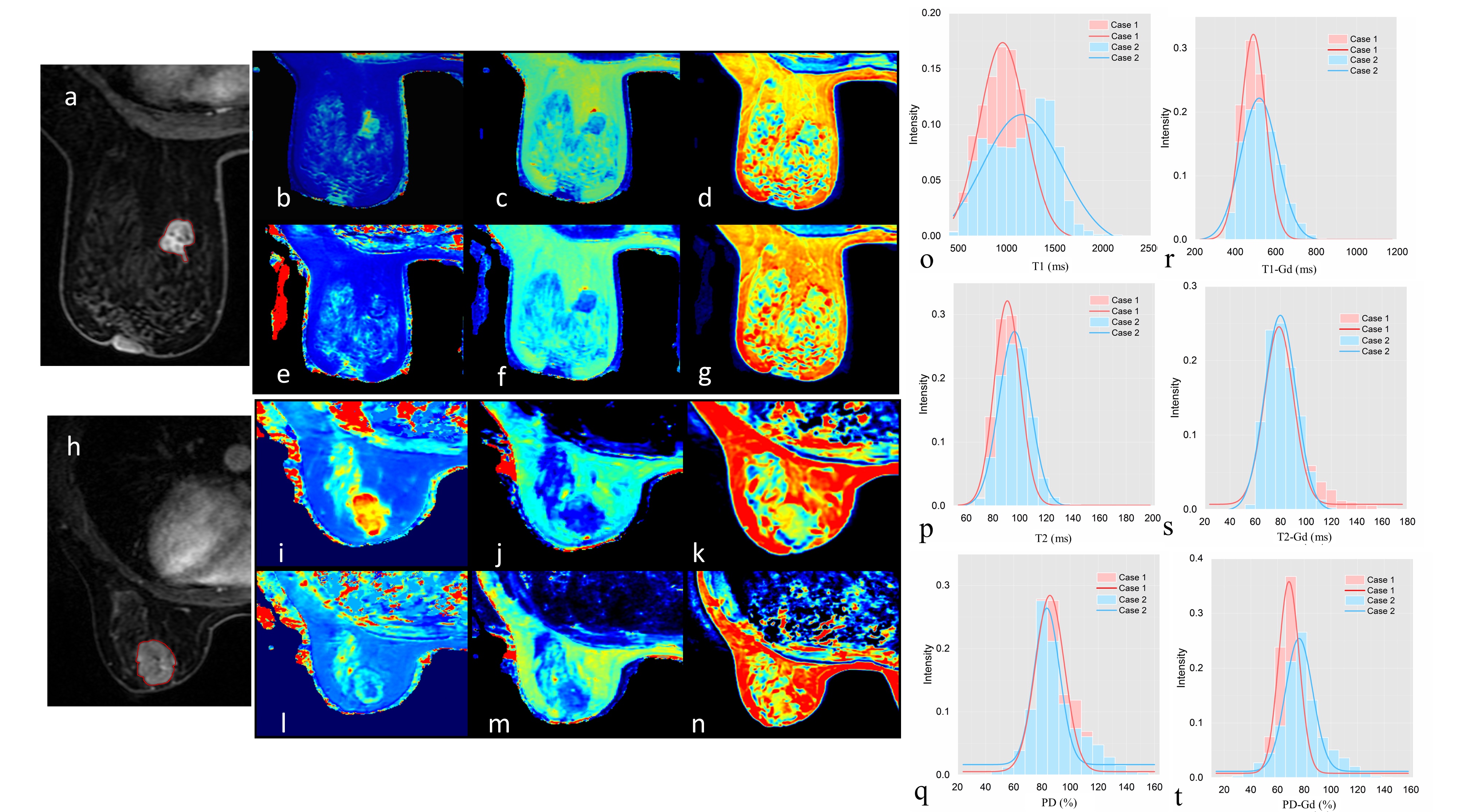
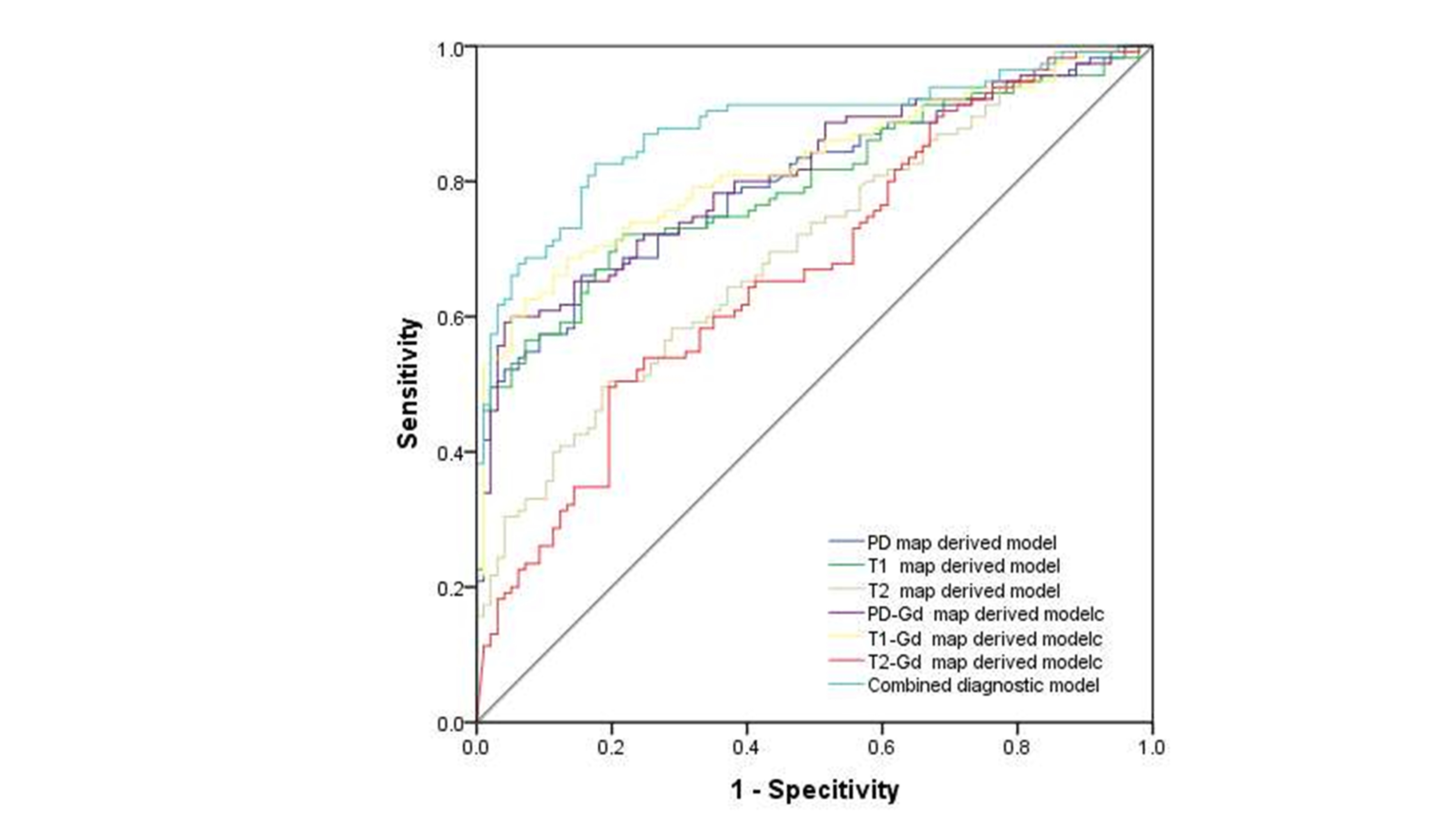
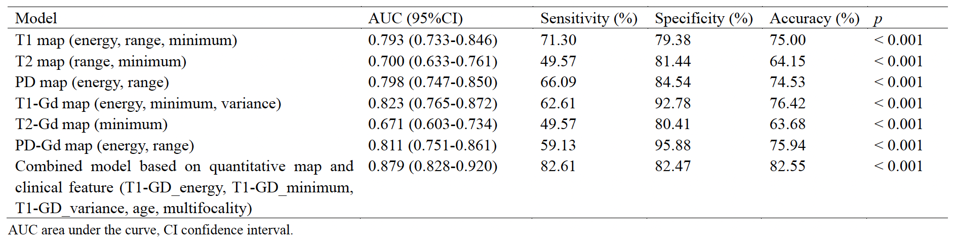
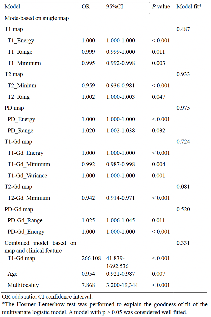
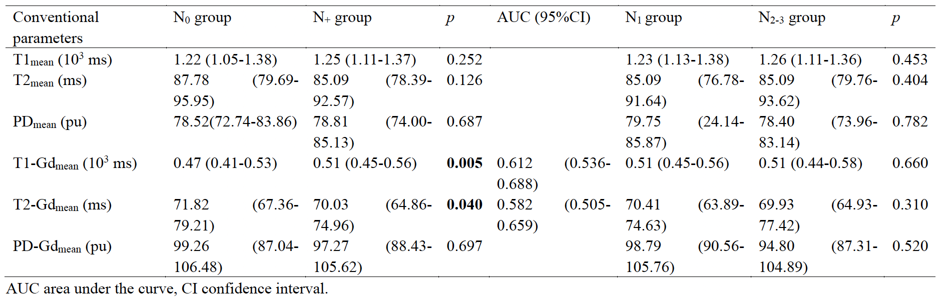
A total of 212 IDC patients were finally included. Among them, 97 cases were in N0 group, while 115 were in N+ group. Among the N+ group, 65 were in N1 group, and 50 were in N2-3 group. Representative examples showing the SyMRI quantitative maps and corresponding histograms are illustrated in Figure 1. For models derived from single quantitative map, T1 model included energy, range, and minimum, with an AUC of 0.793 (Table 1 and Figure 2). T2 model was based on energy and minimum, with an AUC of 0.700. PD model used energy and range and achieved an AUC of 0.798. The T1-Gd model included energy, minimum, and variance, with an AUC of 0.823. The T2-Gd model utilized only the minimum feature, with an AUC of 0.671. The PD-Gd model incorporated energy and range, with an AUC of 0.811. Statistical differences were observed between the N0 and N+ groups in age, clinical T stage, Ki-67 index, and multifocality. A combined model integrating quantitative map and clinical features achieved a significantly higher AUC of 0.879 compared to single diagnostic models. Results for multivariate logistic combined models can be found in Table 2. A total of 27 histogram features from 6 quantitative parametric maps exhibited significant differences between N1 and N2-3 groups. Finally, the diagnostic model selected T2 entropy and PD-Gd energy, with an AUC of 0.722. Regarding the conventional approach, the N0 group displayed significantly lower T1-Gd_mean and higher T2-Gd_mean values than the N+ group, with no significant differences in other conventional parameters between two groups (Table 3). The AUC of T1-Gd_mean and T2-Gd_mean was significantly lower than the T1-Gd and T2-Gd diagnostic models. The N1 and N2-3 groups had similar distributions of all conventional parameters.Discussion
This study successfully demonstrated the efficacy of histogram analysis of SyMRI before and after contrast agent administration as a non-invasive method for predicting ALN status. The logistic regression model combining SyMRI quantitative maps and clinical features yielded a satisfactory AUC of 0.879. Notably, T2-GD_entropy and PD-GD_energy emerged as promising parameters for differentiating ALN between N1 and N2-3 groups. This study represented the first attempt to evaluate SyMRI's predictive ability for ALN status in breast cancer, offering a valuable addition to future clinical practice.Acknowledgements
No acknowledgement found.References
1. Sung H, Ferlay J, Siegel RL et al (2021) Global Cancer Statistics 2020: GLOBOCAN Estimates of Incidence and Mortality Worldwide for 36 Cancers in 185 Countries. CA Cancer J Clin 71:209-249
2. Galimberti V, Cole BF, Viale G et al (2018) Axillary dissection versus no axillary dissection in patients with breast cancer and sentinel-node micrometastases (IBCSG 23-01): 10-year follow-up of a randomised, controlled phase 3 trial. Lancet Oncol 19:1385-1393
3. Giuliano AE, Ballman KV, McCall L et al (2017) Effect of Axillary Dissection vs No Axillary Dissection on 10-Year Overall Survival Among Women With Invasive Breast Cancer and Sentinel Node Metastasis: The ACOSOG Z0011 (Alliance) Randomized Clinical Trial. JAMA 318:918-926
4. Marino MA, Helbich T, Baltzer P, Pinker-Domenig K (2018) Multiparametric MRI of the breast: A review. J Magn Reson Imaging 47:301-315
5. Yu Y, He Z, Ouyang J et al (2021) Magnetic resonance imaging radiomics predicts preoperative axillary lymph node metastasis to support surgical decisions and is associated with tumor microenvironment in invasive breast cancer: A machine learning, multicenter study. EBioMedicine 69:103460
6. Ansari B, Morton MJ, Adamczyk DL et al (2011) Distance of breast cancer from the skin and nipple impacts axillary nodal metastases. Ann Surg Oncol 18:3174-3180
7. Zhao M, Wu Q, Guo L, Zhou L, Fu K (2020) Magnetic resonance imaging features for predicting axillary lymph node metastasis in patients with breast cancer. Eur J Radiol 129:109093
8. Mann RM, Cho N, Moy L (2019) Breast MRI: State of the Art. Radiology 292:520-536
9. Liu Y, Luo H, Wang C et al (2022) Diagnostic performance of T2-weighted imaging and intravoxel incoherent motion diffusion-weighted MRI for predicting metastatic axillary lymph nodes in T1 and T2 stage breast cancer. Acta Radiol 63:447-457
10. Warntjes JB, Dahlqvist O, Lundberg P (2007) Novel method for rapid, simultaneous T1, T2*, and proton density quantification. Magn Reson Med 57:528-537
11. Gao W, Yang Q, Li X et al (2022) Synthetic MRI with quantitative mappings for identifying receptor status, proliferation rate, and molecular subtypes of breast cancer. Eur J Radiol 148:110168
12. Li Q, Xiao Q, Yang M et al (2021) Histogram analysis of quantitative parameters from synthetic MRI: Correlations with prognostic factors and molecular subtypes in invasive ductal breast cancer. Eur J Radiol 139:109697
13. Xie T, Zhao Q, Fu C et al (2019) Differentiation of triple-negative breast cancer from other subtypes through whole-tumor histogram analysis on multiparametric MR imaging. Eur Radiol 29:2535-2544
Figures