4591
Comparison of two deep learning models for contrast agent dose reduction in dynamic contrast enhanced breast MRI1Diagnostic and Interventional Radiology, University Hospital RWTH Aachen, Aachen, Germany, 2Diagnostic and Interventional Radiology, Uniklinik RWTH Aachen University, Aachen, Germany
Synopsis
Keywords: Breast, Machine Learning/Artificial Intelligence
Motivation: In the context of MRI-based breast cancer screening, reducing contrast agent dose is desirable. However, this yields decreased contrast-to-noise ratio in dynamic-contrast-enhanced subtraction images.
Goal(s): This work aimed to compare two deep learning techniques, diffusion probabilistic models (DDPM) and general adversarial networks (GAN), for retrospective contrast enhancement of low-dose breast MRI subtraction images.
Approach: Training and testing was performed on virtual low dose subtraction images, which we generated by subjecting original subtraction images to different amounts of noise.
Results: Both DDPM and GAN could denoise these images; however, neither model was superior over the other across all tested dose levels and evaluation metrics.
Impact: Diffusion probabilistic models and general adversarial networks can retrospectively enhance the signal of virtual low-dose images. They may supplement imaging with reduced doses in the future; yet, further development and validation on real low dose images are warranted.
Introduction
The current guidelines of the European Society of Breast Imaging call for the introduction of MRI-based screening for women with dense breasts1. Breast MRI relies on the dynamic contrast-enhanced (DCE) sequence, which acquires one dynamic phase before and several ones after bolus injection of Gadolinium-based contrast agent (GBCA). Malignant lesions are detectable on the first post-contrast dynamic scan2. Especially in a screening context, less GBCA dose is desirable. Yet, dose reduction decreases contrast-to-noise ratio (CNR) in subtraction images. Deep learning (DL) may help reduce GBCA dose without sacrificing image quality3-5. Here we compare two types of DL models, denoising diffusion probabilistic models (DDPM) and generative adversarial networks (GAN), to retrieve original-dose from low-dose breast MRI subtraction images.Methods
The institutional review board approved this retrospective study on in-house data (EK028/19). 9551 examinations (i.e., 19102 single-sided breast examinations) constituted the training dataset. The test set included 50 single-sided breast examinations with malignant lesions lesions.Axial bilateral breast MRI was performed at 1.5T (Philips Achieva or Philips Ambition, Best, The Netherlands; 4-channel breast coil; breast immobilization along craniocaudal direction). The T1-weighted gradient echo DCE series consisted of 1(4) dynamic scans performed prior to(after) GBCA injection, respectively.
Virtual low-dose subtraction images corresponding to 25%, 10% and 5% dose were simulated by adding white Gaussian noise to the original DCE subtraction images3.
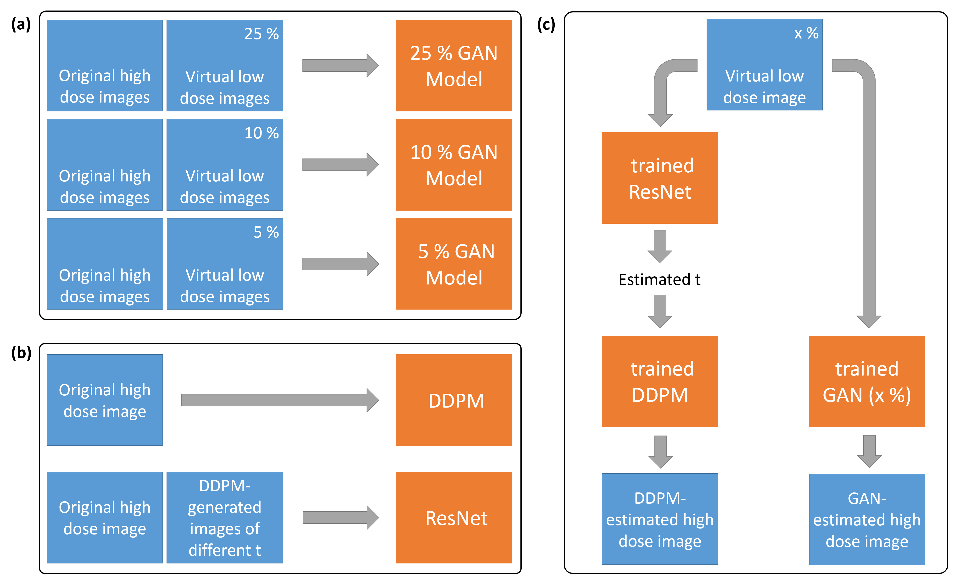
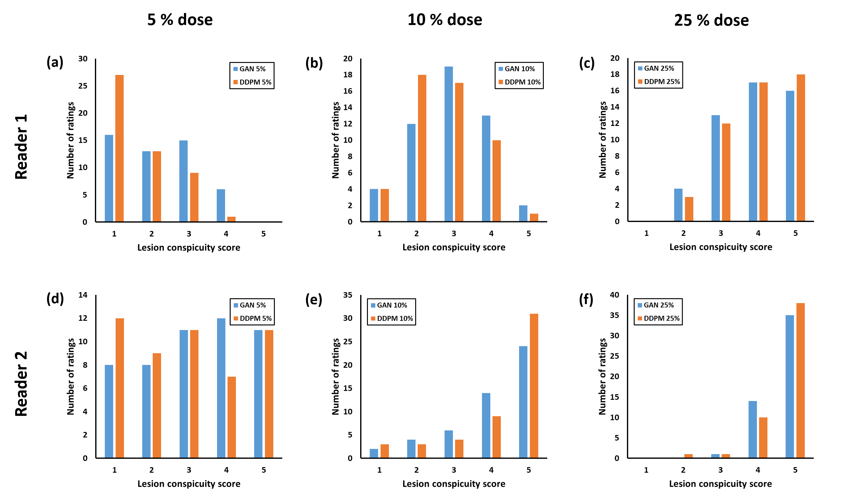
DDPM and GAN were trained for synthetic image generation (Figure 1). DDPMs add noise to an input image in successive time steps t and learn to revert this process step-by-step. For training of the DDPM6, a cosine noise schedule with 1000 steps, L1-loss and the AdamW optimizer (AWO) with a learning rate LR=0.0001 were used. To determine the number of time steps required to retrieve an image’s original dose level, we trained a ResNet-347 separately from the DDPM (AWO, LR=0.0001). Three distinct GAN models were trained (i.e., one per dose level) based on the Pix2PixHD GAN model8 with AWO and LR=0.0001. In total, 150 DDPM- and 150 GAN-reconstructed breast volumes were obtained. Two radiologists (6 and 7 years of experience in breast imaging) indicated their preference for a model when presented with original, GAN- and DDPM-reconstructed images side-by-side. Additionally, they rated lesion conspicuity as compared to the original image from 1=”poor” to 5=”excellent”. Readers were blinded to model type and dose level. Two-sided binomial tests were used to discern their preference from random choice. The effect of dose, model and reader on lesion conspicuity was evaluated by ordinal mixed effects logistic regression followed by Bonferroni-corrected pair-wise post-hoc tests.
Results
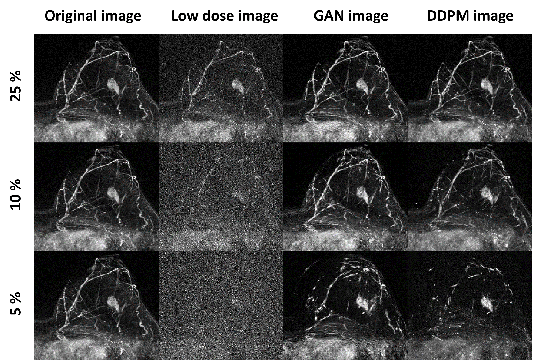
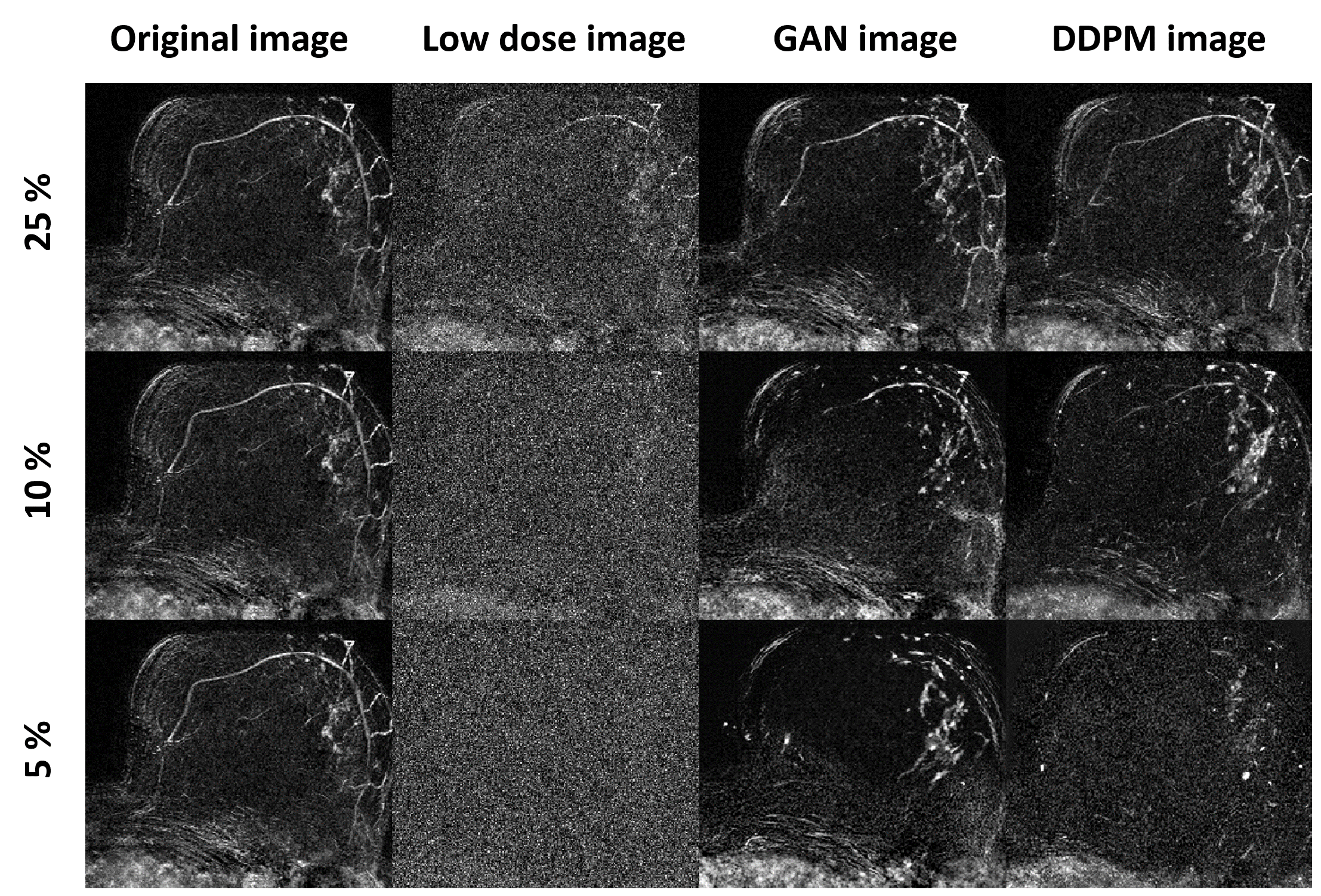
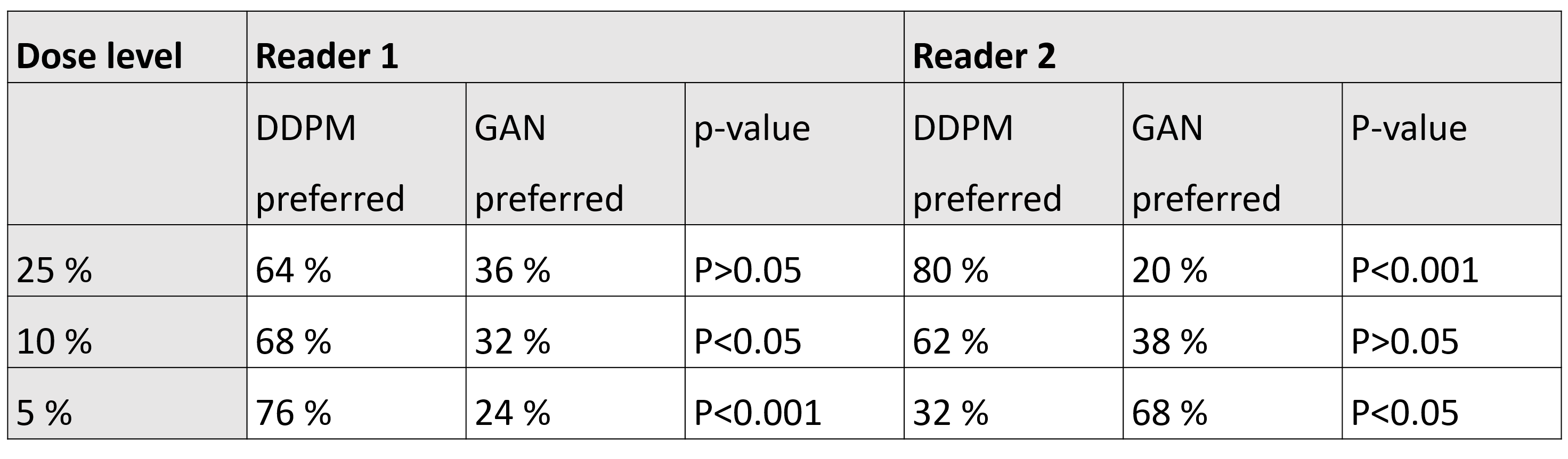
Patients in the training and two test cohorts were 56±10 and 57±10 years old. Figure 2 shows a 48 year-old patient with mass enhancement (invasive breast cancer [NST, G3, pT1c]). Although the lesion was reconstructed even at 5%, fine details of its margin and also the vessels in the breast are no longer discernible. Figure 3 shows a 67-year-old woman with non-mass enhancement (extensive segmental high-grade ductal carcinoma in situ). From 25% to 10%, there is a slight decrease in image quality for both models, which becomes more pronounced at 5%. In the 5% DDPM-image, the enhancement appears fainter and less well defined as compared to the 5% GAN-image. Both readers preferred the DDPM-images at 25% (P=0.06 and P<0.001 for reader 1 and 2), whereas they preferred the GAN-images at 5% (P<0.001 and P=0.01) (Figure 4). Overall higher lesion conspicuity scores were assigned towards higher doses by both readers (Figure 5). At 5%, both readers rated conspicuity higher for GAN than for DDPM (median scores of 2 vs 1 [reader 1] and 3 vs 2 [reader 2]). Ordinal logistic regression revealed dose and reader to have a significant effect on conspicuity (both P<0.001), but not the model type (P=0.13). At 5%, conspicuity scores differed significantly between models (P=0.007).Discussion
While separate GAN models were trained per investigated dose, the DDPM+ResNet architecture could work with arbitrary doses. This is preferable in a multi-institutional context; however, the GAN obtained higher lesion conspicuity and both readers’ preference at 5%. Dedicated training of a GAN model per dose level may have yielded higher sensitivity with regard to lesions at lower dose. In the future, real low dose images are needed for model testing.Conclusion
Both GAN and DDPM show promising results in low-dose image reconstruction. Yet, neither model type was superior over the other for all tested dose levels and evaluation metrics. Further studies are needed to determine which of the two methods is better suited for GBCA reduction. Also, model testing on real low-dose subtraction images is warranted.Acknowledgements
No acknowledgement found.References
1 Mann RM et al. Eur Radiol. 2022;32(6):4036–4045.
2 Kuhl CK et al. Abbreviated Breast Magnetic Resonance Imaging (MRI): First Postcontrast Subtracted Images and Maximum-Intensity Projection - A Novel Approach to Breast Cancer Screening With MRI. JCO. 2014;32(22):2304–2310.
3 Müller-Franzes G et al. Using Machine Learning to Reduce the Need for Contrast Agents in Breast MRI through Synthetic Images. Radiology. 2023;222211.
4 Gong E, Pauly JM, Wintermark M, Zaharchuk G. Deep learning enables reduced gadolinium dose for contrast-enhanced brain MRI: Deep Learning Reduces Gadolinium Dose. J Magn Reson Imaging. 2018;48(2):330–340.
5 Wang P et al. Synthesizing the First Phase of Dynamic Sequences of Breast MRI for Enhanced Lesion Identification. Front Oncol. 2021;11:792516.
6 Ho J et al. Denoising Diffusion Probabilistic Models. arXiv; 2020. doi: https://doi.org/10.48550/arXiv.2006.11239.
7 He et al. Deep Residual Learning for Image Recognition. axXiv; 2015. doi: https://doi.org/10.48550/arXiv.1512.03385
8 Isola P et al. Image-to-Image Translation with Conditional Adversarial Networks. 2017 IEEE Conference on Computer Vision and Pattern Recognition (CVPR). Honolulu, HI. p. 5967–5976.
Figures