4590
High-Resolution Quantitative T2 and T1 Mapping of the Hippocampus1Department of Biomedical Engineering, The University of Arizona, Tucson, AZ, United States, 2Department of Medical Imaging, The University of Arizona, Tucson, AZ, United States, 3Department of Surgery, The University of Arizona, Tucson, AZ, United States, 4Department of Neuroradiology, Barrow Neurological Institute, Phoenix, AZ, United States, 5Department of Electrical and Computer Engineering, The University of Arizona, Tucson, AZ, United States, 6Evelyn F. McKnight Brain Institute, The University of Arizona, Tucson, AZ, United States
Synopsis
Keywords: Quantitative Imaging, Image Reconstruction, Hippocampus
Motivation: The hippocampus is important in memory and is an early target in neurodegenerative diseases. Structural MRI may lack sensitivity to microstructural changes in this region, necessitating more quantitative techniques.
Goal(s): We evaluate reproducibility of new radial MRI methods for T2/T1 mapping in the brain and examine age-related changes.
Approach: Quantitative maps were generated using novel pulse sequences and reconstruction algorithms. We assessed reproducibility and age-related trends.
Results: We observed strong correlations in T2 and T1 measurements between imaging sessions. We observed positive relationships between T2 and T1 measurements and age in white matter and negative relationships in gray matter regions.
Impact: This study introduces novel radial MRI techniques for mapping of the brain and hippocampus, with potential to offer insights into early neurodegenerative changes. Findings on reproducibility and age-related trends contribute to improved understanding of brain health and potential diagnostic applications.
Introduction
The hippocampus, a well-studied brain region crucial for learning and memory, is among the early targets in conditions like Alzheimer's Disease and other neuropsychiatric disorders1. Previous research has mainly centered on using structural T1-weighted MRI to assess hippocampal volume changes as early biomarkers of dementia. However, due to the complex morphology and cytoarchitecture of this region, volumetric MRI techniques may lack the ability to detect subtle changes in microstructure that may precede hippocampal atrophy2. This has led to an increased interest in integrating more quantitative imaging techniques to survey parameters associated with these changes. The challenge in quantitative techniques lies in acquiring multiple data points over extended scan durations for accurate parameter estimation. Here, we investigate reproducibility of two novel radial MRI techniques developed for rapid, high-resolution quantitative T2 and T1 mapping of the hippocampus at 3T and assess relaxation times as a function of age in several regions of the brain.Methods
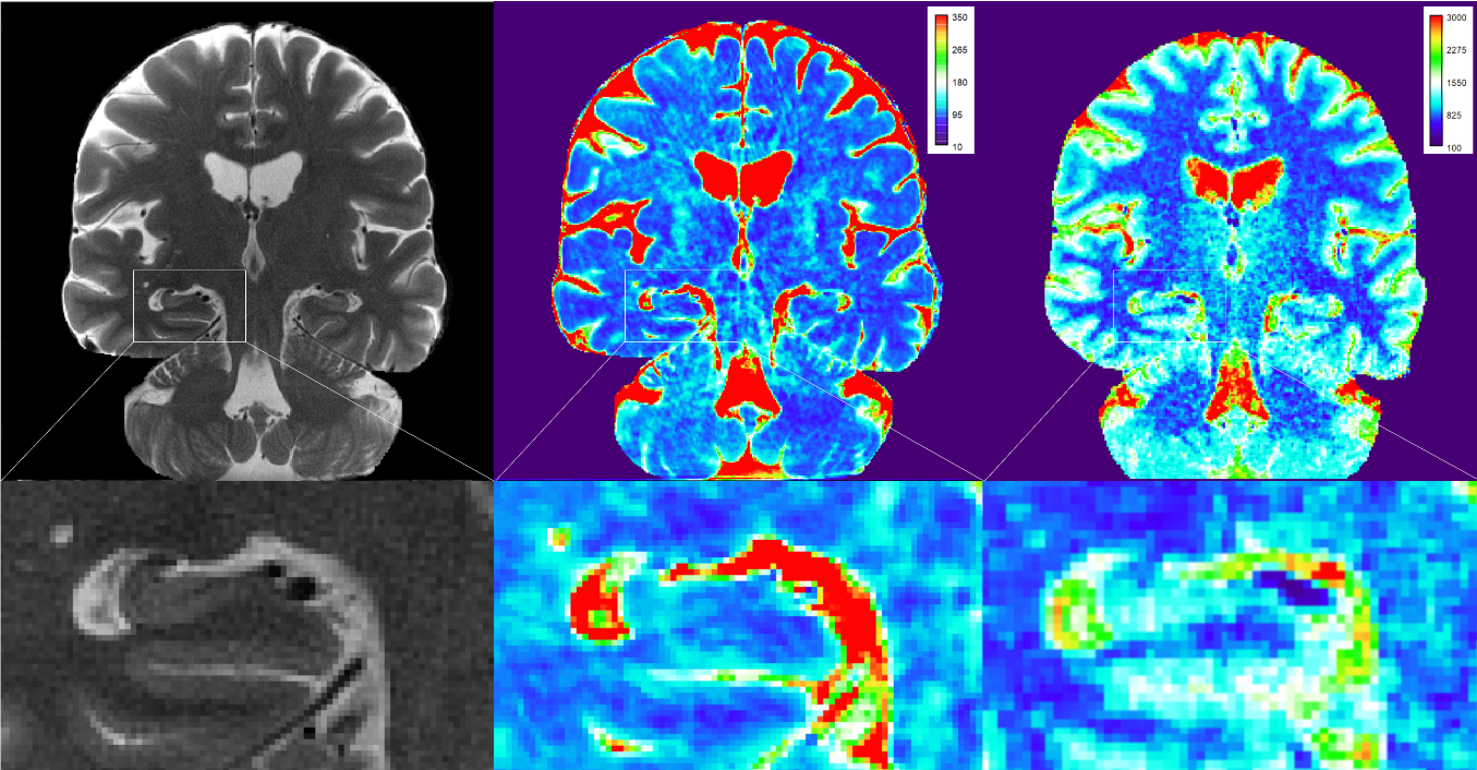
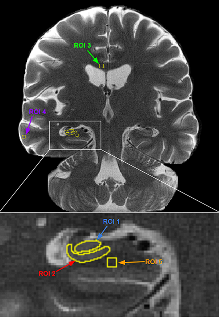
Radial Turbo Spin-Echo (RADTSE; TR=4000ms, TE=15ms, ETL=12, 648 radial views, 0.48x0.48x2 mm3, 35 slices, imaging time= 7:44 min)3,4 and Inversion-Recovery Radial Gradient Echo (IR-RADGRE; TR=8.75ms, TE=4.34ms, 960 radial views, 0.64x0.64x2 mm3, 33 slices, imaging time= 6:17 min)5 quantitative mapping acquisitions were carried out on a 3T MRI scanner (Skyra, Siemens Healthcare, Malvern, PA) with a 32-channel head coil. 16 healthy subjects (9 female, age range: 20-82, mean age: 46.9) were imaged. Eight of the subjects (3 female, age range: 20-82, mean age: 47.5) were imaged twice to assess reproducibility. An in-house iterative reconstruction algorithm6 was used to generate multiple TE and TI images from RADTSE and IR-RADGRE data, from which final quantitative T2 and T1 maps were produced, respectively (Figure 1).Five regions of interest (ROI) were analyzed: the cornu ammonis 4 (CA4) / dentate gyrus (DG), cornu ammonis 1 (CA1), corpus callosum, cortex, and white matter (WM) of the parahippocampal gyrus. ROIs were manually drawn on a single coronal slice of a reconstructed T2 contrast image as anatomical reference (Figure 2). ROIs were then applied to the quantitative T2 and T1 maps wherein mean T2 and T1 values were recorded. To assess reproducibility, mean T2 and T1 values were compared between imaging sessions for the eight subjects via simple linear regression, Pearson’s correlation, and Bland-Altman analyses. Additionally, mean T2 and T1 values from the first imaging session were assessed as a function of age via simple linear regression for all 16 subjects in these five ROI.
Results
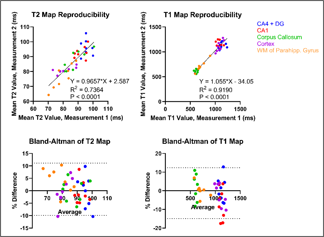
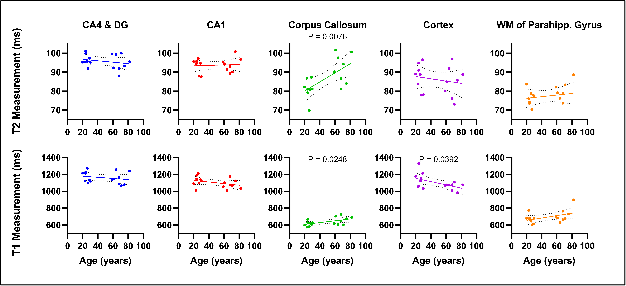
Assessment of reproducibility indicated a statistically significant correlation of T2 measurements between imaging sessions, r(38)=0.86 and P<0.0001, as well as a significant correlation of T1 measurements between sessions, r(38)=0.96 and P<0.0001 (Figure 3). The computed slopes from simple linear regression approximated unity. Bland-Altman analysis found mean and standard deviations of percent differences in T1 measurements to be -1.274 and 6.956, respectively, while those in T2 measurements were found to be 0.5941 and 5.362, respectively (Figure 3). When assessing T2 relaxation as a function of age, a significantly non-zero, positive relationship was observed in white matter, namely the corpus callosum (P=0.0076) (Figure 4). A significantly non-zero, positive relationship was also observed between T1 relaxation and age within the corpus callosum (P=0.0248), contrasting with a significant negative relationship within the cortex (P=0.0392) (Figure 4). In summary, our findings show a negative relationship between T2 and T1 relaxation and age in regions of interest within grey matter, while a positive relationship exists between T2 and T1 relaxation and age in regions of interest within white matter.Conclusion
RADTSE and IR-RADGRE pulse sequences yielded high-quality, high-resolution images at multiple contrasts in ~7 min per sequence. Assessment of reproducibility for these two sequences found concurrence in quantitative T2 and T1 measurements between acquisitions for these five ROI. Results also demonstrated a positive relationship with T2 and T1 relaxation times and age in white matter regions, and a negative relationship in grey matter regions. This is consistent with other reported age-dependent changes7,8. Future research will focus on the potential utility of slice super-resolution neural networks to further reduce scan times for these acquisitions, implementing automated hippocampal subfield segmentation for enhanced precision in region of interest analysis, and examining the relationship between relaxation times and hippocampal subfield volumes and cognitive scores.Acknowledgements
This study was supported by the Arizona Alzheimer’s Consortium, Arizona Biomedical Research Centre (CTR056039), and RO1 AG070987.References
[1] Anand, K. S., & Dhikav, V. (2012). Hippocampus in health and disease: An overview. Annals of Indian Academy of Neurology, 15(4), 239–246. https://doi.org/10.4103/0972-2327.104323[2] ] Fox, N. C., Warrington, E. K., Freeborough, P. A., Hartikainen, P., Kennedy, A. M., Stevens, J. M., & Rossor, M. N. (1996). Presymptomatic hippocampal atrophy in Alzheimer's disease. A longitudinal MRI study. Brain : a journal of neurology, 119 ( Pt 6), 2001–2007. https://doi.org/10.1093/brain/119.6.2001
[3] Huang, C., Bilgin, A., Barr, T. and Altbach, M.I. (2013), T2 relaxometry with indirect echo compensation from highly undersampled data. Magn. Reson. Med., 70: 1026-1037. https://doi.org/10.1002/mrm.24540
[4] Altbach, M.I., Bilgin, A., Li, Z., Clarkson, E.W., Trouard, T.P. and Gmitro, A.F. (2005), Processing of radial fast spin-echo data for obtaining T2 estimates from a single k-space data set. Magn. Reson. Med., 54: 549-559. https://doi.org/10.1002/mrm.20611
[5] Li, Z., Bilgin, A., Johnson, K., Galons, J.-P., Vedantham, S., Martin, D.R. and Altbach, M.I. (2019), Rapid high-resolution T1 mapping using a highly accelerated radial steady-state free-precession technique. J. Magn. Reson. Imaging, 49: 239-252. https://doi.org/10.1002/jmri.26170
[6] Mandava, S., Keerthivasan, M. B., Martin, D. R., Altbach, M. I., & Bilgin, A. (2021). Improving subspace constrained radial fast spin echo MRI using block matching driven non-local low rank regularization. Physics in medicine and biology, 66(4), 04NT03. https://doi.org/10.1088/1361-6560/abd4b8
[7]Hagiwara, A., Fujimoto, K., Kamagata, K., Murata, S., Irie, R., Kaga, H., Someya, Y., Andica, C., Fujita, S., Kato, S., Fukunaga, I., Wada, A., Hori, M., Tamura, Y., Kawamori, R., Watada, H., & Aoki, S. (2021). Age-Related Changes in Relaxation Times, Proton Density, Myelin, and Tissue Volumes in Adult Brain Analyzed by 2-Dimensional Quantitative Synthetic Magnetic Resonance Imaging. Investigative radiology, 56(3), 163–172. https://doi.org/10.1097/RLI.0000000000000720
[8] Knight, M. J., McCann, B., Tsivos, D., Couthard, E., & Kauppinen, R. A. (2016). Quantitative T1 and T2 MRI signal characteristics in the human brain: different patterns of MR contrasts in normal ageing.
Magma (New York, Magma (New York, N.Y.), 29(6), 833–842. https://doi.org/10.1007/s10334-016-0573-0
Figures