4589
Accelerated Whole-Brain Mapping of Venous Cerebral Blood Volume Using Velocity-Selective Venous-Spin-Labeling With 3D GRASE Encoding1Electronic and electrical engineering, Kyungpook National University, Daegu, Korea, Republic of, 2Medical Device Development Center, Daegu–Gyeongbuk Medical Innovation Foundation (K-MEDI Hub), Daegu, Korea, Republic of
Synopsis
Keywords: Quantitative Imaging, Quantitative Imaging
Motivation: The VS venous-spin-labeling-prepared 3D TSE method has recently shown promise in whole-brain venous CBV mapping with relative immunity to field variations. Nevertheless, the method’s relatively long scan times limits its application to functional studies involving short-term stimuli. We aimed to develop a highly accelerated technique for CBVv mapping across the entire brain.
Goal(s): To reduce imaging time while mitigating inherent artifacts in GRASE imaging.
Approach: We employed GRASE encoding with variable refocusing flip angles while subsampling k-space data, followed by compressed sensing based image reconstruction.
Results: The proposed technique yields comparable CBVv values across the entire brain, with highly accelerated scan times.
Impact: We introduce a scan-time efficient CBVv mapping strategy by means of GRASE encoding with sparse k-space sampling. Upon further evaluation of these issues, the present GRASE-based CBVv mapping method is expected to find various applications in neuroimaging studies.
Introduction
Venous cerebral blood volume (CBVV) is one of important parameters for understanding the underlying mechanism of BOLD MRI signals1. Yet, imaging-based methods enabling voxel-wise quantification of CBVV are sparse2, due primarily to a very small portion of venous blood in tissues and difficulty in targeting the venous compartment exclusively. Recently, the velocity-selective venous-spin-labeling (VS-VSL)-prepared 3D turbo spin echo (TSE) method3 has shown promise with that regard, achieving whole-brain CBVV mapping based purely on endogenous contrast mechanism. Nevertheless, the method’s relatively long scan times (~3 minutes) limits its application to functional studies involving short-term stimuli. Thus, in this work we aimed to develop a highly accelerated technique for CBVV mapping across the entire brain.Methods
Sequence configuration: Figure 1 shows a timing diagram of the proposed pulse sequence, which consists of three consecutive modules: 1) magnetization preparations (Fig. 1a) leading to selective nulling of arterial blood and cerebrospinal fluid signals, 2) velocity-selective (VS) RF pulse train (Fig.1b) for exclusive labeling of venous blood spins, and 3) 3D gradient-and-spin-echo (GRASE) encoding (Fig. 1c). To accelerate data acquisitions relative to the parent method3, a particular focus in this work was given to the GRASE module. Specifically, variable flip angles (Fig. 2a), calculated based on a prescribed two-step signal evolution (Fig. 2b), were applied in the refocusing RF pulse train so as to achieve a long echo train length and thus short scan times. Phase-encoding views are sampled in a sparse elliptical ky-kz space, leading to further scan acceleration. Here, a segmented linear, center-out view-ordering scheme4 was employed (Figs. 2c, 2d) to minimize a loss of efficiency in venous blood spin labeling, while preventing image artifacts potentially resulting from inter-echo signal inconsistency.CBVV mapping: Control (VS gradients off) and tag (VS gradients on) data acquired with sparse and incoherent sampling in k-space were individually processed using the standard compressed sensing image reconstruction technique6. Based on the assumption that T2 values of brain tissues and venous blood are in the same range at 3 T field strength, CBVV quantification can be simplified to the following equation3:
$$ \frac{S_{control} - S_{tag}}{S_{control}} = CBV_V $$
Experiments and data analysis: Experiments were performed at a 3T scanner (Siemens Skyra) in four healthy subjects using VS-VSL-GRASE with the following imaging parameters: field-of-view: 220 x 220 x 120 mm3, reconstruction matrix size: 72 x 72 x 60, sagittal orientation, echo train length = 36, GRASE factor = 3, RF spacing = 5.2 ms, and gradient-echo spacing = 1.2 ms. To ascertain the method’s maximally achievable acceleration factor without loss of quantification accuracy, data were acquired with three different sampling ratios: full elliptical ky-kz sampling, 50 %, and 30 %, leading to scan times of 185, 123, and 74 seconds, respectively. Additional data were collected in each subject using high-resolution MP-RAGE for brain segmentation. Whole-brain VS-VSL images along with derived CBVV maps obtained with full elliptical k-space sampling were reformatted in sagittal, coronal, and axial orientations. Whole-brain CBVV maps for the three cases with different sampling ratios were also presented. Furthermore, regional averages of CBVV were computed in gray and white matter for all experiments performed.
Results
Figure 3 shows whole-brain 3D images in the three orthogonal planes in a study subject: VS-VSL control (Fig. 3a), tag (Fig. 3b), control/tag difference (Fig. 3c), and CBVV (Fig. 3d). The difference image highlighting large veins suggests that the venous blood spins are properly labeled as intended, while CBVV maps exhibit the expected contrast between gray and white matter regions. CBVV maps in Figure 4 obtained with different data subsampling ratios are visually comparable. Table 1 lists CBVV averaged across the entire gray and white matter voxels in the four study participants scanned at various sampling rates.Discussion and Conclusion
We introduce a scan-time efficient CBVV mapping strategy by means of GRASE encoding with sparse k-space sampling. Compared with the parent method based on TSE readout, the presented technique achieves scan accelerations up to approximately three-fold. While resulting CBVV maps depict physiologically known contrast within plausible ranges (group averages: gray matter ~ 2% and white matter ~ 1%), the values are somewhat slightly lower than those in previous reports3, 5. Thus, we are currently focused on probing the cause of this difference while optimizing the sequence and scan parameters to explore a possibility of further reduction of imaging time. Upon further evaluation of these issues, the present GRASE-based CBVV mapping method is expected to find various applications in neuroimaging studies.Acknowledgements
No acknowledgement found.References
1. Emma Biondetti, Junghun Cho, Hyunyeol Lee, Cerebral oxygen metabolism from MRI susceptibility, Neuroimage, Volume 276, https://doi.org/10.1016/j.neuroimage.2023.120189
2. Jun Hua, Peiying Liu, Tae Kim, Manus Donahue, Swati Rane, J Jean Chen, Qin Qin, Seong-Gi Kim, MRI techniques to measure arterial and venous cerebral blood volume, Neuroimage, Volume 187, Pages 17-31, https://doi.org/10.1016/j.neuroimage.2018.02.027
3. Hyunyeol Lee, Felix W. Wehrli, Venous cerebral blood volume mapping in the whole brain using venous-spin-labeled 3D turbo spin echo, Magn Reson Med, Volume 84, Issue 4, October 2020, Pages 1991-2003, https://doi.org/10.1002/mrm.28262
4. A. Cristobal‐Huerta, Poot DH, Vogel MW, Krestin GP, Hernandez‐ Tamames JA. Compressed Sensing 3D‐GRASE for faster High‐Resolution MRI. Magn Reson Med, Volume 82, Issue 3, September 2019,Pages 984-999. https://doi.org/10.1002/mrm.27789
5. Nicholas P Blockley, Valerie E M Griffeth, Michael A Germuska, Daniel P Bulte, Richard B Buxton, An analysis of the use of hyperoxia for measuring venous cerebral blood volume: comparison of the existing method with a new analysis approach, Neuroimage, Volume 72, 15 May 2013, Pages 33-40, https://doi.org/10.1016/j.neuroimage.2013.01.039
6. Lustig M, Donoho D, Pauly JM. Sparse MRI: The application of compressed sensing for rapid MR imaging. Magn Reson Med. 2007;58:1182–1195
Figures
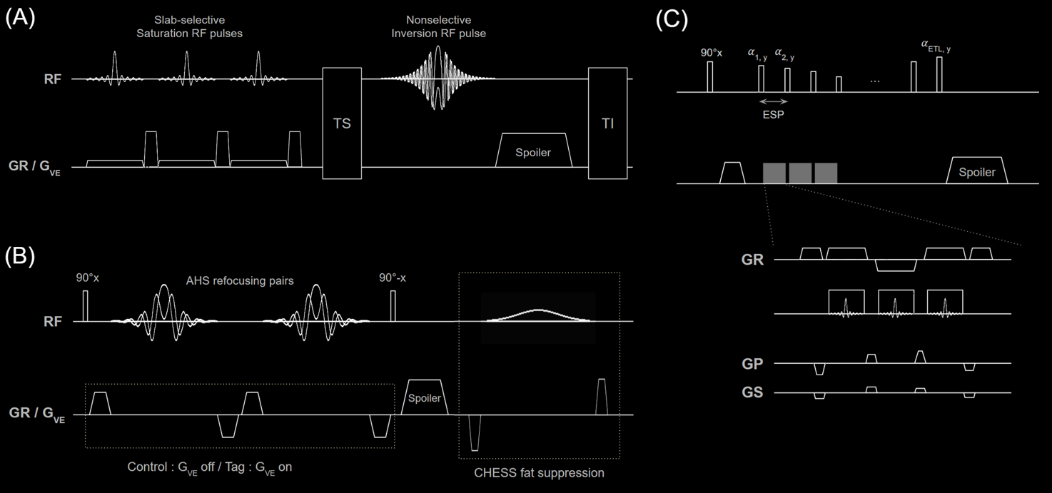
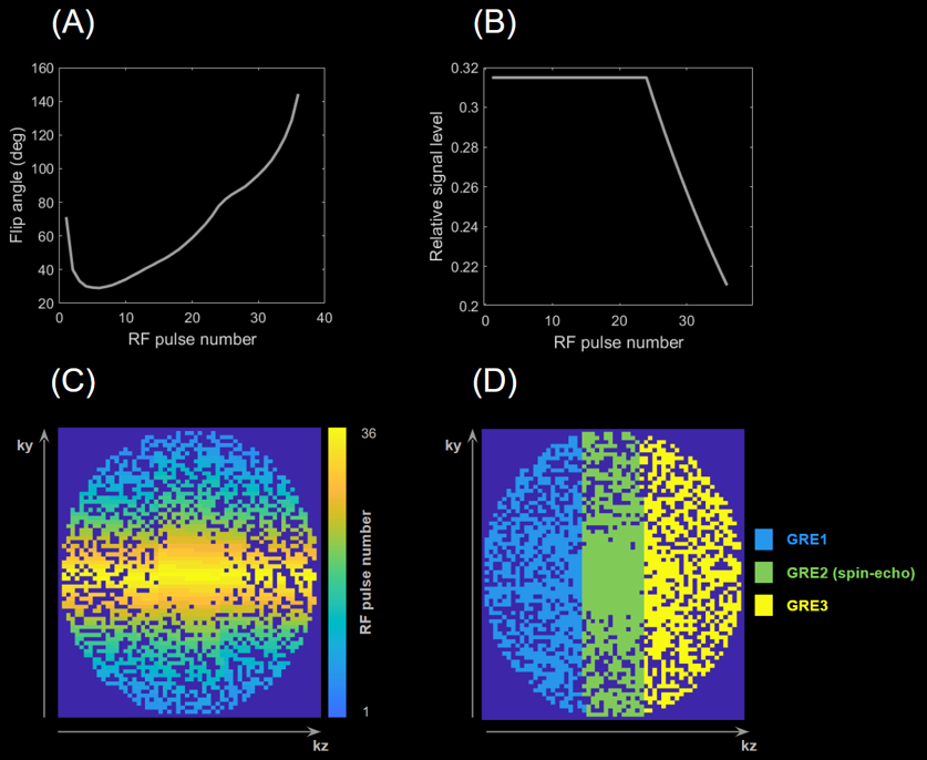
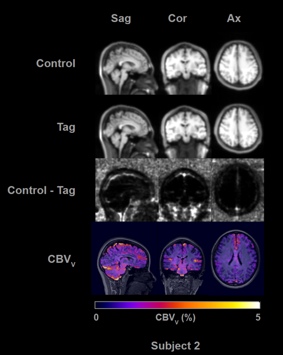
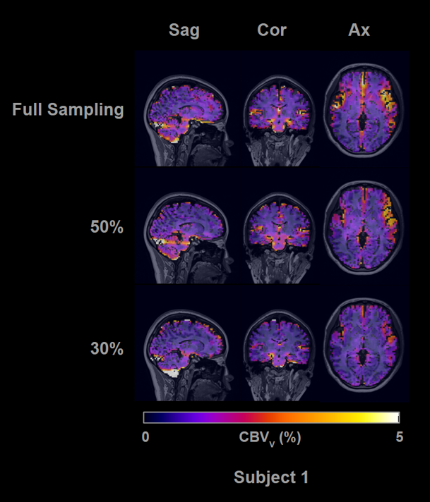
Figure 4. Whole-brain CBVV maps in three orthogonal planes, obtained with three different data sampling ratios.
